leishmania.pptx by Manoj Mahato Clinical Microbiologist
•Download as PPTX, PDF•
0 likes•5 views

Manoj Mahato

Recommended
More Related Content
Similar to leishmania.pptx by Manoj Mahato Clinical Microbiologist
Similar to leishmania.pptx by Manoj Mahato Clinical Microbiologist (20)
More from Manoj Mahato
More from Manoj Mahato (20)
Recently uploaded
Recently uploaded (20)
leishmania.pptx by Manoj Mahato Clinical Microbiologist
- 1. LEISHMANIA DONOVANI MR.MANOJ MEHTA -CLINICALMICROBIOLOGIST
- 2. The parasite was named by Ronald Ross in 1903 after the Scottish pathologist William Boog Leishman. In 1901, Leishman identified the organism in smears taken from the spleen of a patient who had died from ‘dum-dum fever’. This disease was similar to what Indian physicians called kala-azar (black fever). Initially, these organisms were considered to be trypanosomes. In 1903, Captain Charles Donovan described them as being new. These new microorganisms were given the name ‘Leishman-Donovan bodies’ After that taxonomically designated Leishmania donovani HISTORY
- 3. Most of the affected countries are in the tropics and subtropics. More than 90 percent of the world's cases of visceral leishmaniasis are in India, Bangladesh, Nepal, Sudan, and Brazil. Leishmaniasis is found in Asia (not Southeast Asia), Central America, South America (not in Uruguay, Chile, or Canada), Southern Europe (not common in travelers to southern Europe), The Middle East, and Africa (particularly East and North Africa GEOGRAPHICAL DISTRIBUTIONS
- 4. Ninety per cent of Visceral Leishmaniasis cases are found in Bangladesh, Brazil, India, Nepal and Sudan Currently, leishmaniasis occurs in 4 continents and is considered to be endemic in 88 countries, 72 of which are developing countries. Found on every continent except Australia and Antarctica. Annual incidence of disease= 600,000 cases per year. People infected worldwide=12 million. People at risk=350 million 90 % of cases with Cutaneous Leishmaniasis occur in Afghanistan, Algeria, Brazil, Iran, Peru, Saudi Arabia and Syria. EPIDEMIOLOGY
- 5. The major risk factor is being exposed to infected sand flies. Infection is more common in adventure travelers, Corps workers, soldiers WHO IS AT RISK ?
- 6. Kingdom: Protista Subkingdom: Sarcomastigophora Phylum: Protozoa Subphylum: Mastigophora Class: zoomastigophora Order: Kinetplastida Genus: Leishmania Species: donovani, tropica, Mexicana,braziliensis, etc CLASSIFICATION
- 7. Leishmaniasis is a vector borne disease caused by protozoan parasites of to the genus Leishmania and is transmitted by the bite of sand fly. This disease is also known as kala azar, black fever, sandfly disease, Dum-Dum fever. Kala-azar means dark pigmentation which is characteristic of cases of visceral leishmaniasis. It is caused by Leishmania donovani bodies and may be present either in endemic, epidemic or sporadic forms. It iswidely prevalent in India in epidemic form in states of Bihar, Assam and Bengal,Tarai of Nepal. Kala azar found in East and North Africa is a disease of young children and young adults, being more common in males as compared to females. KALA AZAR
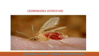
- 8. Mainly by the bite of Female Sand Fly (Phlebotomus argentipus) -Genus Phlebotomus(in old world) -Genus Lutzomyia(in new world) Blood transfusion Accidental inoculation from culture MODE OF TRAMSMISSION
- 9. The parasite exists in 2 forms;- 1. Amastigotes: a flagellar stage 2. Promastigotes: flagellar stage MORPHOLOGY
- 10. A flagellar stage Infective stage for sand fly. Occurs in the vertebrate host Habitat: Found in R.E. system of vertebrate host (Man, Dog & hamster). divides by binary fission at 37oC. Shape: Round /Oval Size: 2-4 mm, along longitudinal axis. Has following components: Cell membrane Nucleus relatively larger and situated centrally. Kinetoplast situated right angle to nucleus. AMASTIGOTE STAGE(LEISHMANIAL FORM)
- 11. Flagellar stage Infective stage for Man. Occurs in the Sandflies and Culture divides by binary fission at 27oC. They are spindle shaped ;15-20 μm in length & 1-2μm in width. Nucleus smaller and situated in the middle of the cell or along the side of cell-wall. Kinetoplast lies transversely near the anterior end. Free anterior flagellum Kinetoplast at the anterior end of the body. There is no undulating membrane. PROMASTIGOTE: (LEPTOMONAD STAGE)
- 12. TYPES OF LEISHMANIASIS Visceral leishmaniasis Kala-azar, Black fever Dum-Dum fever, Sahib’s disease Kala Dukh White leprosy Cutaneous leishmaniasis Aleppo boil, Baghdad boil, Delhi boil, Kandahar sore, Lahore sore, Oriental sore, Mucocutaneous Leishmaniasis Breda's disease bosch yaws, bush yaws forest yaws
- 13. Life Cycle Stages: Amastigote and Promastigote Host: Two Hosts oHuman(definitive): Amastigote forms found in man reticuloendothelial cells of spleen, bone marrow, Liver, intestinal mucosa,mesentric lymph node. Sand Fly(intermediate): Promastigote forms found in sand fly Vector: Sand Fly Reservoir Host: Dog(China&Brazil),Man(India),Rodents(Africa)andJackals(Russia). Infective Form: Promastigote Pathogenic Form: Amastigote Route of Infection: Penetration through skin by the bite of female sandflies. Site of Localization: Mononuclear phagocyte system/R.E.system. LIFE CYCLE OF LEISHMANIA DONOVANI
- 14. LIFE CYCLE OF LEISHMANIA DONOVANI
- 15. LIFE CYCLE OF LEISHMANIA DONOVANI
- 16. -Promastigote is the infective form which is introduced into human body by bite of sand fly in which the promastigote has been developed. -Some of the promastigotes are destroyed by body’s immune system. Some others take refuge inside the cells of Re-system ,where they are changed into amastigote form and undergo multiplication by binary fission. -When the cells become packed with parasites, they rupture releasing the parasites into circulation from where they are again engulfed by or invade cells of RE system. -The cycle is repeated and entire RE system is progressively infected. Some of the free parasites in circulation are engulfed by neutrophils and monocytes(Macrophage). -During blood meal, the vector(sand fly) draws free as well as those parasites inside the monocyte. LIFE CYCLE OF LEISHMANIA DONOVANI
- 17. -In sand fly, the amastigotes are changed into promastigote sand multiply enormously by binary fission in them id gut and forwards to the pharynx and buccal cavity. Between 6th to 9th day of infection, heavy pharyngeal infection is observed. This type of development is known as anterior station development. -When this sand fly bites a man, transmission is effected. LIFE CYCLE OF LEISHMANIA DONOVANI
- 18. Low grade fever continuous / intermittent Hepato-splenomegaly RE system affected Bone marrow hyperplasia Anemia , Leucopenia Hypergammaglobulinnemia Epistaxis , Proteinuria, Hematuria If left untreated ....Fatal 75-95% Complications:- pneumonia, TB, dysentery, uncontrolled haemorrhage Prognosis:- With an early treatment, cure rate >90% If not treated, death occurs within 2 years SIGN AND SYMPTOMS OF VISCERAL LEISHMANIASIS
- 19. Incubation Period : 3-6 months Inoculation of Promastigotes in skin Ingestion of Promastigote by Neutrophils Attraction of macrophages Ingestion of infected neutrophil, Amastigote formation in Macrophage. Invasion by Plasma cells and lymphocytes. Amastigotes liberated in circulation after rupture RE cells proliferate, heavily parasitized PATHOGENESIS OF LEISHMANIA DONOVANI
- 20. LABORATORY DIAGNOSIS OF LEISHMANIA DONOVANI
- 21. Microscopy: slit-skin smear, splenic aspirate, liver biopsy or bone marrow biopsy. - -Examination of Giemsa and Leishman stained slides of the relevant tissue is still the technique most commonly used to detect the parasite. Peripheral blood by thick film method. LABORATORY DIAGNOSIS OF LEISHMANIA DONOVANI Amastigotes in a macrophage
- 22. Culture: Novy Mac Neal, later modified by Nicolle (N.N.N.)medium -The aspirates can be cultured in NNN. In culture the amastigote stage converts to the promastigote stage. 2 parts of salt agar , 1 part defibrinated Rabbit blood, Ascorbic acid and Haematin Specimen inoculated in water of condensation 22°C – 24°C, This is not a rapid technique, as the parasites may take from 10 - 21 days to grow. Least sensitive, Long time LABORATORY DIAGNOSIS OF LEISHMANIA DONOVANI Promastigote Promastigote from culture in NNN medium
- 23. Serodiagnosis: VL produces large amounts of specific IgG which can be used for diagnosis. Currently the most used sero diagnostic tests Enzyme Linked Immunosorbent Assay (ELISA). Molecular techniques: PCR, such technique, however, are not readily available in general diagnostic laboratories LABORATORY DIAGNOSIS OF LEISHMANIA DONOVANI
- 24. Blood count : Neutropenia, Relative Lymphocytosis, Leucopenia,Anaemia (raised ESR) Serological Tests : Aldehyde ( Formal gel) Test (Napier): To detect gamma globulin Antimony test : Increase in globulin levels ( Not used now) Complement Fixation Test : UtilisesW.K.K. antigen Klinenstein and Kuhn:Early diagnosis ( Not used now ) LABORATORY DIAGNOSIS OF LEISHMANIA DONOVANI
- 25. rK39 Rapid dipstick test Based on the recombinant k39 protein, a 39-amino acid cloned in Escherichia coli, from the C terminus of the kinesin protein of Leishmania major in India LABORATORY DIAGNOSIS OF LEISHMANIA DONOVANI
- 26. LABORATORY DIAGNOSIS OF LEISHMANIA DONOVANI DIRECT AGGLUTINATION TEST (DAT) o Semi-quantitative test o Microtitre plates : increasing dilutions of patient's serum or blood is mixed with stained killed L. donovani promastigotes. o Agglutination visible after 18 hours o A titre greater than 1:3200 is positive
- 28. Education in the community about the causes and modes of transmission of leishmaniasis Reduction of sand fly population by insecticides mainly DDT,dieldrin, malathion Reduction of reservoir by killing all the infected dogs in the cases of zoonotic kala-azar Prevention of exposure to sand fly using insect repellent, bed nets and window mess as needed Wearing protective clothing There are no vaccines or drugs that prevent leishmaniasis PREVENTION AND CONTROL