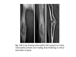
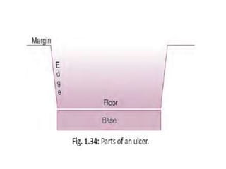
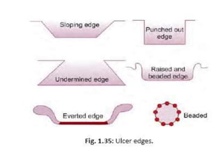
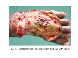
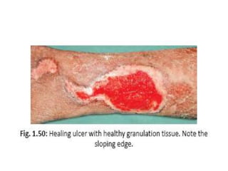
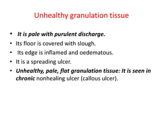
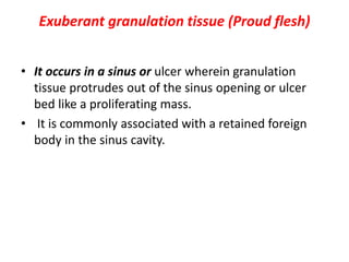
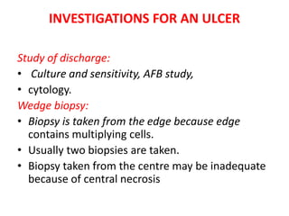
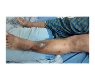
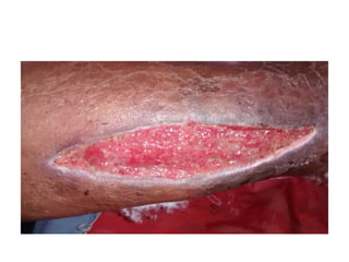
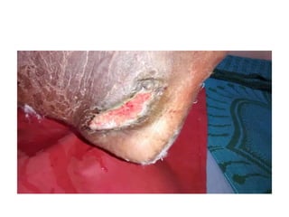
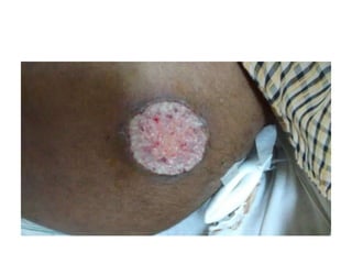
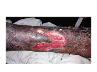
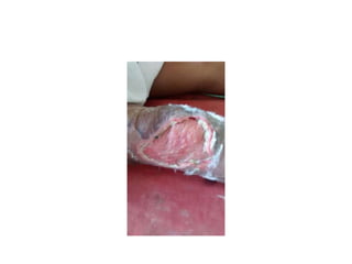
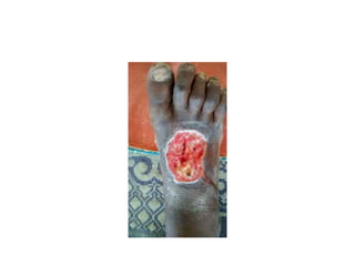
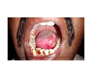
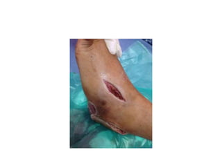
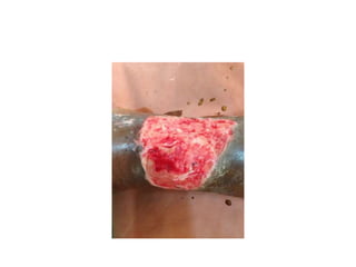
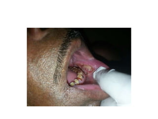
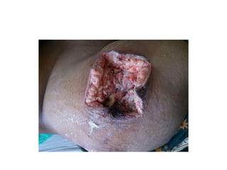
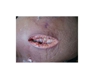
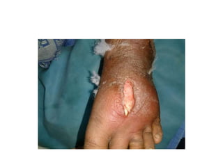
This document discusses ulcers, including their definition, parts, classifications, investigations, and management. It defines an ulcer as a break in the skin or mucous membrane epithelium due to cell death. The key parts of an ulcer are the margin, edge, floor, and base. Ulcers can be classified clinically based on healing status, duration, or etiology. Investigations may include wound cultures, biopsies, and imaging. Management involves treating the underlying cause, wound cleaning, debridement, dressings, and therapies like vacuum-assisted closure or maggot debridement to promote healing.
Examination of Swelling in a patient is always a task for MBBS students. This PPT provides the students, how to elicit a history & also the easy way to examine a swelling.
Colles fracture is the fracture at the distal end of radius, at its
cortico cancellous junction(about 2cm from the distal articular
surface).
It is not just the fracture of distal radius but the fracture
dislocation of the inferior radio-ulnar joint.
Explanation of what splenomegaly is in relation to its dimension deviation from normal spleen.Classification of splenomegaly according to it's size in adult and pediatric. The causes of splenomegaly along with the symptom that would manifest as a result of this anomaly. Lastly, diagnosis of splenomegaly
Anti ulcer drugs and their Advance pharmacology ||
Anti-ulcer drugs are medications used to prevent and treat ulcers in the stomach and upper part of the small intestine (duodenal ulcers). These ulcers are often caused by an imbalance between stomach acid and the mucosal lining, which protects the stomach lining.
||Scope: Overview of various classes of anti-ulcer drugs, their mechanisms of action, indications, side effects, and clinical considerations.
- Video recording of this lecture in English language: https://youtu.be/lK81BzxMqdo
- Video recording of this lecture in Arabic language: https://youtu.be/Ve4P0COk9OI
- Link to download the book free: https://nephrotube.blogspot.com/p/nephrotube-nephrology-books.html
- Link to NephroTube website: www.NephroTube.com
- Link to NephroTube social media accounts: https://nephrotube.blogspot.com/p/join-nephrotube-on-social-media.html
The prostate is an exocrine gland of the male mammalian reproductive system
It is a walnut-sized gland that forms part of the male reproductive system and is located in front of the rectum and just below the urinary bladder
Function is to store and secrete a clear, slightly alkaline fluid that constitutes 10-30% of the volume of the seminal fluid that along with the spermatozoa, constitutes semen
A healthy human prostate measures (4cm-vertical, by 3cm-horizontal, 2cm ant-post ).
It surrounds the urethra just below the urinary bladder. It has anterior, median, posterior and two lateral lobes
It’s work is regulated by androgens which are responsible for male sex characteristics
Generalised disease of the prostate due to hormonal derangement which leads to non malignant enlargement of the gland (increase in the number of epithelial cells and stromal tissue)to cause compression of the urethra leading to symptoms (LUTS
Flu Vaccine Alert in Bangalore Karnatakaaddon Scans
As flu season approaches, health officials in Bangalore, Karnataka, are urging residents to get their flu vaccinations. The seasonal flu, while common, can lead to severe health complications, particularly for vulnerable populations such as young children, the elderly, and those with underlying health conditions.
Dr. Vidisha Kumari, a leading epidemiologist in Bangalore, emphasizes the importance of getting vaccinated. "The flu vaccine is our best defense against the influenza virus. It not only protects individuals but also helps prevent the spread of the virus in our communities," he says.
This year, the flu season is expected to coincide with a potential increase in other respiratory illnesses. The Karnataka Health Department has launched an awareness campaign highlighting the significance of flu vaccinations. They have set up multiple vaccination centers across Bangalore, making it convenient for residents to receive their shots.
To encourage widespread vaccination, the government is also collaborating with local schools, workplaces, and community centers to facilitate vaccination drives. Special attention is being given to ensuring that the vaccine is accessible to all, including marginalized communities who may have limited access to healthcare.
Residents are reminded that the flu vaccine is safe and effective. Common side effects are mild and may include soreness at the injection site, mild fever, or muscle aches. These side effects are generally short-lived and far less severe than the flu itself.
Healthcare providers are also stressing the importance of continuing COVID-19 precautions. Wearing masks, practicing good hand hygiene, and maintaining social distancing are still crucial, especially in crowded places.
Protect yourself and your loved ones by getting vaccinated. Together, we can help keep Bangalore healthy and safe this flu season. For more information on vaccination centers and schedules, residents can visit the Karnataka Health Department’s official website or follow their social media pages.
Stay informed, stay safe, and get your flu shot today!
Ozempic: Preoperative Management of Patients on GLP-1 Receptor Agonists Saeid Safari
Preoperative Management of Patients on GLP-1 Receptor Agonists like Ozempic and Semiglutide
ASA GUIDELINE
NYSORA Guideline
2 Case Reports of Gastric Ultrasound
TEST BANK for Operations Management, 14th Edition by William J. Stevenson, Ve...kevinkariuki227
TEST BANK for Operations Management, 14th Edition by William J. Stevenson, Verified Chapters 1 - 19, Complete Newest Version.pdf
HOT NEW PRODUCT! BIG SALES FAST SHIPPING NOW FROM CHINA!! EU KU DB BK substit...GL Anaacs
Contact us if you are interested:
Email / Skype : kefaya1771@gmail.com
Threema: PXHY5PDH
New BATCH Ku !!! MUCH IN DEMAND FAST SALE EVERY BATCH HAPPY GOOD EFFECT BIG BATCH !
Contact me on Threema or skype to start big business!!
Hot-sale products:
NEW HOT EUTYLONE WHITE CRYSTAL!!
5cl-adba precursor (semi finished )
5cl-adba raw materials
ADBB precursor (semi finished )
ADBB raw materials
APVP powder
5fadb/4f-adb
Jwh018 / Jwh210
Eutylone crystal
Protonitazene (hydrochloride) CAS: 119276-01-6
Flubrotizolam CAS: 57801-95-3
Metonitazene CAS: 14680-51-4
Payment terms: Western Union,MoneyGram,Bitcoin or USDT.
Deliver Time: Usually 7-15days
Shipping method: FedEx, TNT, DHL,UPS etc.Our deliveries are 100% safe, fast, reliable and discreet.
Samples will be sent for your evaluation!If you are interested in, please contact me, let's talk details.
We specializes in exporting high quality Research chemical, medical intermediate, Pharmaceutical chemicals and so on. Products are exported to USA, Canada, France, Korea, Japan,Russia, Southeast Asia and other countries.
Prix Galien International 2024 Forum ProgramLevi Shapiro
June 20, 2024, Prix Galien International and Jerusalem Ethics Forum in ROME. Detailed agenda including panels:
- ADVANCES IN CARDIOLOGY: A NEW PARADIGM IS COMING
- WOMEN’S HEALTH: FERTILITY PRESERVATION
- WHAT’S NEW IN THE TREATMENT OF INFECTIOUS,
ONCOLOGICAL AND INFLAMMATORY SKIN DISEASES?
- ARTIFICIAL INTELLIGENCE AND ETHICS
- GENE THERAPY
- BEYOND BORDERS: GLOBAL INITIATIVES FOR DEMOCRATIZING LIFE SCIENCE TECHNOLOGIES AND PROMOTING ACCESS TO HEALTHCARE
- ETHICAL CHALLENGES IN LIFE SCIENCES
- Prix Galien International Awards Ceremony
micro teaching on communication m.sc nursing.pdfAnurag Sharma
Microteaching is a unique model of practice teaching. It is a viable instrument for the. desired change in the teaching behavior or the behavior potential which, in specified types of real. classroom situations, tends to facilitate the achievement of specified types of objectives.
2. Definition
• An ulcer is a break in the continuity of the
covering epithelium, either skin or mucous
membrane due to molecular death.
Parts of an Ulcer
a. Margin: It may be regular or irregular. It may
be rounded or oval.
b. Edge: Edge is the one which connects floor of
the ulcer to the margin.
3. Different edges are:
• Sloping edge. It is seen in a healing ulcer.
• Its inner part is red because of red, healthy
granulation tissue.
• Its outer part is white due to scar/fibrous
tissue.
• Its middle part is blue due to epithelial
proliferation.
4. • Undermined edge is seen in a tuberculous ulcer
• Punched out edge is seen in a gummatous (syphilitic)
ulcer and trophic ulcer.
It is due to endarteritis.
• Raised and beaded edge (pearly white) is seen in a
rodent ulcer (BCC).
Beads are due to proliferating active cells.
• Everted edge (rolled out edge): It is seen in a
carcinomatous ulcer due to spill of the proliferating
malignant tissues over the normal skin.
5. c. Floor:
• It is the one which is seen. Floor may contain
discharge, granulation tissue or slough.
d. Base:
Base is the one on which ulcer rests. It may be bone or
soft tissue.
6.
7.
8. Induration of an Ulcer
• Induration is a clinical palpatory sign which means a
specific type of hardness in the diseased tissue.
• It is obvious in well-differentiated carcinomas.
• It is better felt in squamous cell carcinoma.
• It is also observed in long standing ulcer with
underlying fibrosis
• Brawny induration is a feature of an abscess.
9. • Induration is felt at edge, base and surrounding area
of an ulcer.
• Induration at surrounding area signifies the extent of
disease (tumour).
• Outermost part of the indurated area is taken as the
point from where clearance of wide excision is
planned.
10. Classifications
Classification I (Clinical)
1. Spreading ulcer:
• Here edge is inflamed, irregular and oedematous.
• It is an acute painful ulcer; floor does not contain
healthy granulation tissue (or granulation tissue is
absent) but with profuse purulent discharge and
slough; surrounding area is red and edematous.
11.
12. 2. Healing ulcer
• Edge is sloping with healthy pink/red healthy
granulation tissue with scanty/minimal serous
discharge in the floor;
• slough is absent;
• regional lymph nodes may or may not be enlarged
but when enlarged always non-tender.
• Surrounding area does not show any signs of
inflammation or induration; base is not indurated.
13. Three zones are observed in healing ulcer.
• Innermost red zone of healthy granulation
tissue;
• middle bluish zone of growing epithelium;
• outer whitish zone of fibrosis and scar
formation.
14.
15. 3. Non-healing ulcer
• It may be a chronic ulcer depending on the cause of
the ulcer; here edge will be depending on the
cause—punched out (trophic), undermined
(tuberculous), rolled out (carcinomatous ulcer),
beaded (rodent ulcer);
• floor contains unhealthy granulation tissue and
slough, and serosanguineous/purulent/bloody
discharge;
• Regional draining lymph nodes may be enlarged but
non-tender.
16.
17. 4. Callous (stationary) ulcer
• It is also a chronic non-healing ulcer; floor contains
pale unhealthy, flabby, whitish yellow granulation
tissue and thin scanty serous discharge or often with
copious serosanguinous discharge, with indurated
nontender edge; base is indurated, nontender and
often fixed.
• Ulcer does not show any tendency to heal.
• It lasts for many months to years.
• Tissue destruction is more with absence of or only
minimal regeneration.
18. • Induration and pigmentation may be seen in the
surrounding area.
• There is no/less discharge.
• Regional lymph nodes may be enlarged; are firm/
hard and nontender.
• It is callousness towards healing; wordcallous
means—insensitive and cruel; and also it means—
hard skinned.
19.
20. Classification II (Based on Duration)
1. Acute ulcer
duration is less than 2 weeks.
2. Chronic ulcer
duration is more than 2 weeks (long).
21. Classification III (Pathological)
1. Specific ulcers:
• Tuberculous ulcer.
• Syphilitic ulcer: It is punched out, deep, with “wash-
leather” slough in the floor and with indurated base.
• Actinomycosis.
• Meleney’s ulcer.
22. 2. Malignant ulcers:
• Carcinomatous ulcer
• Rodent ulcer.
• Melanotic ulcer.
3. Non-specific ulcers:
• Traumatic ulcer: It may be mechanical, physical,
chemical— common.
• Arterial ulcer: Atherosclerosis, TAO
23. • Venous ulcer: Gravitational ulcer, post-phlebitic ulcer.
• Trophic ulcer/Pressure sore.
• Infective ulcers: Pyogenic ulcer.
• Tropical ulcers: It occurs in tropical countries. It is
callous type of ulcer, e.g. Vincent’s ulcer.
• Ulcers due to chilblains and frostbite (cryopathic
ulcer).
• Martorell’s hypertensive ulcer.
24. • Bazin’s ulcer.
• Diabetic ulcer.
• Ulcers due to leucaemia, polycythemia, jaundice,
collagen diseases, lymphoedema.
• Cortisol ulcers are due to long-time application of
cortisol (steroid) creams to certain skin diseases.
• These ulcers are callous ulcers last for long time and
require excision and skin grafting.
25. Wagner’s Grading/Classification of Ulcer
• Grade 0 – Preulcerative lesion/healed ulcer
• Grade 1 – Superficial ulcer
• Grade 2 – Ulcer deeper to subcutaneous tissue
exposing soft tissues or bone
• Grade 3 – Abscess formation
underneath/osteomyelitis
• Grade 4 – Gangrene of part of the tissues/limb/foot
• Grade 5 – Gangrene of entire one area/foot
26.
27. GRANULATION TISSUE
• It is proliferation of new capillaries and
fibroblasts intermingled with red blood cells
and white blood cells with thin fibrin cover
over it.
28.
29. Unhealthy granulation tissue
• It is pale with purulent discharge.
• Its floor is covered with slough.
• Its edge is inflamed and oedematous.
• It is a spreading ulcer.
• Unhealthy, pale, flat granulation tissue: It is seen in
chronic nonhealing ulcer (callous ulcer).
30. Exuberant granulation tissue (Proud flesh)
• It occurs in a sinus or ulcer wherein granulation
tissue protrudes out of the sinus opening or ulcer
bed like a proliferating mass.
• It is commonly associated with a retained foreign
body in the sinus cavity.
31.
32. INVESTIGATIONS FOR AN ULCER
Study of discharge:
• Culture and sensitivity, AFB study,
• cytology.
Wedge biopsy:
• Biopsy is taken from the edge because edge
contains multiplying cells.
• Usually two biopsies are taken.
• Biopsy taken from the centre may be inadequate
because of central necrosis
33. • X-ray of the part to look for
periostitis/osteomyelitis.
• FNAC of the lymph node.
• Chest X-ray, Mantoux test in suspected case of
tuberculous ulcer.
• Haemoglobin, ESR, total WBC count, serum
protein estimation (albumin).
34.
35. MANAGEMENT OF AN ULCER
• Cause should be found and treated.
• Correction of the anaemia, deficiencies like of
protein and vitamins.
• Proper investigation as needed.
• Transfusion of the blood if required.
• Control the pain and infection.
• Rest, immobilization, elevation, avoidance of
repeated trauma.
36. • Care of the ulcer by debridement, ulcer cleaning and
dressing.
• Desloughing is done either mechanically or
chemically. Mechanically it is done using scissor by
excising the slough.
• Hydrogen peroxide which releases nascent oxygen is
used as chemical agent.
• Acriflavine is antiseptic and irritant and so desloughs
the area and promotes granulation tissue formation.
37. • Eusol (Edinburgh University Solution) which contains
sodium hypochlorite releases nascent chlorine which
forms a water soluble complex with slough to
dissolve it.
• Use of povidone iodine in ulcer cleaning is
controversial (open wound is not suitable; it is mainly
for cleaning the surgical field prior to incision).
38. • Maggots if present in the wound will cause crawling
sensation and are removed using turpentine
solution.
• Removal of the exuberant granulation tissue is also
required when present.
• Ulcer cleaning and dressing is done daily or twice
daily or once in 2–3 days depending on the type of
ulcer and type of dressing used.
39. • Normal saline is ideal for ulcer cleaning.
• Various dressings are available.
• Films (opsite/semipermeable polyurethane),
hydrocolloids (duoderm), hydrogels (polyethylene
oxide with water), hydroactives (nonpectin-based
polyurethane matrix), foams.
40.
41.
42.
43. EUSOL bath.
• Dilute EUSOL solution in a basin is used wherein
ulcer foot is dipped and kept in place for 20–30
minutes.
• EUSOL removes the slough and cleans the ulcer bed.
• Hydrogen peroxide releases nascent oxygen and
helps in removing necrotic material.
• Povidone iodine is not used for open wound; it is only
a surface antiseptic
44. Vacuum assisted closure (VAC) therapy
• It is by creation of negative pressure (25–200 mmHg),
continuous or intermittent over the wound surface; it
causes reduced fluid in the interstitial space, reduces
oedema, increases the cell proliferation and protein
matrix synthesis, promotes formation of healthy
granulation tissue.
• Sterile foam is placed over the ulcer bed covering
widely; tube drain with multiple holes is kept within it
and end of the tube comes out significantly away; foam
is sealed airtight using a sterile adhesive film.
45. • Tube is connected to suction system.
• Suction is maintained initially continuously
later intermittently.
• Redressing is done only after 4–7 days.
• Therapy using infrared/short wave/ultraviolet
rays to decrease the ulcer size is often used
but their benefits are not proved.
46.
47. Maggot debridement therapy
• It is used as biotherapy (but not commonly) by
placing cultured live disinfected maggots.
• Maggots are larvae of the green bottle fly, also
known as the green blowfly (Lucilia sericata).
• They act by dissolving and engulfing dead necrotic
tissues; they may reduce the bacterial content in the
wound.
• They can inhibit many bacteria including MRSA
(methicillin resistant bacteria), anaerobic and aerobic
bacteria.
48. • They secrete proteolytic enzymes to have mechanical
effects; secretion of ammonia alters the pH in the
ulcer bed which inhibits bacterial growth.
• They increase the granulation tissue formation also.
• Once ulcer granulates, defect is closed with
secondary suturing, skin graft or flaps
49. TRAUMATIC ULCER
• Such ulcer occurs after trauma. It may be
mechanical—dental ulcer along the margin of the
tongue due to tooth injury; physical like by electrical
burn; chemical like by alkali injury.
• Such ulcer is acute, superficial, painful and tender.
• Secondary infection or poor blood supply of the area
make it chronic and deep.
50. • Footballer’s ulcer is a traumatic ulcer occurring over
the shin of males due to direct knocks on the shin. It
is staphylococcal infection with a chronic and deep
ulcer.
• Traumatic ulcers can occur anywhere in the body due
to trauma
51.
52. • Trauma causes infection, necrosis, fasciitis, crush
injury, endarteritis of the skin leading into formation
of large/deep nonhealing ulcer.
• Treatment depends on size and extent of ulcer.
• Regular dressing,
• later skin grafting .
53. TROPHIC ULCER (PRESSURE SORE/DECUBITUS
ULCER)
• Pressure sore is tissue necrosis and ulceration due to
prolonged pressure.
• Blood flow to the skin stops once external pressure
becomes more than 30 mmHg (more than capillary
occlusive pressure) and this causes tissue hypoxia,
necrosis and ulceration.
• It is more prominent between bony prominence and
an external surface.
54.
55.
56. It is due to:
• Impaired nutrition.
• Defective blood supply.
• Neurological deficit.
57. Sites
• Over the ischial tuberosity.
• Sacrum.
• In the heel.
• In relation to heads of metatarsals.
• Buttocks.
• Over the shoulder.
• Occiput.
58. • Due to the presence of neurological deficit, trophic
ulcer is also called as neurogenic ulcer/neuropathic
ulcer.
• Initially it begins as callosity due to repeated trauma
and pressure, under which suppuration occurs and
gives way through a central hole which extends down
into the deeper plane up to the underlying bone as
perforating ulcer (penetrating ulcer).
• Bedsores are trophic ulcers.
59.
60.
61. Clinical Features
• Occurs in 5% of all hospitalised patients.
• Painless ulcer which is punched out.
• Ulcer is non-mobile with base formed by bone.
62. Investigations
• Study of discharge, blood sugar, biopsy from the
edge, X-ray of the part, X-ray spine
63. Treatment
• Cause should be treated.
• Nutritional supplementation.
• Rest, antibiotics, slough excision, regular dressings.
• Vacuum-assisted closure (VAC): It is the creation of
intermittent negative pressure of minus 125 mmHg
to promote formation of healthy granulation tissue.
64. • Negative pressure reduces tissue oedema, clears the
interstitial fluid and improves the perfusion,
increases the cell proliferation and so promotes the
healing.
• A perforated drain is kept over the foam dressing
covered over the pressure sore.
• It is sealed with a transparent adhesive sheet.
65. • Drain is connected to required vacuum apparatus.
• Once ulcer granulates well, flap cover or skin grafting
is done.
• Excision of the ulcer and skin grafting.
• Flaps—local rotation or other flaps (transposition
flaps).
• Cultured muscle interposition.
66. • Proper care: Change in position once in 2 hours;
lifting the limb upwards for 10 seconds once in 10
minutes; nutrition; use of water bed/air bed/air-fluid
floatation bed and pressure dispersion cushions to
the affected area; urinary and faecal care; hygiene;
psychological counselling.
• Regular skin observation; keeping skin clean and dry
(using regular use of talcum powder); oil massaging
of the skin and soft tissues using clean, absorbent
porous clothing; control and prevention of sepsis
helps in the management.
67. ULCER DUE TO FROSTBITE
• It is due to exposure of a part to wet cold
below the freezing point (cold wind).
• There is arteriolar spasm, denaturation of
proteins and cell destruction.
• It leads to gangrene of the part.
• Ulcers here are always deep.