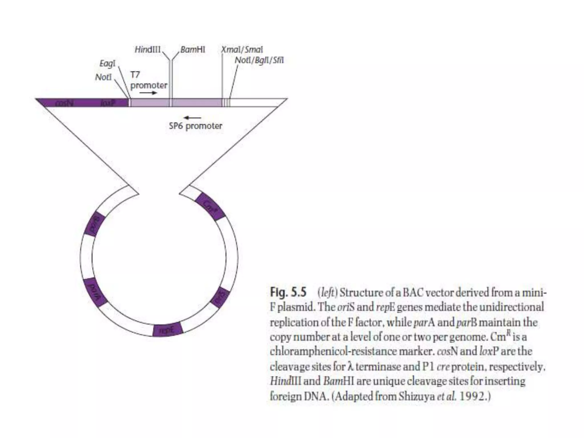
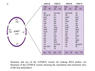
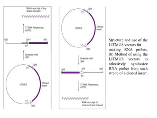
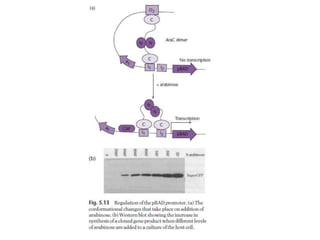
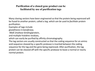
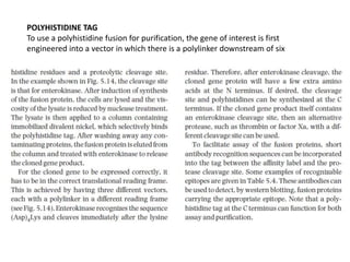
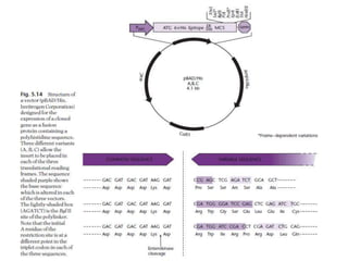
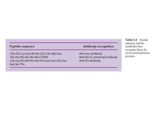
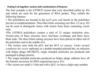
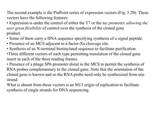
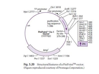
•The strength of a promoter, i.e. how many RNA copies are synthesized per unit time per
enzyme molecule, depends on how close its sequence is to the consensus.
•The distance between the −35 and −10 regions is also important. In all cases examined,
the promoter was weaker when the spacing was increased or decreased from 17 bp.
Bacterial DNA has two types of transcription termination site: factor-
independent and factor dependent.
The factor independent transcription terminators are easy to recognize
because they have similar sequences: an inverted repeat followed by a
string of A residues.
The factor-dependent transcription terminators have very little sequence
in common with each other. Rather, termination involves interaction with
one of the three known E. coli termination factors, Rho (ρ), Tau (τ), and
NusA.
Although single-stranded DNA can be used as a sequence probe in hybridization
experiments, RNA probes are preferred. The reasons for this are that The rate of
hybridization and the stability are far greater for RNA–DNA hybrids compared with
DNA–DNA hybrids. To make an RNA probe, the relevant gene sequence is cloned in a
plasmid vector such that it is under the control of a phage promoter. After purification, the
plasmid is linearized with a suitable restriction enzyme and then incubated with the phage
RNA polymerase and the four ribonucleoside triphosphates (Fig. 5.9). No transcription
terminator is required because the RNA polymerase will fall off the end of the linearized
plasmid.
Specialist vectors have been developed that facilitate the production of RNA
probes and interfering RNA
There are three reasons for using a phage promoter.
First, such promoters are very strong, enabling large amounts of RNA to be made in vitro.
Secondly, the phage promoter is not recognized by the E. coli RNA
polymerase and so no transcription will occur inside the cell. This minimizes any
selection of variant inserts.
Thirdly, the RNA polymerases encoded by phages such as SP6, T7, and T3 are much
simpler molecules to handle than the E. coli enzyme, since the active enzyme is a single
polypeptide.
Structure and use of the LITMUS vectors for making RNA probes. (a)
Structure of the LITMUS vectors showing the orientation and restriction sites
of the four polylinkers.
Structure and use of the
LITMUS vectors for
making RNA probes.
(b) Method of using the
LITMUS vectors to
selectively synthesize
RNA probes from each
strand of a cloned insert.
Vectors with strong, controllable promoters
are used to maximize synthesis of cloned
gene products
When maximizing gene expression it is not enough to select the strongest
promoter possible: the effects of overexpression on the host cell also need to
be considered. Many gene products can be toxic to the host cell even when
synthesized in small amounts.
when overexpression of a protein is not toxic to the host cell, high-level
synthesis exerts a metabolic drain on the cell. This leads to slower.
To minimize the problems associated with high-level expression, it is usual to
use a vector in which the cloned gene is under the control of a regulated
promoter.
Many different vectors have been constructed for regulated expression of gene
inserts but most of those in current use contain one of the following controllable
promoters: λ PL, T7, trc (tac), or BAD. Table 5.3 shows the different levels of
expression that can be achieved when the gene for chloramphenicol
transacetylase (CAT) is placed under the control of three of these promoters. The
trc and tac promoters are hybrid promoters derived from the lac and trp
promoters (Brosius 1984). They are stronger than either of the two parental
promoters because their sequences are more like the consensus sequence. Like
lac, the trc and tac promoters are inducibile by lactose and isopropyl-β-d-
thiogalactoside (IPTG). Vectors using these promoters also carry the lacO
operator and the lacI gene, which encodes the repressor.
The λ PL promoter system combines very tight transcriptional control with
high levels of gene expression. This is achieved by putting the cloned gene
under the control of the PL promoter carried on a vector, while the PL
promoter is controlled by a cI repressor gene in the E. coli host. This cI gene
is itself under the control of the tryptophan (trp) promoter (Fig. 5.12). In the
absence of exogenous tryptophan, the cI gene is transcribed and the cI
repressor binds to the PL promoter, preventing expression of the cloned gene.
Upon addition of tryptophan, the trp repressor binds to the cI gene,
preventing synthesis of the cI repressor. In the absence of cI repressor, there
is a high level of expression from the very strong PL promoter.
The pBAD vectors, like the ones based on PL promoter, offer extremely tight
control of expression of cloned genes (Guzman et al. 1995). The pBAD vectors
carry the promoter of the araBAD (arabinose) operon and the gene encoding the
positive and negative regulator of this promoter, araC. AraC is a transcriptional
regulator that forms a complex with l-arabinose. In the absence of arabinose, AraC
binds to the O2 and I1 half-sites of the araBAD operon, forming a 210 bp DNA loop
and thereby blocking transcription (Fig. 5.13). As arabinose is added to the growth
medium, it binds to AraC, thereby releasing the O2 site. This in turn causes AraC to
bind to the I2 site adjacent to the I1 site. This releases the DNA loop and allows
transcription to begin. Binding of AraC to I1 and I2 is activated in the presence of
cAMP activator protein (CAP) plus cyclic adenosine monophosphate (cAMP). If
glucose is added to the growth medium, this will lead to a repression of cAMP
synthesis, thereby decreasing expression from the araBAD promoter. Thus one can
titrate the level of cloned gene product by varying the glucose and arabinose
content of the growth medium (Fig. 5.13). According to Guzman et al. (1995), the
pBAD vectors permit fine-tuning of gene expression. All that is required is to
change the sugar composition of the medium.
Purification of a cloned gene product can be
facilitated by use of purification tags
Many cloning vectors have been engineered so that the protein being expressed will
be fused to another protein, called a tag, which can be used to facilitate protein
purification.
Examples of tags include
glutathione-S-transferase,
MalE (maltose-binding)protein,
and multiple histidine residues,
which can easily be purified by affinity chromatography.
The tag vectors are usually constructed so that the coding sequence for an amino
acid sequence cleaved by a specific protease is inserted between the coding
sequence for the tag and the gene being expressed. After purification, the tag
protein can be cleaved off with the specific protease to leave a normal or nearly
normal protein.
POLYHISTIDINE TAG
To use a polyhistidine fusion for purification, the gene of interest is first
engineered into a vector in which there is a polylinker downstream of six
BIOTIN TAG
Biotin is an essential cofactor for a number of carboxylases important in cell
metabolism. The biotin in these enzyme complexes is covalently attached at a specific
lysine residue of the biotin carboxylase carrier protein. Fusions made to a segment of
the carrier protein are recognized in E. coli by biotin ligase, the product of the birA gene,
and biotin is covalently attached in an ATP-dependent reaction. The expressed fusion
protein can be purified using streptavidin affinity chromatography (Fig. 5.15). E. coli
expresses a single endogenous biotinylated protein, but it does not bind to streptavidin
in its native configuration, making the affinity purification highly specific for the
recombinant fusion protein. The presence of biotin on the fusion protein has an
additional advantage: its presence can be detected with enzymes coupled to
streptavidin.
The affinity purification systems described above suffer from the disadvantage that a
protease is required to separate the target protein from the affinity tag.
Also, the protease has to be separated from the protein of interest. Chong et al. (1997,
1998) have described a unique purification system that has
neither of these disadvantages. The system utilizes a protein splicing element, an intein,
from the Saccharomyces cerevisiae VMA1 gene (see Box 5.2).
The intein is modified such that it undergoes a self-cleavage reaction at its N terminus at
low temperatures in the presence of thiols, such as cysteine,
dithiothreitol, or β-mercaptoethanol. The gene encoding the target protein is inserted
into a multiple cloning site (MCS) of a vector to create a fusion
between the C terminus of the target gene and the
N terminus of the gene encoding the intein. DNA
encoding a small (5 kDa) chitin-binding domain
from Bacillus circulans was added to the C terminus of
the intein for affinity purification (Fig. 5.16).
The above construct is placed under the control of
an IPTG-inducible T7 promoter. When crude extracts
from induced cells are passed through a chitin column,
the fusion protein binds and all contaminating
proteins are washed through. The fusion is then induced to undergo intein-mediated self-
cleavage on the column by incubation with a thiol. This releases
the target protein, while the intein chitin-binding
domain remains bound to the column.
One of the problems associated with the overproduction of proteins in E. coli
is the sequestration of the product into insoluble aggregates or “inclusion
bodies” (Fig. 5.17).
Two parameters that can be manipulated to reduce inclusion-body formation
are temperature and growth rate.
There are a number of reports which show that lowering the temperature of
growth increases the yield of correctly folded, soluble protein
Media compositions and pH values that reduce the growth rate also reduce
inclusion-body formation.
Renaturation of misfolded proteins can sometimes be achieved following
solubilization in guanidinium hydrochloride
Vectors are available that promote solubilization of expressed proteins
Three “genetic” methods of preventing inclusion body formation have been
described.
In the first of these, the host cell is engineered to overproduce a
chaperon (e.g. DnaK, GroEL, or GroES proteins) in addition to the protein of
interest (Van Dyk et al. 1989, Blum et al. 1992, Thomas et al. 1997).
Castanie et al. (1997) have developed a series of vectors which are compatible
with pBR322-type plasmids and which encode the overproduction of chaperons
(proteins whose function is to assist with the folding and refolding of other
proteins). These vectors can be used to test the effect of chaperons on the
solubilization of heterologous gene products. Even with excess chaperon there is
no guarantee of proper folding.
The second method involves making minor changes to the amino acid sequence
of the target protein. For example, cysteine-to-serine changes in fibroblast growth
factor minimized inclusion-body formation (Rinas et al. 1992).
The third method is derived from the observation that many proteins produced as
insoluble aggregates in their native state are synthesized in soluble form as
thioredoxin fusion proteins (La Vallie et al. 1993). More recently, Davis et al.
(1999) have shown that the NusA and GrpE proteins, as well as bacterioferritin,
are even better than thioredoxin at solubilizing proteins expressed at a high level.
Kapust and Waugh (1999) have reported that the maltose-binding protein is also
much better than thioredoxin.
Proteins that are synthesized with signal sequences are exported from the cell
The Gateway® system is a highly efficient method for transferring DNA
fragments to a large number of different vectors
The traditional method for moving a gene fragment from one vector
to another would involve restriction enzyme digestion and would
include some or all of
the following steps:
• restriction endonuclease digestion of the donor plasmid;
• purification of the gene insert;
• restriction digestion of the target vector;
• ligation of the gene insert with the digested target vector;
• transformation of E. coli and selection of the new recombinant
plasmid;
• isolation of the new plasmid and confirmation by endonuclease
digestion and gel electrophoresis that it has the correct properties.
The Gateway system is designed to replace all these steps by using the
phage λ site-specific recombinase (Box 3.3, p. 51) in a simple two-step
procedure (Fig. 5.18).
To use the Gateway system, the gene of interest is cloned by
conventional means in a Gateway entry vector. This vector carries two
att sites that are recognized by the λ site-specific recombinase and the
cloned gene should lie between them. Moving this cloned gene to
another vector (destination vector) is very simple. The entry vector
containing the cloned gene is mixed with the destination vector and λ
recombinase in vitro and after a short incubation period the desired
recombinant is selected by transformation (Fig. 5.18). The beauty of the
Gateway system is that after the initial entry clone is made the
gene of interest can be transferred to many other vectors (Fig. 5.19)
while maintaining orientation and reading frame with high efficiencies
(>99%).
Putting it all together: vectors with combinations of features
The first example is the LITMUS vectors that were described earlier (p. 83)
and which are used for the generation of RNA probes. They exhibit the
following features:
• The polylinkers are located in the lacZ′ gene and inserts in the polylinker
prevent α-complementation. Thus blue/white screening (see Box 3.2 on p. 45)
can be used to distinguish clones with inserts from those containing vector
only.
•The LITMUS polylinkers contain a total of 32 unique restriction sites.
Twenty-nine of these enzymes leave four-base overhangs and three leave
blunt ends. The three blunt cutting enzymes have been placed at either end of
the polylinker and in the middle of it.
• The vectors carry both the pUC and the M13 ori regions. Under normal
conditions the vector replicates as a double-stranded plasmid but, on infection
with helper phage (M13KO7), single stranded molecules are produced and
packaged in phage protein.
• The single-stranded molecules produced on helper phage addition have all
the features necessary for DNA sequencing (see p. 81).
• The vectors are small (<3 kb) and with a pUC ori have a high copy number.
The second example is the PinPoint series of expression vectors (Fig. 5.20). These
vectors have the following features:
• Expression is under the control of either the T7 or the tac promoter, allowing the
user great flexibility of control over the synthesis of the cloned gene
product.
• Some of them carry a DNA sequence specifying synthesis of a signal peptide.
• Presence of an MCS adjacent to a factor-Xa cleavage site.
• Synthesis of an N-terminal biotinylated sequence to facilitate purification.
•Three different vectors of each type permitting translation of the cloned gene
insert in each of the three reading frames.
• Presence of a phage SP6 promoter distal to the MCS to permit the synthesis of
RNA probes complementary to the cloned gene. Note that the orientation of the
cloned gene is known and so the RNA probe need only be synthesized from one
strand.
What is absent from these vectors is an M13 origin of replication to facilitate
synthesis of single strands for DNA sequencing.
Lecture 2a cosmids

Lecture 2a cosmids

  • 5.
    •The strength ofa promoter, i.e. how many RNA copies are synthesized per unit time per enzyme molecule, depends on how close its sequence is to the consensus. •The distance between the −35 and −10 regions is also important. In all cases examined, the promoter was weaker when the spacing was increased or decreased from 17 bp.
  • 6.
    Bacterial DNA hastwo types of transcription termination site: factor- independent and factor dependent. The factor independent transcription terminators are easy to recognize because they have similar sequences: an inverted repeat followed by a string of A residues. The factor-dependent transcription terminators have very little sequence in common with each other. Rather, termination involves interaction with one of the three known E. coli termination factors, Rho (ρ), Tau (τ), and NusA.
  • 7.
    Although single-stranded DNAcan be used as a sequence probe in hybridization experiments, RNA probes are preferred. The reasons for this are that The rate of hybridization and the stability are far greater for RNA–DNA hybrids compared with DNA–DNA hybrids. To make an RNA probe, the relevant gene sequence is cloned in a plasmid vector such that it is under the control of a phage promoter. After purification, the plasmid is linearized with a suitable restriction enzyme and then incubated with the phage RNA polymerase and the four ribonucleoside triphosphates (Fig. 5.9). No transcription terminator is required because the RNA polymerase will fall off the end of the linearized plasmid. Specialist vectors have been developed that facilitate the production of RNA probes and interfering RNA There are three reasons for using a phage promoter. First, such promoters are very strong, enabling large amounts of RNA to be made in vitro. Secondly, the phage promoter is not recognized by the E. coli RNA polymerase and so no transcription will occur inside the cell. This minimizes any selection of variant inserts. Thirdly, the RNA polymerases encoded by phages such as SP6, T7, and T3 are much simpler molecules to handle than the E. coli enzyme, since the active enzyme is a single polypeptide.
  • 10.
    Structure and useof the LITMUS vectors for making RNA probes. (a) Structure of the LITMUS vectors showing the orientation and restriction sites of the four polylinkers.
  • 11.
    Structure and useof the LITMUS vectors for making RNA probes. (b) Method of using the LITMUS vectors to selectively synthesize RNA probes from each strand of a cloned insert.
  • 12.
    Vectors with strong,controllable promoters are used to maximize synthesis of cloned gene products When maximizing gene expression it is not enough to select the strongest promoter possible: the effects of overexpression on the host cell also need to be considered. Many gene products can be toxic to the host cell even when synthesized in small amounts. when overexpression of a protein is not toxic to the host cell, high-level synthesis exerts a metabolic drain on the cell. This leads to slower. To minimize the problems associated with high-level expression, it is usual to use a vector in which the cloned gene is under the control of a regulated promoter.
  • 13.
    Many different vectorshave been constructed for regulated expression of gene inserts but most of those in current use contain one of the following controllable promoters: λ PL, T7, trc (tac), or BAD. Table 5.3 shows the different levels of expression that can be achieved when the gene for chloramphenicol transacetylase (CAT) is placed under the control of three of these promoters. The trc and tac promoters are hybrid promoters derived from the lac and trp promoters (Brosius 1984). They are stronger than either of the two parental promoters because their sequences are more like the consensus sequence. Like lac, the trc and tac promoters are inducibile by lactose and isopropyl-β-d- thiogalactoside (IPTG). Vectors using these promoters also carry the lacO operator and the lacI gene, which encodes the repressor.
  • 15.
    The λ PLpromoter system combines very tight transcriptional control with high levels of gene expression. This is achieved by putting the cloned gene under the control of the PL promoter carried on a vector, while the PL promoter is controlled by a cI repressor gene in the E. coli host. This cI gene is itself under the control of the tryptophan (trp) promoter (Fig. 5.12). In the absence of exogenous tryptophan, the cI gene is transcribed and the cI repressor binds to the PL promoter, preventing expression of the cloned gene. Upon addition of tryptophan, the trp repressor binds to the cI gene, preventing synthesis of the cI repressor. In the absence of cI repressor, there is a high level of expression from the very strong PL promoter.
  • 17.
    The pBAD vectors,like the ones based on PL promoter, offer extremely tight control of expression of cloned genes (Guzman et al. 1995). The pBAD vectors carry the promoter of the araBAD (arabinose) operon and the gene encoding the positive and negative regulator of this promoter, araC. AraC is a transcriptional regulator that forms a complex with l-arabinose. In the absence of arabinose, AraC binds to the O2 and I1 half-sites of the araBAD operon, forming a 210 bp DNA loop and thereby blocking transcription (Fig. 5.13). As arabinose is added to the growth medium, it binds to AraC, thereby releasing the O2 site. This in turn causes AraC to bind to the I2 site adjacent to the I1 site. This releases the DNA loop and allows transcription to begin. Binding of AraC to I1 and I2 is activated in the presence of cAMP activator protein (CAP) plus cyclic adenosine monophosphate (cAMP). If glucose is added to the growth medium, this will lead to a repression of cAMP synthesis, thereby decreasing expression from the araBAD promoter. Thus one can titrate the level of cloned gene product by varying the glucose and arabinose content of the growth medium (Fig. 5.13). According to Guzman et al. (1995), the pBAD vectors permit fine-tuning of gene expression. All that is required is to change the sugar composition of the medium.
  • 19.
    Purification of acloned gene product can be facilitated by use of purification tags Many cloning vectors have been engineered so that the protein being expressed will be fused to another protein, called a tag, which can be used to facilitate protein purification. Examples of tags include glutathione-S-transferase, MalE (maltose-binding)protein, and multiple histidine residues, which can easily be purified by affinity chromatography. The tag vectors are usually constructed so that the coding sequence for an amino acid sequence cleaved by a specific protease is inserted between the coding sequence for the tag and the gene being expressed. After purification, the tag protein can be cleaved off with the specific protease to leave a normal or nearly normal protein.
  • 20.
    POLYHISTIDINE TAG To usea polyhistidine fusion for purification, the gene of interest is first engineered into a vector in which there is a polylinker downstream of six
  • 23.
    BIOTIN TAG Biotin isan essential cofactor for a number of carboxylases important in cell metabolism. The biotin in these enzyme complexes is covalently attached at a specific lysine residue of the biotin carboxylase carrier protein. Fusions made to a segment of the carrier protein are recognized in E. coli by biotin ligase, the product of the birA gene, and biotin is covalently attached in an ATP-dependent reaction. The expressed fusion protein can be purified using streptavidin affinity chromatography (Fig. 5.15). E. coli expresses a single endogenous biotinylated protein, but it does not bind to streptavidin in its native configuration, making the affinity purification highly specific for the recombinant fusion protein. The presence of biotin on the fusion protein has an additional advantage: its presence can be detected with enzymes coupled to streptavidin.
  • 25.
    The affinity purificationsystems described above suffer from the disadvantage that a protease is required to separate the target protein from the affinity tag. Also, the protease has to be separated from the protein of interest. Chong et al. (1997, 1998) have described a unique purification system that has neither of these disadvantages. The system utilizes a protein splicing element, an intein, from the Saccharomyces cerevisiae VMA1 gene (see Box 5.2). The intein is modified such that it undergoes a self-cleavage reaction at its N terminus at low temperatures in the presence of thiols, such as cysteine, dithiothreitol, or β-mercaptoethanol. The gene encoding the target protein is inserted into a multiple cloning site (MCS) of a vector to create a fusion between the C terminus of the target gene and the N terminus of the gene encoding the intein. DNA encoding a small (5 kDa) chitin-binding domain from Bacillus circulans was added to the C terminus of the intein for affinity purification (Fig. 5.16). The above construct is placed under the control of an IPTG-inducible T7 promoter. When crude extracts from induced cells are passed through a chitin column, the fusion protein binds and all contaminating proteins are washed through. The fusion is then induced to undergo intein-mediated self- cleavage on the column by incubation with a thiol. This releases the target protein, while the intein chitin-binding domain remains bound to the column.
  • 28.
    One of theproblems associated with the overproduction of proteins in E. coli is the sequestration of the product into insoluble aggregates or “inclusion bodies” (Fig. 5.17). Two parameters that can be manipulated to reduce inclusion-body formation are temperature and growth rate. There are a number of reports which show that lowering the temperature of growth increases the yield of correctly folded, soluble protein Media compositions and pH values that reduce the growth rate also reduce inclusion-body formation. Renaturation of misfolded proteins can sometimes be achieved following solubilization in guanidinium hydrochloride Vectors are available that promote solubilization of expressed proteins
  • 30.
    Three “genetic” methodsof preventing inclusion body formation have been described. In the first of these, the host cell is engineered to overproduce a chaperon (e.g. DnaK, GroEL, or GroES proteins) in addition to the protein of interest (Van Dyk et al. 1989, Blum et al. 1992, Thomas et al. 1997). Castanie et al. (1997) have developed a series of vectors which are compatible with pBR322-type plasmids and which encode the overproduction of chaperons (proteins whose function is to assist with the folding and refolding of other proteins). These vectors can be used to test the effect of chaperons on the solubilization of heterologous gene products. Even with excess chaperon there is no guarantee of proper folding. The second method involves making minor changes to the amino acid sequence of the target protein. For example, cysteine-to-serine changes in fibroblast growth factor minimized inclusion-body formation (Rinas et al. 1992). The third method is derived from the observation that many proteins produced as insoluble aggregates in their native state are synthesized in soluble form as thioredoxin fusion proteins (La Vallie et al. 1993). More recently, Davis et al. (1999) have shown that the NusA and GrpE proteins, as well as bacterioferritin, are even better than thioredoxin at solubilizing proteins expressed at a high level. Kapust and Waugh (1999) have reported that the maltose-binding protein is also much better than thioredoxin.
  • 31.
    Proteins that aresynthesized with signal sequences are exported from the cell
  • 32.
    The Gateway® systemis a highly efficient method for transferring DNA fragments to a large number of different vectors The traditional method for moving a gene fragment from one vector to another would involve restriction enzyme digestion and would include some or all of the following steps: • restriction endonuclease digestion of the donor plasmid; • purification of the gene insert; • restriction digestion of the target vector; • ligation of the gene insert with the digested target vector; • transformation of E. coli and selection of the new recombinant plasmid; • isolation of the new plasmid and confirmation by endonuclease digestion and gel electrophoresis that it has the correct properties.
  • 33.
    The Gateway systemis designed to replace all these steps by using the phage λ site-specific recombinase (Box 3.3, p. 51) in a simple two-step procedure (Fig. 5.18). To use the Gateway system, the gene of interest is cloned by conventional means in a Gateway entry vector. This vector carries two att sites that are recognized by the λ site-specific recombinase and the cloned gene should lie between them. Moving this cloned gene to another vector (destination vector) is very simple. The entry vector containing the cloned gene is mixed with the destination vector and λ recombinase in vitro and after a short incubation period the desired recombinant is selected by transformation (Fig. 5.18). The beauty of the Gateway system is that after the initial entry clone is made the gene of interest can be transferred to many other vectors (Fig. 5.19) while maintaining orientation and reading frame with high efficiencies (>99%).
  • 36.
    Putting it alltogether: vectors with combinations of features The first example is the LITMUS vectors that were described earlier (p. 83) and which are used for the generation of RNA probes. They exhibit the following features: • The polylinkers are located in the lacZ′ gene and inserts in the polylinker prevent α-complementation. Thus blue/white screening (see Box 3.2 on p. 45) can be used to distinguish clones with inserts from those containing vector only. •The LITMUS polylinkers contain a total of 32 unique restriction sites. Twenty-nine of these enzymes leave four-base overhangs and three leave blunt ends. The three blunt cutting enzymes have been placed at either end of the polylinker and in the middle of it. • The vectors carry both the pUC and the M13 ori regions. Under normal conditions the vector replicates as a double-stranded plasmid but, on infection with helper phage (M13KO7), single stranded molecules are produced and packaged in phage protein. • The single-stranded molecules produced on helper phage addition have all the features necessary for DNA sequencing (see p. 81). • The vectors are small (<3 kb) and with a pUC ori have a high copy number.
  • 37.
    The second exampleis the PinPoint series of expression vectors (Fig. 5.20). These vectors have the following features: • Expression is under the control of either the T7 or the tac promoter, allowing the user great flexibility of control over the synthesis of the cloned gene product. • Some of them carry a DNA sequence specifying synthesis of a signal peptide. • Presence of an MCS adjacent to a factor-Xa cleavage site. • Synthesis of an N-terminal biotinylated sequence to facilitate purification. •Three different vectors of each type permitting translation of the cloned gene insert in each of the three reading frames. • Presence of a phage SP6 promoter distal to the MCS to permit the synthesis of RNA probes complementary to the cloned gene. Note that the orientation of the cloned gene is known and so the RNA probe need only be synthesized from one strand. What is absent from these vectors is an M13 origin of replication to facilitate synthesis of single strands for DNA sequencing.