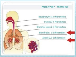
PULMONARY DEFENSE MECHANISMS
Dr. Dalia Abdallah El-Shafei
Lecturer, Community medicine department, Zagazig
University
PARTICLES
Inhalable particles are small droplets or solids -
organic or inorganic, viable or nonviable- that
can become airborne and penetrate into the
oral or nasal airways.
FACTORS AFFECTING PARTICLES’
EFFECTS:
 Diameter
 Shape
 Density
 Composition
 Concentration
 Dimensions of Air passages
 Pattern of air flow
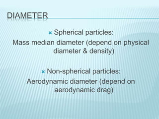
DIAMETER
 Spherical particles:
Mass median diameter (depend on physical
diameter & density)
 Non-spherical particles:
Aerodynamic diameter (depend on
aerodynamic drag)
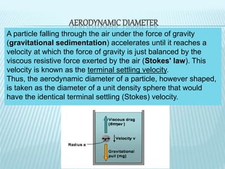
A particle falling through the air under the force of gravity
(gravitational sedimentation) accelerates until it reaches a
velocity at which the force of gravity is just balanced by the
viscous resistive force exerted by the air (Stokes' law). This
velocity is known as the terminal settling velocity.
Thus, the aerodynamic diameter of a particle, however shaped,
is taken as the diameter of a unit density sphere that would
have the identical terminal settling (Stokes) velocity.
AERODYNAMIC DIAMETER
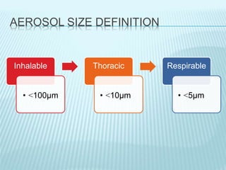
AEROSOL SIZE DEFINITION
Inhalable
• >100µm
Thoracic
• >10µm
Respirable
• >5µm
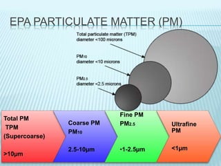
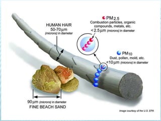
EPA PARTICULATE MATTER (PM)
Total PM
TPM
(Supercoarse)
>10µm
Coarse PM
PM10
2.5-10µm
Fine PM
PM2.5
•1-2.5µm
Ultrafine
PM
<1µm
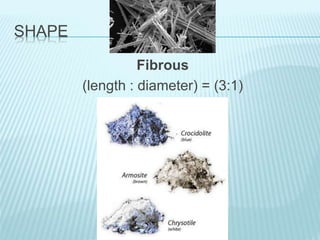
SHAPE
Fibrous
(length : diameter) = (3:1)
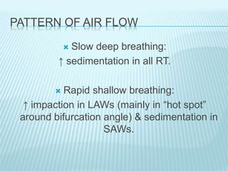
PATTERN OF AIR FLOW
 Slow deep breathing:
↑ sedimentation in all RT.
 Rapid shallow breathing:
↑ impaction in LAWs (mainly in “hot spot”
around bifurcation angle) & sedimentation in
SAWs.
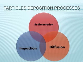
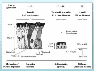
PARTICLES DEPOSITION PROCESSES
Perfect Lung
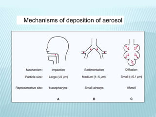
Mechanisms of deposition of aerosol
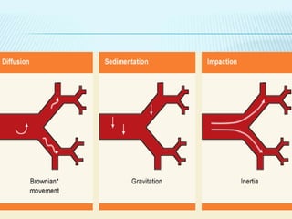
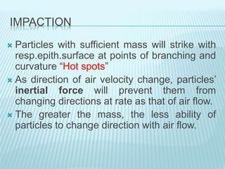
IMPACTION
 Particles with sufficient mass will strike with
resp.epith.surface at points of branching and
curvature “Hot spots”
 As direction of air velocity change, particles’
inertial force will prevent them from
changing directions at rate as that of air flow.
 The greater the mass, the less ability of
particles to change direction with air flow.
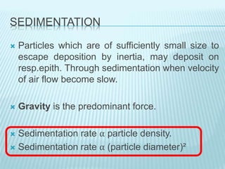
SEDIMENTATION
 Particles which are of sufficiently small size to
escape deposition by inertia, may deposit on
resp.epith. Through sedimentation when velocity
of air flow become slow.
 Gravity is the predominant force.
 Sedimentation rate α particle density.
 Sedimentation rate α (particle diameter)²
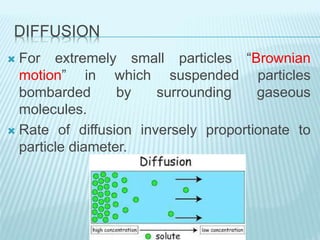
DIFFUSION
 For extremely small particles “Brownian
motion” in which suspended particles
bombarded by surrounding gaseous
molecules.
 Rate of diffusion inversely proportionate to
particle diameter.
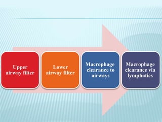
LUNG DEFENSE MECHANISMS
Upper
airway filter
Lower
airway filter
Macrophage
clearance to
airways
Macrophage
clearance via
lymphatics
UPPER AIRWAY FILTER
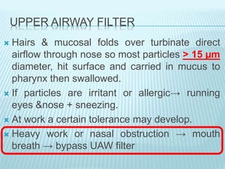
 Hairs & mucosal folds over turbinate direct
airflow through nose so most particles > 15 µm
diameter, hit surface and carried in mucus to
pharynx then swallowed.
 If particles are irritant or allergic→ running
eyes &nose + sneezing.
 At work a certain tolerance may develop.
 Heavy work or nasal obstruction → mouth
breath → bypass UAW filter
LOWER AIRWAY FILTER
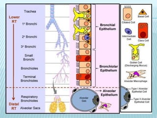
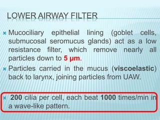
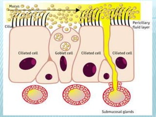
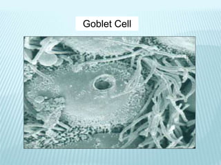
 Mucociliary epithelial lining (goblet cells,
submucosal seromucus glands) act as a low
resistance filter, which remove nearly all
particles down to 5 µm.
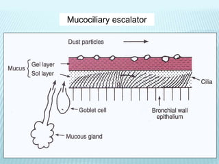
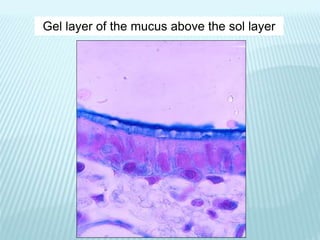
 Particles carried in the mucus (viscoelastic)
back to larynx, joining particles from UAW.
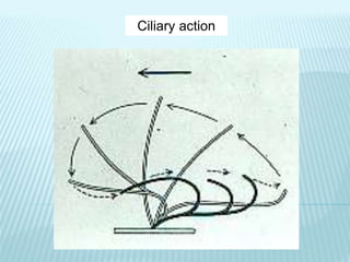
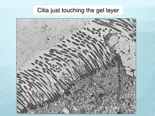
 200 cilia per cell, each beat 1000 times/min in
a wave-like pattern.
Mucociliary escalator
Goblet Cell
Ciliary action
Cilia just touching the gel layer
Gel layer of the mucus above the sol layer
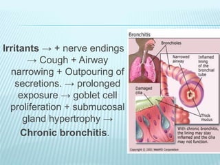
Irritants → + nerve endings
→ Cough + Airway
narrowing + Outpouring of
secretions. → prolonged
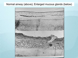
exposure → goblet cell
proliferation + submucosal
gland hypertrophy →
Chronic bronchitis.
Normal airway (above); Enlarged mucous glands (below)
IMPAIRMENT OF NORMAL MUCOCILIARY
FUNCTION
Mucus: too much, or change in composition, e.g.
chronic bronchitis, cystic fibrosis, asthma
Cilia: paralysis by toxic gases
bronchial epithelium destroyed
congenital defect of ciliary motion
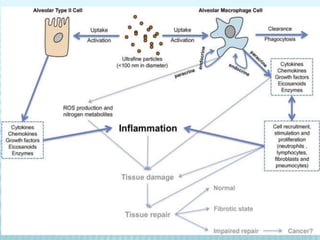
MACROPHAGE CLEARANCE TO AIRWAYS
 Particles getting beyond mucociliary clearance
called “Respirable or Alveolar dust”
 Very small particles (<0.5 µm) sediment so slowly
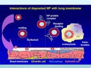
→ + type II cells → surfactant → coat particles.
 Macrophages move out from wall →engulf coated
particles → some moving back in when fully
loaded →many of dust particles dissolve by
lysosomal enzymes → when they are fully laden
with insoluble material →migrate through
interstitium to centers of lobules →enter mucus-
lined airways →carried by mucus to larynx with
rest of dust.
 By this mechanisms, lungs can clear most
retained dusts as results of regular exposure
at work up to 4mg/m³ of respirable particles
provided that macrophages not damaged.
 Above this level →system become
overloaded →dust accumulate in lung
IMPAIRMENT OF NORMAL MACROPHAGE
FUNCTION
 Inhaled gases (ozone, cigarette smoke)
 Toxic particles (silica)
 Alveolar hypoxia
 Radiation
 Corticosteroids
 Alcohol ingestion
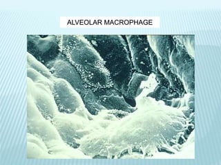
ALVEOLAR MACROPHAGE
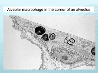
Alveolar macrophage in the corner of an alveolus
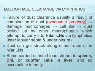
MACROPHAGE CLEARANCE VIA LYMPHATICS
 Failure of dust clearance usually a result of
combination of dust (overload + property) →
damage macrophages → cell die → dust
picked up by other macrophages which
attempt to carry it to Hilar LNs via lymphatics
(inter lobular septa & under pleura)
 Dust can get struck along either route or in
hilar LNs
 Some carried on into blood stream to spleen,
BM, or kupffer cells in liver, and so
accumulate in body.
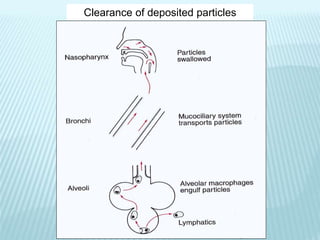
Clearance of deposited particles
LUNG IMMUNE DEFENSES AGAINST
ENVIRONMENTAL AGENTS
PATHOGENS
Pathogens enter the airways from two major
sources:
 Inhalation of bioaerosols in the environment
 Aspiration of nasopharyngeal secretions.
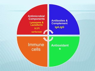
Antimicrobial
Components
Lysozyme &
Lactoferrin
SLPI
surfactant
Antibodies &
Complement
IgA,IgG
Antioxidant
s
Immune
cells
ANTIMICROBIAL COMPONENTS
Two of the most abundant antimicrobial
proteins of airway secretions are:
 Lysozyme & Lactoferrin (0.1 - 1 mg/ml).
 Secretory leukoprotease inhibitor (SLPI)
 surfactant
 Lysozyme:
- Enzymatic lysis of bacterial cell walls, can also kill
bacteria non-enzymatically.
- Highly active against many Gram +ve species but
is relatively ineffective against Gram -ve bacteria.
- Produced by both epithelial cells & leukocytes.
- It is about 10-fold more abundant in the initial
"airway" aliquot than in later samples of BAL.
 Lactoferrin:
- Iron-binding protein highly abundant in the specific
granules of human neutrophils & epithelial
secretions.
- Inhibits microbial respiration & growth, by
sequestering essential iron.
- Can also be directly microbicidal.
 Secretory leukoprotease inhibitor (SLPI)
- Antimicrobial activity against in vitro Gram - ve
& +ve bacteria.
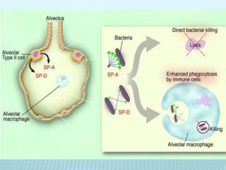
 Surfactant
- At the alveolar level, there are 2 components of the
surfactant layer: Surfactant proteins A & D.
- Bind to microorganisms & enhance adhesion &
phagocytosis of microorganisms by agglutination
& opsonization.
- Also directly antimicrobial.
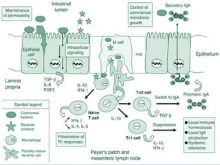
ANTIBODIES AND COMPLEMENT
 Potent immune system molecules present in airway
& alveolar lining fluid.
 Mainly immunoglobulin A (IgA) & G (IgG).
 IgA “UAW” is predominantly found along the
nasopharyngeal mucosa & in large airway; its
relative concentration decreases progressively from
larger to smaller airways.
 IgG “LAW” is the major antibody found in
alveolar fluid.
ANTIOXIDANTS
 The first line of defense against inhaled oxidant
gases (& particles) “O3 & NO2”, normally present
in lung lining fluid.
 Glutathione & Ascorbate, Uric acid, & α-
tocopherol.
 Achieving toxicity through intermediates formed
when antioxidant defenses are overwhelmed.
SURVEILLANCE BY CELLULAR FIRST
RESPONDERS
Macrophages
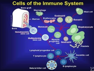
 Include subsets in distinct anatomic compartments
(Alveolar, interstitial, & airway macrophages).
 The most numerous & well studied is alveolar
macrophage (AM). Normal adult lungs contain about
20*109 Ams (BAL routinely yields 10 to 20*106 ).
 AMs are ultimately derived from BM hematopoiesis.
 Injury → influx & differentiation of blood monocytes
→ Increases in macrophage number.
 Life span of AMs in normal individuals range from
one to several months.
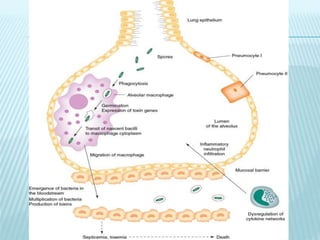
 The main function of the AM is phagocytosis &
clearance of inhaled material.
 The AMs can ingest, but fail to kill, certain
microorganisms, as (Mycobacterium, Nocardia &
Legionella) which are then capable of replicating
intracellularly. Ultimate eradication of these
pathogens requires the development of CMI.
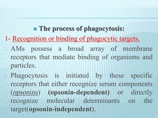
 The process of phagocytosis:
1- Recognition or binding of phagocytic targets.
- AMs possess a broad array of membrane
receptors that mediate binding of organisms and
particles.
- Phagocytosis is initiated by these specific
receptors that either recognize serum components
(opsonins) (opsonin-dependent) or directly
recognize molecular determinants on the
target(opsonin-independent).
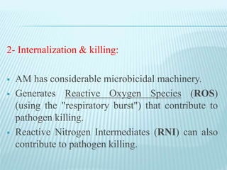
2- Internalization & killing:
 AM has considerable microbicidal machinery.
 Generates Reactive Oxygen Species (ROS)
(using the "respiratory burst") that contribute to
pathogen killing.
 Reactive Nitrogen Intermediates (RNI) can also
contribute to pathogen killing.
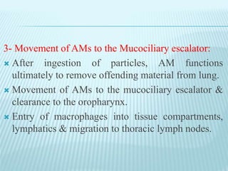
3- Movement of AMs to the Mucociliary escalator:
 After ingestion of particles, AM functions
ultimately to remove offending material from lung.
 Movement of AMs to the mucociliary escalator &
clearance to the oropharynx.
 Entry of macrophages into tissue compartments,
lymphatics & migration to thoracic lymph nodes.
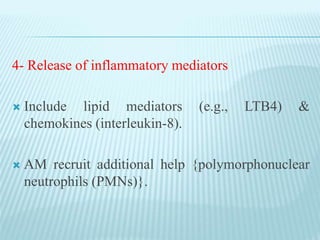
4- Release of inflammatory mediators
 Include lipid mediators (e.g., LTB4) &
chemokines (interleukin-8).
 AM recruit additional help {polymorphonuclear
neutrophils (PMNs)}.
Ciliated Epithelial Cells
 Integral part of the mucociliary clearance system.
 Produce important components of the lining fluid in
airway & alveolus {Mucus, Surfactant,
Complement, Lysozyme}.
 Some direct antibacterial function.
 Secrete a large array of cytokines & other
molecules (e.g., IL-1, -5, -6, -8, GM-CSF) which
chemoattract & activate cells of the innate &
adaptive immune system, which, in turn,
immobilize and kill microorganisms.
Polymorphonuclear Neutrophils
- The major 2nd cellular defense.
- Up to 40% of blood PMNs are marginated or in
transit through the lung, facilitating recruitment when
needed.
- Rapid & large movement of PMNs into the alveoli is
achieved by influence of several chemotactic factors
released by AMs & other lung cells (e.g., IL-8,
leukotrienes, complement fragments).
- Killing of ingested microorganisms by generation of
NADPH oxidase-dependent ROS (e.g., superoxide,
hydrogen peroxide) & by phagolysosomal fusion.
Mast Cells
 In intraepithelial locations or around blood vessels
& bronchioles.
 Produce Tumor Necrosis Factor (TNF) & a wide
range of cytokines & chemokines & important lipid
mediators, such as LTC4 and LTB4.
Natural Killer Cells (NK)
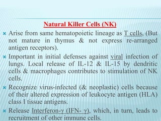
 Arise from same hematopoietic lineage as T cells, (But
not mature in thymus & not express re-arranged
antigen receptors).
 Important in initial defenses against viral infection of
lungs. Local release of IL-12 & IL-15 by dendritic
cells & macrophages contributes to stimulation of NK
cells.
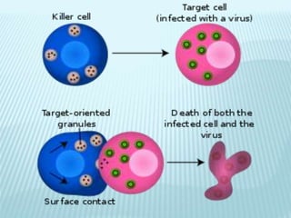
 Recognize virus-infected (& neoplastic) cells because
of their altered expression of leukocyte antigen (HLA)
class I tissue antigens.
 Release Interferon-γ (IFN- γ), which, in turn, leads to
recruitment of other immune cells.
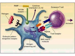
Dendritic Cells (DCs)
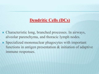
 Characteristic long, branched processes. In airways,
alveolar parenchyma, and thoracic lymph nodes.
 Specialized mononuclear phagocytes with important
functions in antigen presentation & initiation of adaptive
immune responses.
 Acting as sentinels in airways, they sample incoming
pathogens and antigens through by phagocytosis.
When this is accompanied by a second, "danger"
signal, they undergo a phenotypic & functional change
from their basal immature state.
 This maturation promotes the processing of antigen
and its presentation on the cell surface and the
migration of the dendritic cell to T-cell rich areas of
nearby lymph nodes. Here they can initiate or amplify
adaptive immune responses by triggering proliferation
and activation of antigen-specific T lymphocytes.
CYTOKINES
 Critical for successful orchestration of defense
mechanisms against environmental agents & are
also mediators of untoward outcomes such as
acute injury & chronic inflammation & fibrosis.
 TNF & chemokines which function in both acute
and chronic phases of these processes.
Tumor Necrosis Factor
 TNF- αis a protein.
 Monocytes express at least 5 different molecular forms
of TNF-α.
 Macrophages are considered the most prolific sources.
 lymphoid cells, mast cells, endothelial cells,
fibroblasts, and neuronal tissue.
 Two types of TNF receptors, TNF-R1 & TNF-R2, are
present on virtually all cells except RBCs.
 Present during acute response to acute
inflammatory responses to toxic environmental
agents{silica, asbestos, air pollution particles,
welding fumes, ozone}.
Chemokines
 Plays a critical role in this process of recruitment
& maintenance of inflammatory cells in the lungs
after environmental exposures.
 Produced by Ams, monocytes, neutrophils, T & B
lymphocytes, NK cells, epithelial cells,
fibroblasts, smooth muscle cells, mesothelial
cells, and endothelial cells.
ADAPTIVE IMMUNITY
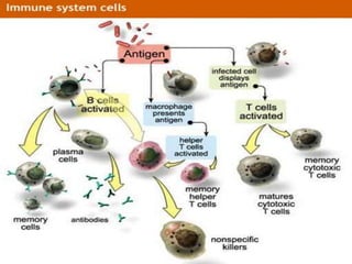
 The adaptive immune response to pulmonary
pathogens includes (humoral & cellular components).
 Both B & T lymphocytes are present in normal lung.
 B cells are predominantly found in airway lymphoid
aggregates, where they outnumber the T cells.
 In normal lavage samples, approximately 5-10% of
cells are lymphocytes, which, in turn, can be further
divided into functionally important subsets, for
example, CD4+ T helpers & CD8+ cytotoxic T cells.
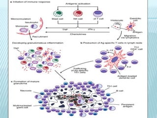
GRANULOMATOUS INFLAMMATION
 In response to certain infectious agents & persistent
foreign material, & as part of a disease of unknown
etiology (e.g., sarcoidosis).
 Chronic inflammation, dominated by mononuclear
phagocytes “macrophages, epithelioid cells, &
multinucleated giant cells”.
 Typically, these cells congregate & form well-
demarcated focal lesions called granulomas.
 Admixture of other cells,(lymphocytes, plasma cells,
& fibroblasts).
 Granuloma formation typically ends in fibrosis.
Fibrosis serves to wall off the granuloma contents and
limit spread of infection and organ damage.
Pulmonary defense mechanisms