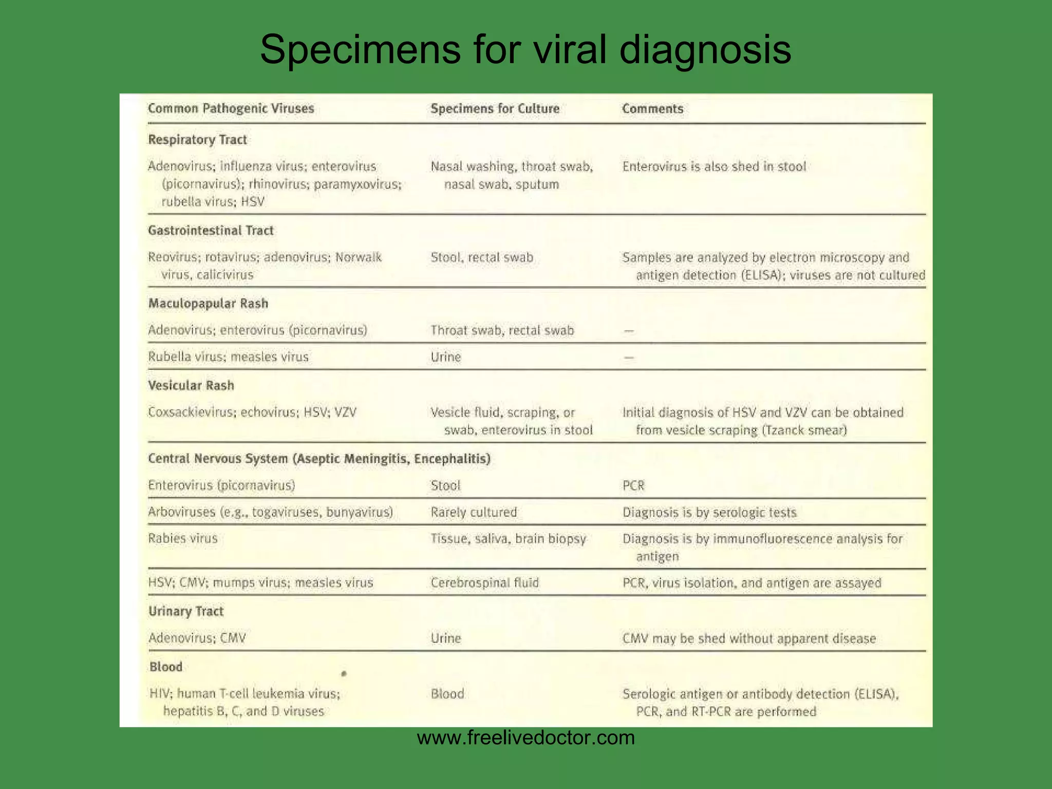
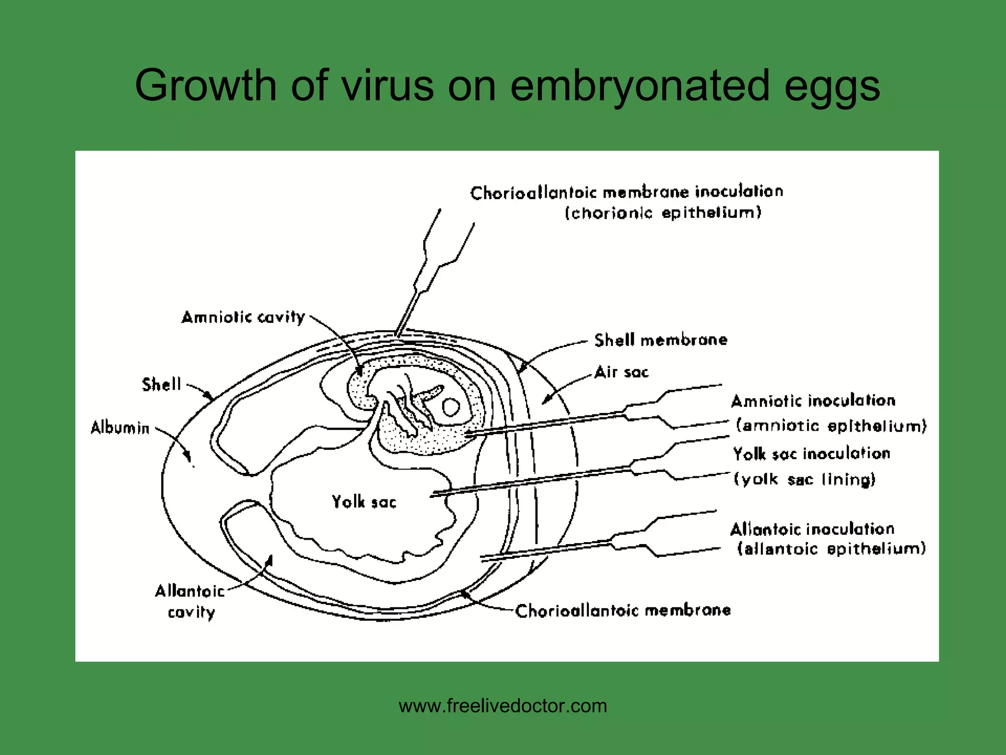
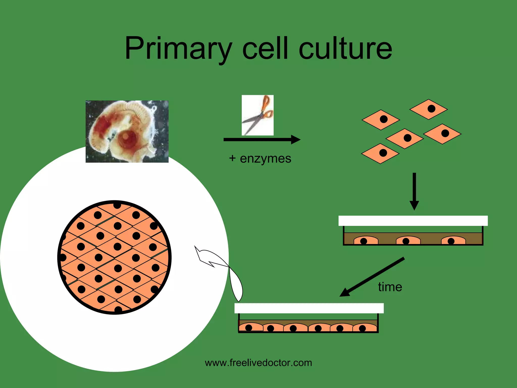
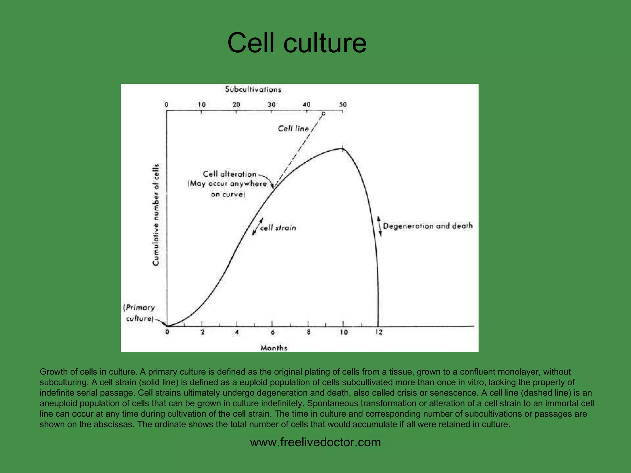
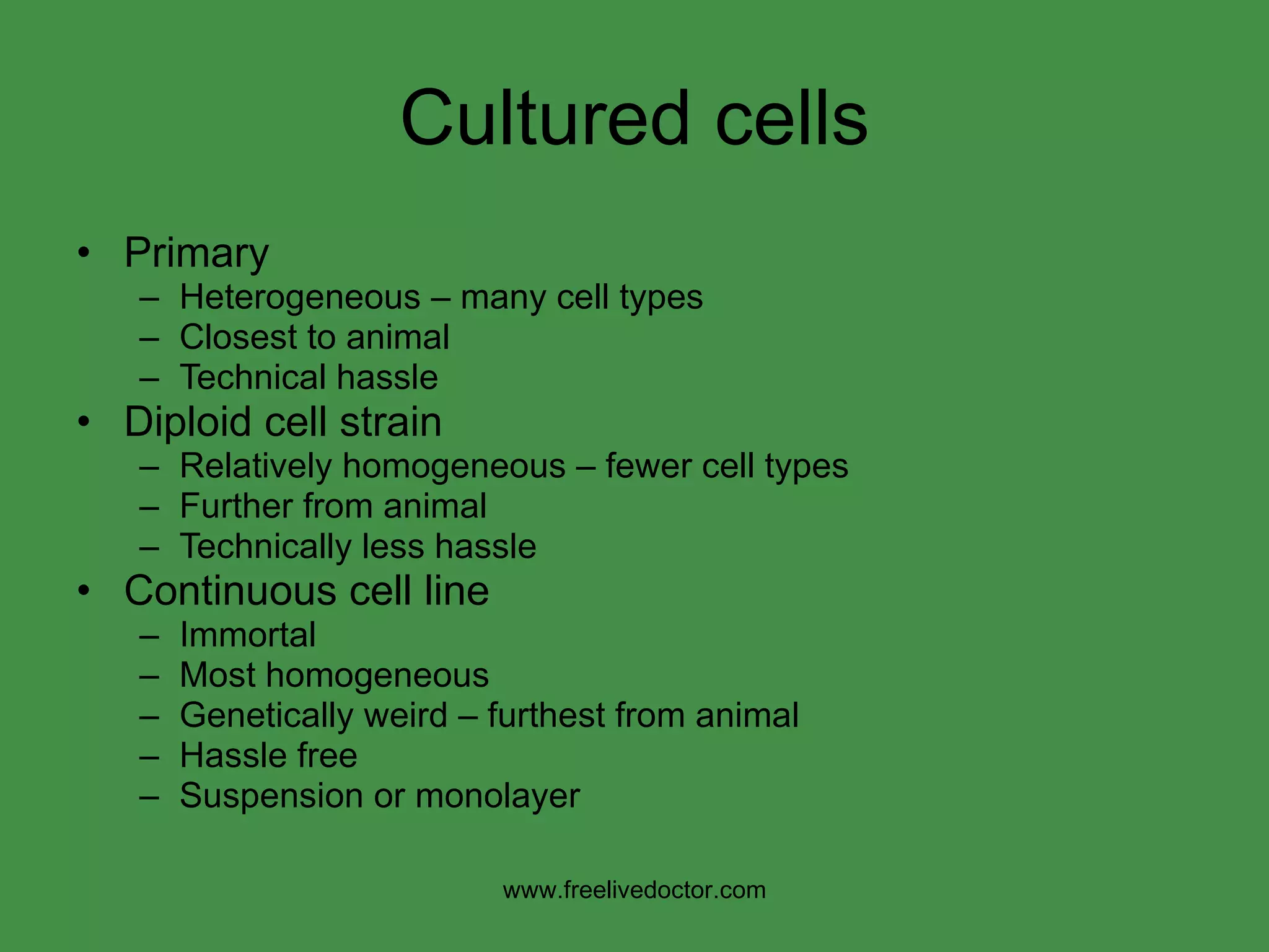
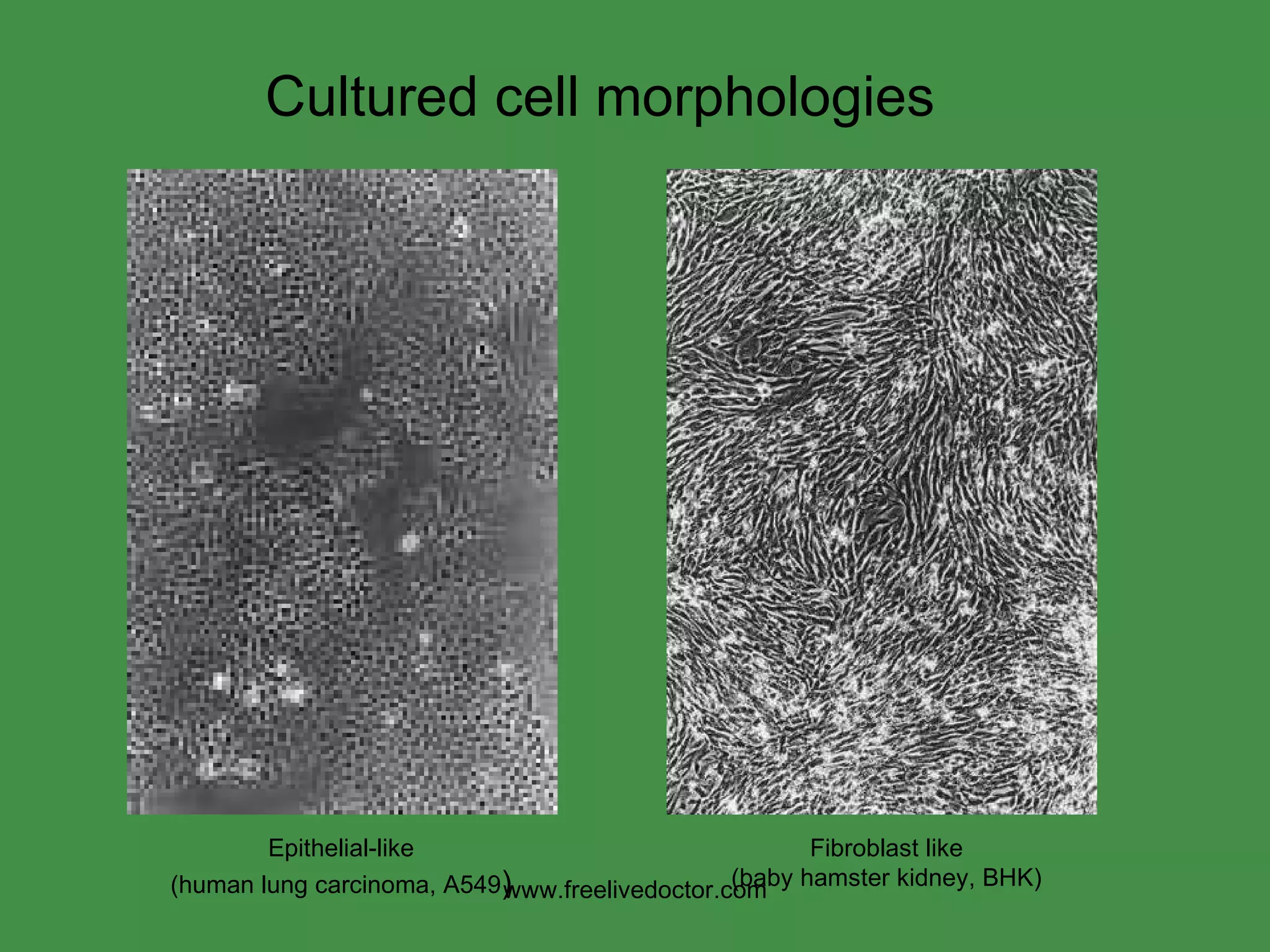
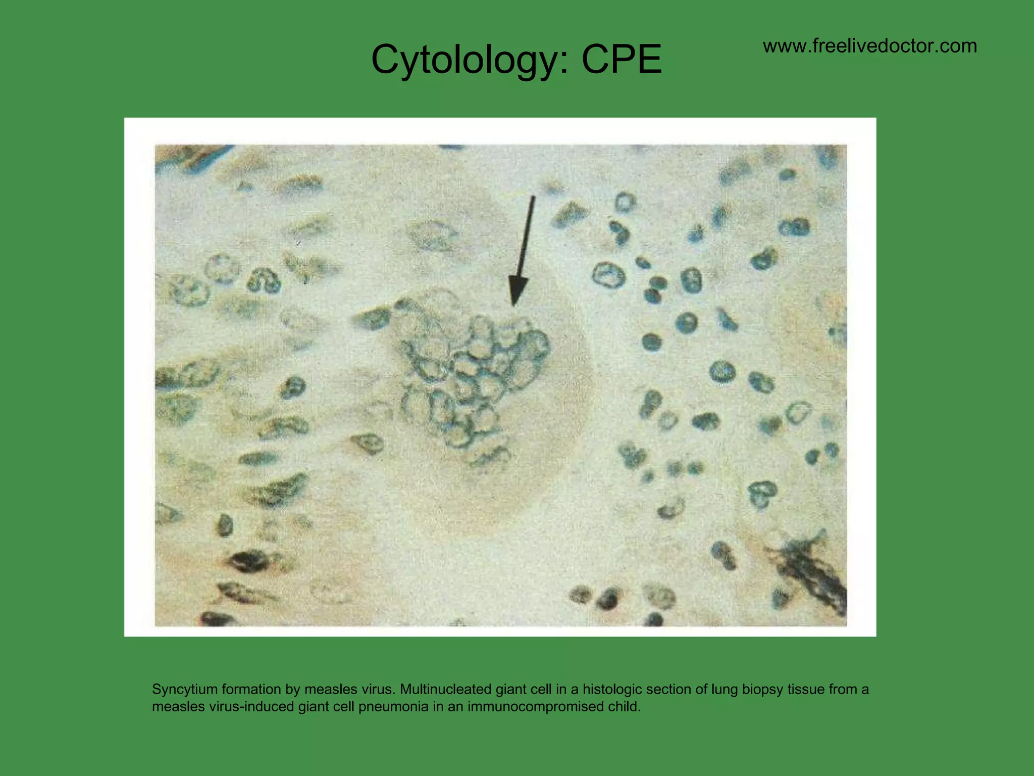
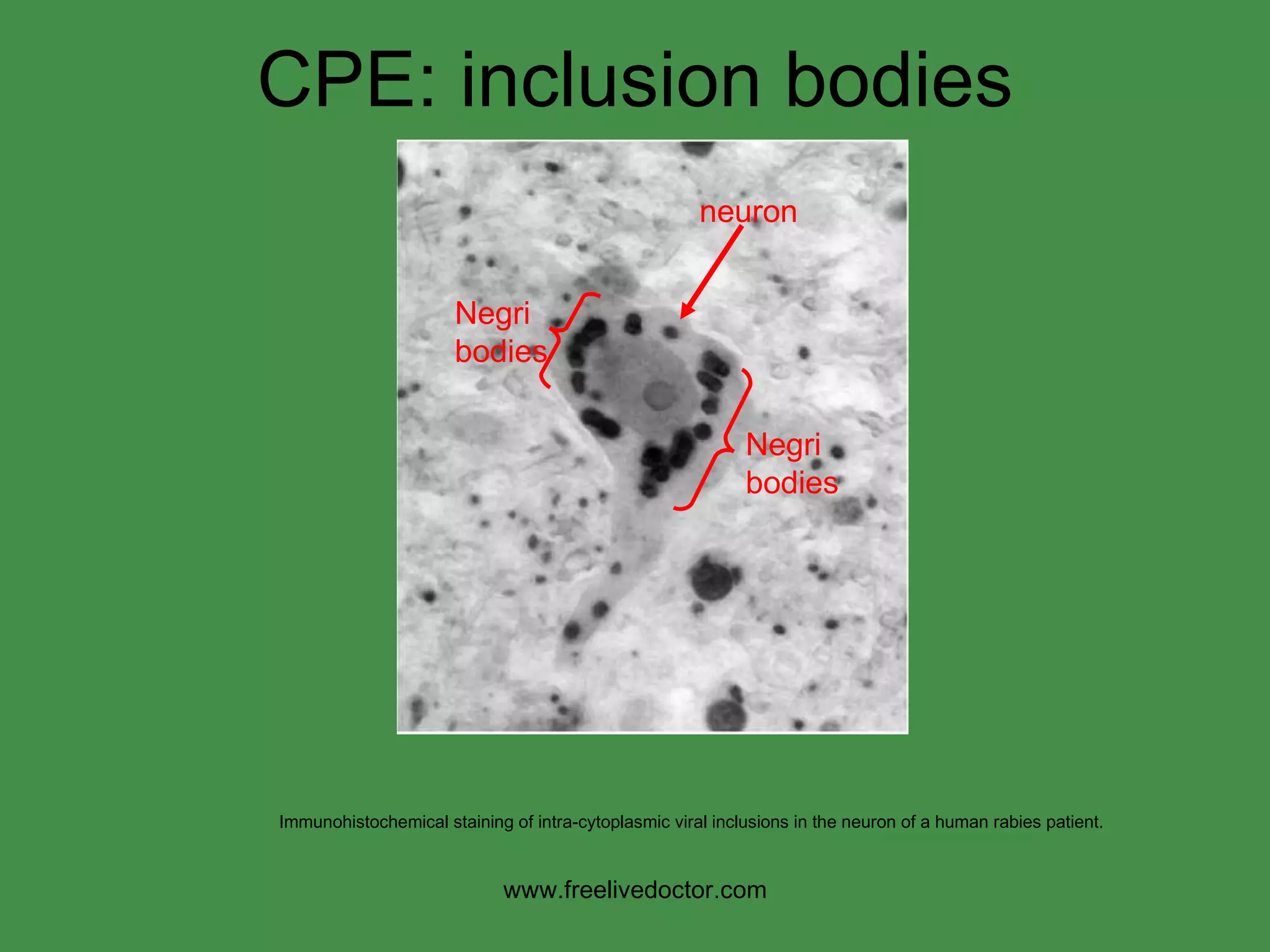
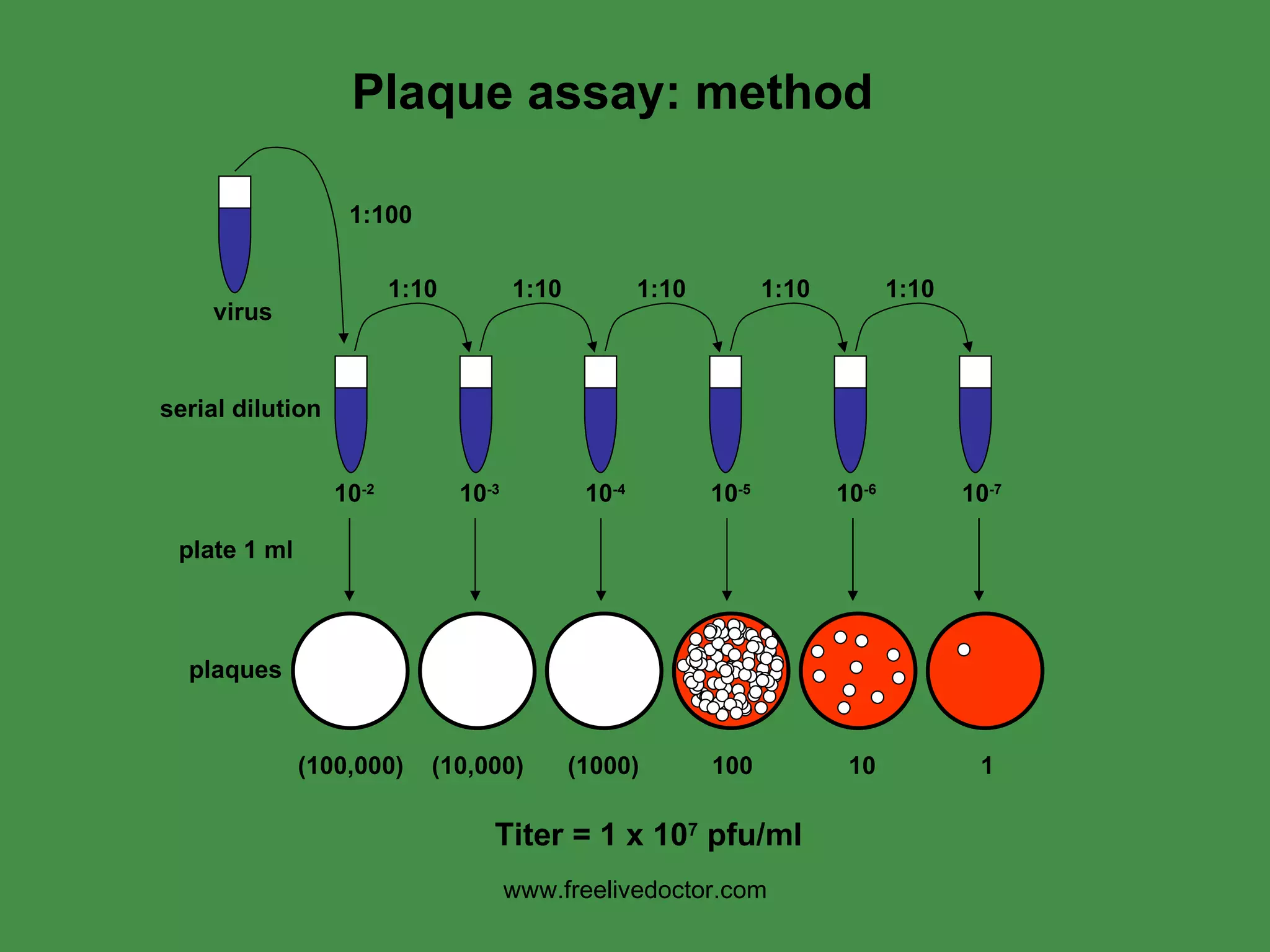
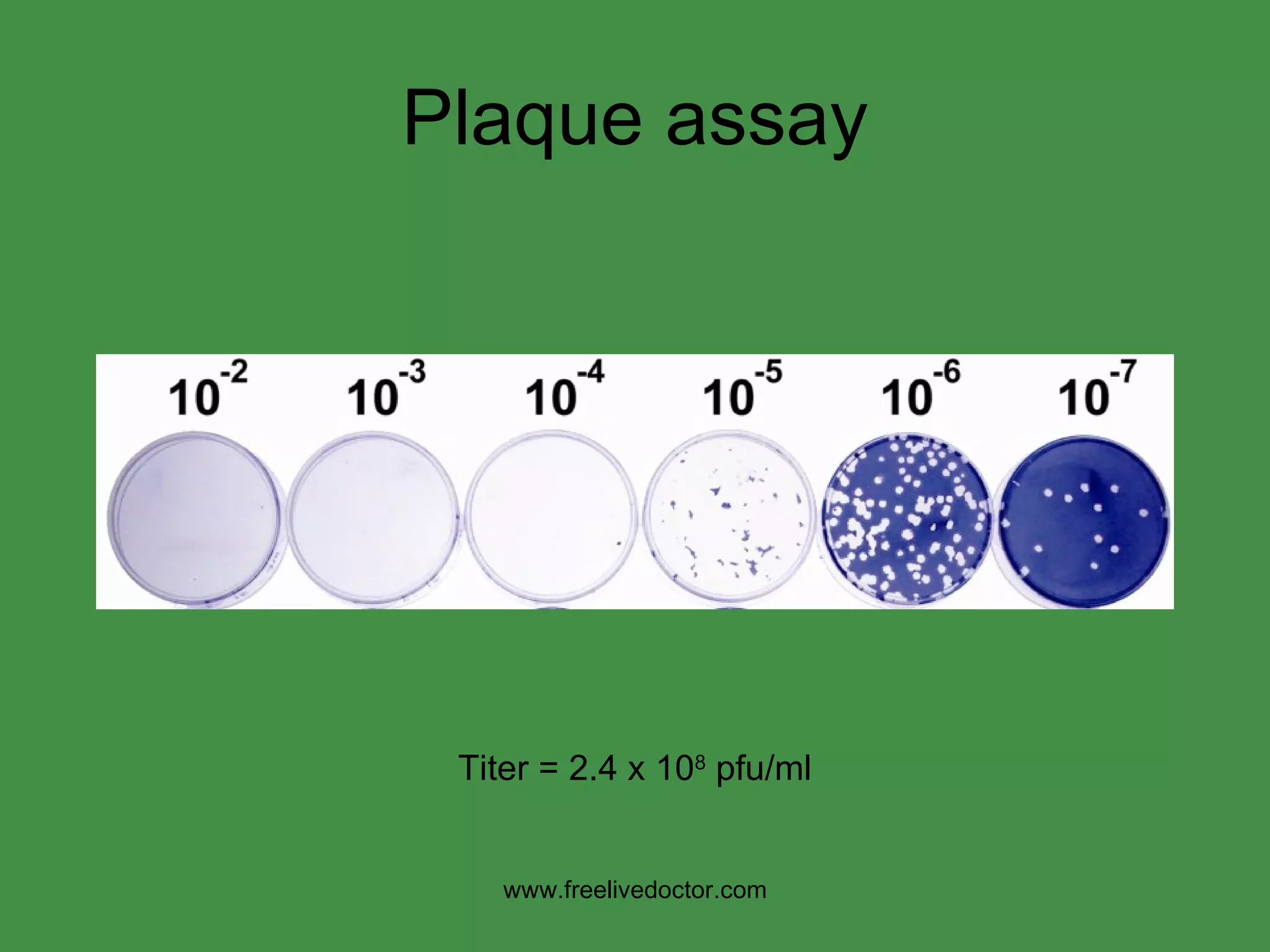
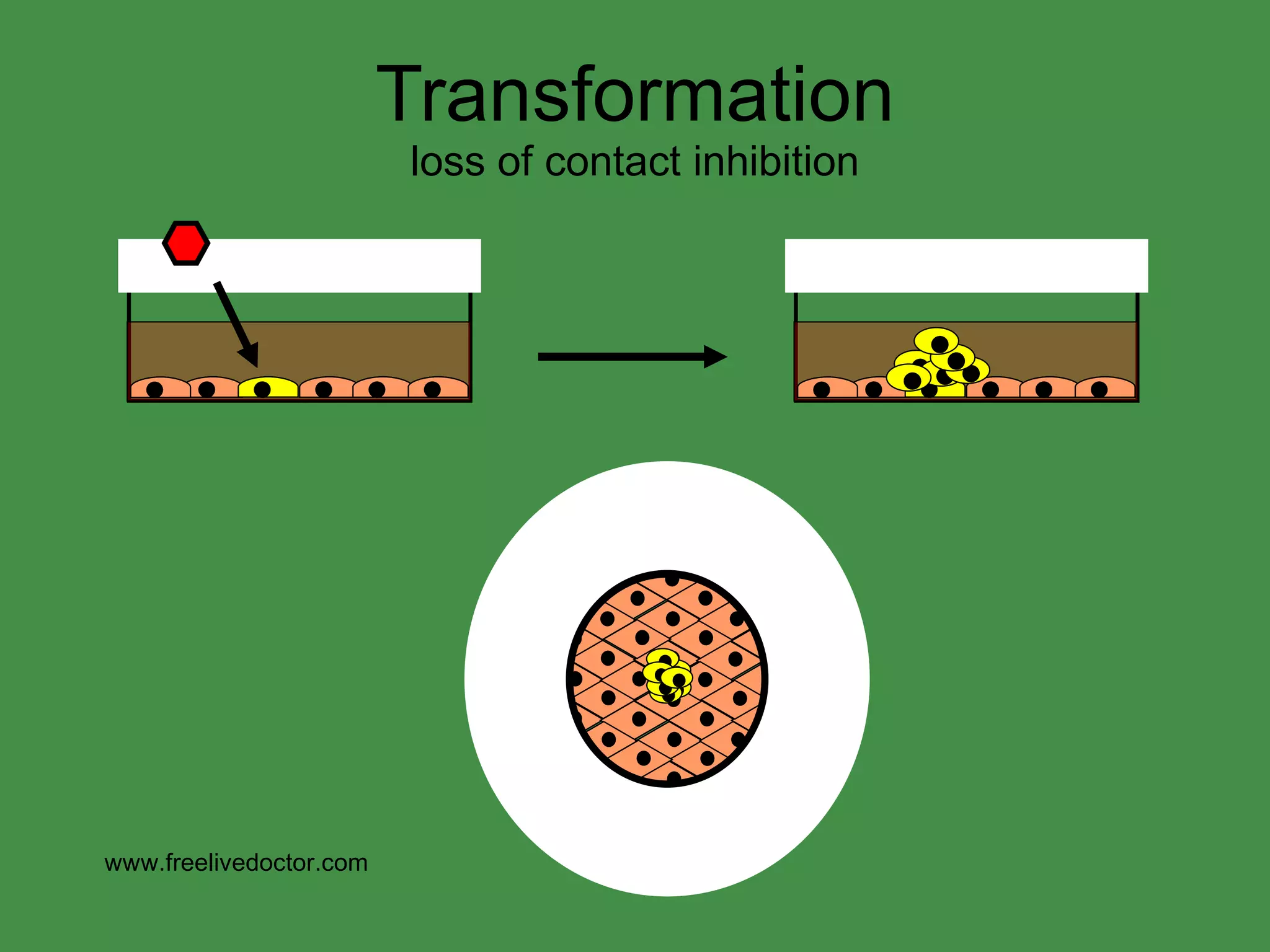
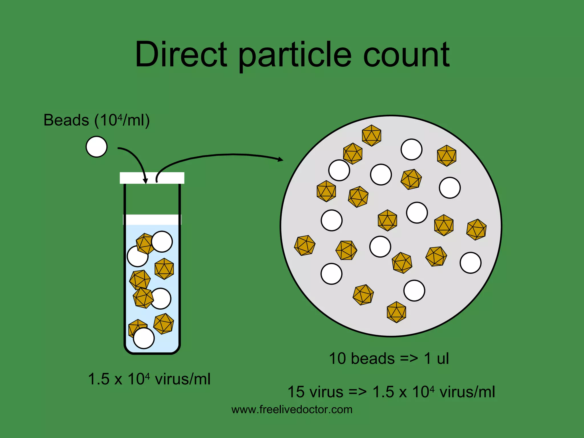
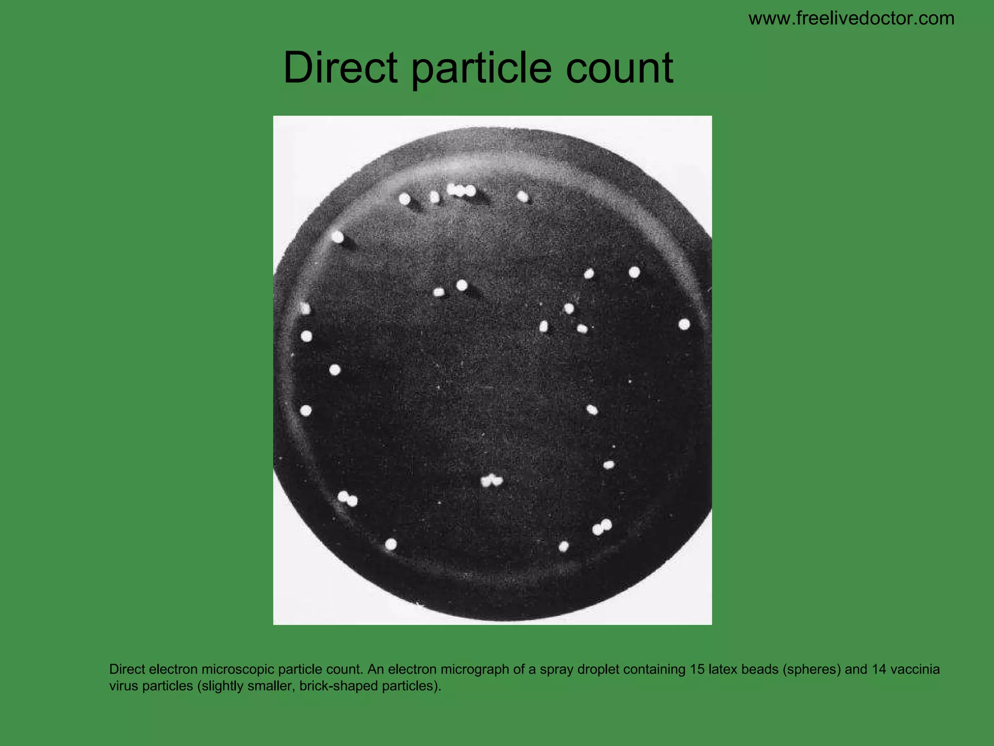
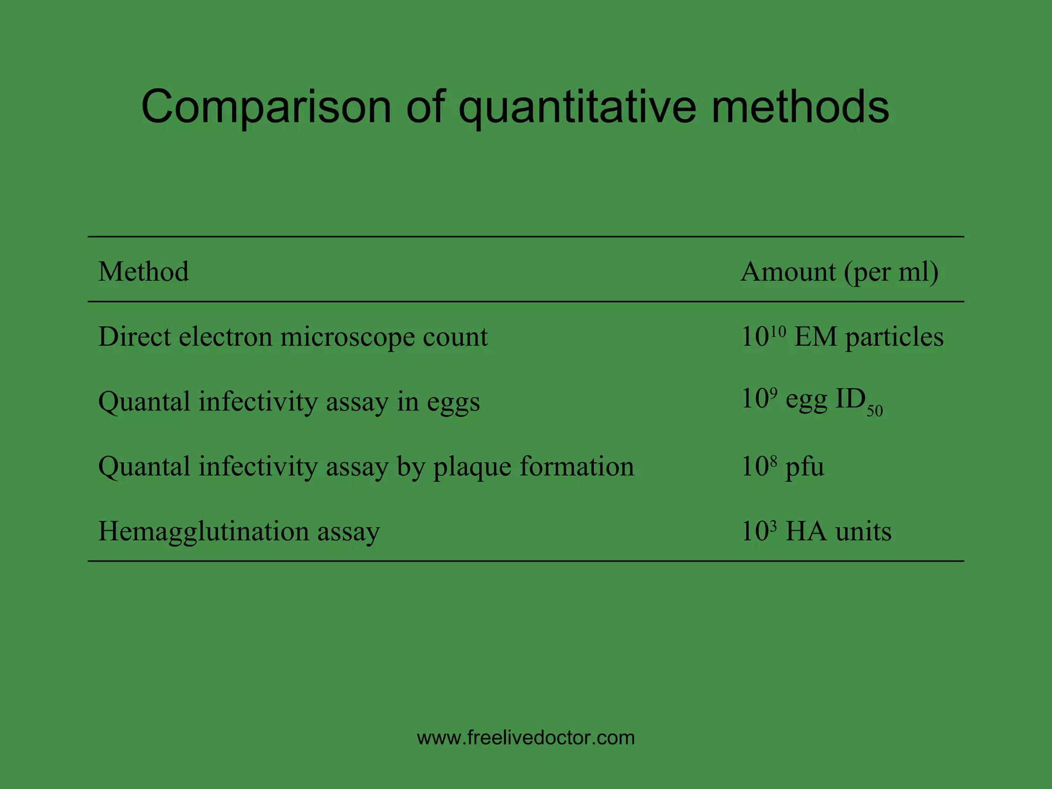
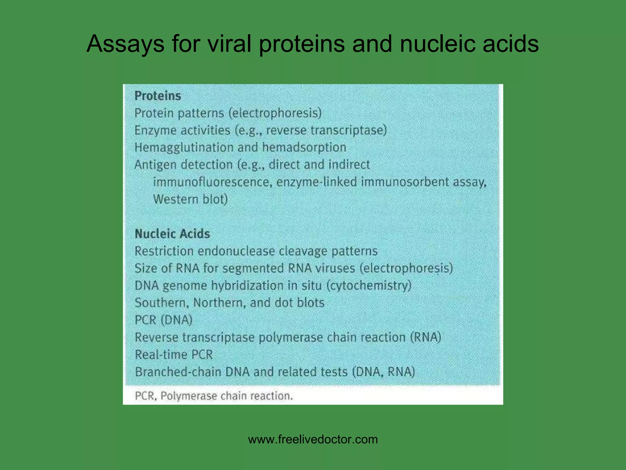
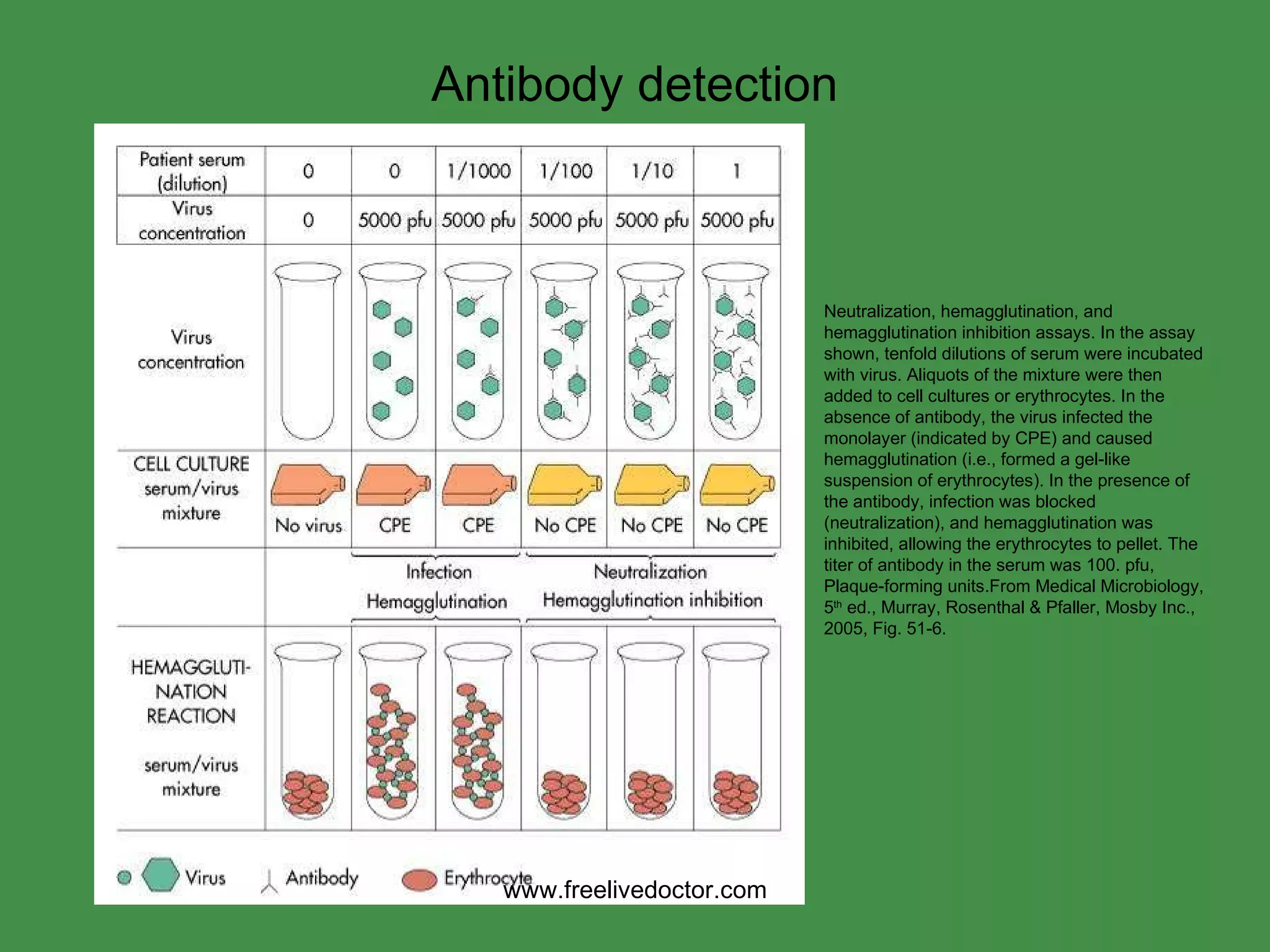
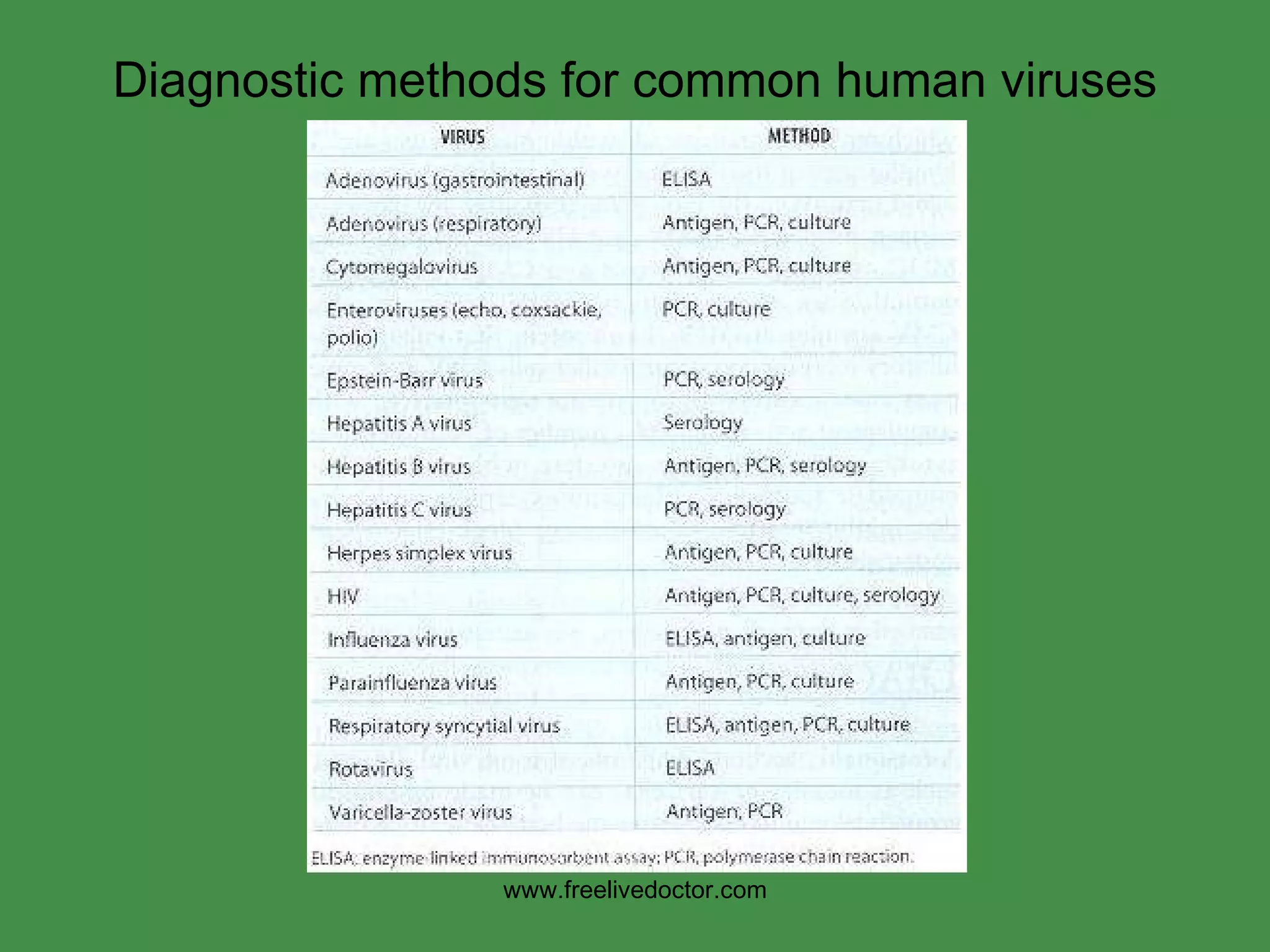
A picornavirus is a virus belonging to the family Picornaviridae, a family of viruses in the order Picornavirales. Vertebrates, including humans, serve as natural hosts. Picornaviruses are nonenveloped viruses that represent a large family of small, cytoplasmic, plus-strand RNA viruses with a 30-nm icosahedral capsid.
lab diagnosis of viral infections - mayuri.pptxDrmayuribhise
T.M. River, 1937
Modified from Koch’s Postulates (proof of bacterial diseases)
Isolate virus from diseased hosts.
Cultivation of virus in host cells.
Proof of filterability.
Production of a comparable disease when the cultivated virus is used to infect experimental animals.
Reisolation of the same virus from the infected experimental animal.
Detection of a specific immune response to the virus.
Much more expensive and difficult to study animal viruses than bacteriophages
Cultivation in host cells
Living animal
Embryonated chicken eggs
Cell or tissue culture (= in vitro)
Over 60% of all infectious disease cases seen by a physician are due to viral infections.
Quality of patient specimens and their transport to the laboratory is importantViral Diagnostics in the Clinical Laboratory
Types of specimens:-
Respiratory tract infections: Nasal and bronchial washings, throat and nasal swabs, sputum
Eye infections: throat and Conjunctival swab/scraping
Gastrointestinal tract infections: stool and rectal swabs
Vesicular rash: vesicle fluid, skin scrapings
Maculopapular rash: throat, stool, and rectal swabs
CNS (encephalitis and meningitis cases): stool, tissue, saliva, brain biopsy, cerebrospinal fluid
Genital infections: vesicle fluid or swab
Urinary tract infections: urine
Blood borne infections: blood
Sterile, leak proof container
Minimal interval
Transport media
Viral infusion broth (VIB)
Sucrose-phosphate-glutamate (SPG)
Storage temperature:
4 deg C for up to 96 hours
Minus 70 deg C beyond 96 hours
Repeated cycles of freezing and thawing to be avoided
106 virus particles per ml required for visualization,
50,000 - 60,000 magnification normally used.Specimens are negatively stained by Potassium phosphotungstate and scanned under EM
Viruses may be detected in the following specimens.
Virus particles are detected and identified on the basis of morphology.
A) Shape
Rabiesvirus –bullet shaped
Rotavirus –Cart wheel
Coronavirus –petal shaped peplomers
Adenovirus –space vehicle shaped
Astrovirus ---Star shaped
B) Direct detection from specimens
For viruses that are difficult to cultivate ,EM can be used as primary tool for diagnosis
Faeces Rotavirus, Adenovirus
Norwalk like viruses
Astrovirus, Calicivirus
Vesicle Fluid HSV
VZV
Skin scrapings papillomavirus, orf
molluscum contagiosum
As an alternative to tissue culture
As tissues culture is time consuming and technically demanding ,EM is used as an alternative :-
1) Vesicular rashes –HSV and VZV detection from vesicular fluid
2) Meningitis—Detection of enterovirus and mumps from CSF.
Virus detection from tissue cultures EM can be used for detection of viral growth in tissue culture
The sensitivity and specificity of EM can be improved by adding specific antiviral antibody to the specimen to aggregate the virus particles which can be centrifuged
The sediment is negatively stained and viewed under EM
Direct immumofluroscence
Titration and isolation of viruses using cell culturesShadia Omar
There are several methods to quantify and isolate viruses, isolation of viral pathogens in cell cultures. This approach is often slow and requires considerable technical expertise, however, it is still considered as the “gold standard” for the laboratory diagnosis of viral disease. In this presentation, I will describe one of these methods which is TCID50 (Tissue culture infective dose 50 )
Poxviruses are brick or oval-shaped viruses with large double-stranded DNA genomes. Poxviruses exist throughout the world and cause disease in humans and many other types of animals. Poxvirus infections typically result in the formation of lesions, skin nodules, or disseminated rash.
Adenoviridae is a group of medium sized, non-enveloped, double stranded DNA viruses that replicate and produce disease in the eye and in the respiratory, gastrointestinal and urinary tracts;
BIOTECHNOLOGY IS
CHALLENGING SUBJECT TO TEACH AND UNDERSTAND ......
ITS A VERY INTERESTING TO LEARN ABOUT HYBRIDOMA TECHNOLOGY .. THEIR PRODUCTION AND
APPLICATION ALSO ....
Viruses are obligate intracellular parasites which means they can only grow or reproduce inside a host cell.
The primary purpose of virus cultivation:
To isolate and identify viruses in clinical samples.
To do research on the viral structure, replication, genetics, and effects on the host cell.
To prepare viruses for vaccine production.
Isolation of the virus is always considered a gold standard for establishing the viral origin of the disease
topics covered
CULTIVATION OF VIRUSES
Animal inoculation
Embryonated eggs
CAM
Allantoic cavity
Amniotic cavity
Yolk sac
Tissue culture
Organ culture
Explant culture
Cell culture
Primary cell culture
diploid cell culture
Continues cell lines
What are the specific linkages among immune surveillance, clonal sel.pdfmanjan6
What are the specific linkages among immune surveillance, clonal selection, and lymph nodes?
Solution
Immune Surveillance - A hypothesis that the immune system perceives and pulverizes tumor
cells that are continually emerging amid the life of the person. A body component by which early
growths are recognized as being strange and are assaulted and typically pulverised. Immune
Surveillance is a process in which a T cell-intervened handle without which tumor would be
much ordinary.
The immune sytem can distinguish and crush rising cancer cells since it perceives strange
antigens on the cell surface as \'foreign substance\' or outsider. Since outside substances are
typically risky to the body, the immune system is customised to demolish them. This consistent
checking of the body for little tumors is immune surveillance. The immune surveillance of living
body is known to work in the dismissal of tumor cells in people with inherited nonpolyposis
colon growth, likewise called Lynch disorder. Those people acquire a broken DNA confound
repair framework and as an outcome deliver numerous mutant proteins. At the point when such
mutant proteins show up on the surface of tumor cells, they are perceived as foriegn and rejected.
Tumors that do rise are those that have figured out how to sidestep the body\'s immune system.
Lymph nodes are Bean Shaped and encapsulated antigen gathering tissues that channel lymph
and permit lymphocytes to test antigens gained from skin, GI tract and respiratory tract
- deliberately situated all through body, bolstered by reticular system of follicular DCs and
macrophages
- lymphocytes enter from blood crosswise over high endothelial venules and from the
interconnected lymphatic vessels; B cells relocate to the cortex and immune system
microorganisms move to paracortical area; plasma move to medulla and emit counter acting
agent
- DCs with antigen enter from lymphatic vessel; T cells and DCs reach each other the T cell tests
the antigens in setting of the MHC on the DC; enacted T cells leaves lymph hubs and enter the
blood to site of contamination.
Clonal Selection - Clonal selecction is a hypothesis clarifying how the cells of the immune
system deliver vast amounts of the correct counter acting agent at the ideal time, in the sense, at
the point when the proper antigen is experienced. It suggests that there is a previous pool of
lymphocytes i.e B cells comprising of various little subsets. Every subset conveys a one of a kind
arrangement of surface counter acting agent atoms with its own specific restricting qualities. In
the event that a cell experiences and ties the comparing antigen it is `selected\' – empowered to
partition more than once and deliver a substantial clone of indistinguishable cells, all emitting the
counter acting agent. The contribution of aide T cells is basic for actuation of the B cell. A type
of clonal determination is additionally summoned to clarify the improvement of immunological
resistance.
6. Growth of cells in culture. A primary culture is defined as the original plating of cells from a tissue, grown to a confluent monolayer, without subculturing. A cell strain (solid line) is defined as a euploid population of cells subcultivated more than once in vitro, lacking the property of indefinite serial passage. Cell strains ultimately undergo degeneration and death, also called crisis or senescence. A cell line (dashed line) is an aneuploid population of cells that can be grown in culture indefinitely. Spontaneous transformation or alteration of a cell strain to an immortal cell line can occur at any time during cultivation of the cell strain. The time in culture and corresponding number of subcultivations or passages are shown on the abscissas. The ordinate shows the total number of cells that would accumulate if all were retained in culture. Cell culture www.freelivedoctor.com
13. Hemadsorption of erythrocytes to cells infected with influenza viruses, mumps virus, parainfluenza viruses, or togaviruses. These viruses express a hemagglutinin on their surfaces, which bind erythrocytes of selected animal species. Hemadsorption www.freelivedoctor.com
14. Syncytium formation by measles virus. Multinucleated giant cell in a histologic section of lung biopsy tissue from a measles virus-induced giant cell pneumonia in an immunocompromised child. Cytolology: CPE www.freelivedoctor.com
15. CPE: inclusion bodies Immunohistochemical staining of intra-cytoplasmic viral inclusions in the neuron of a human rabies patient. www.freelivedoctor.com Negri bodies Negri bodies neuron
19. Focus formation by transforming viruses Focus assay. Monolayers of the NIH3T3 mouse fibroblast cell line were infected with Maloney murine sarcoma virus. The top two panels show photomicrographs of uninfected cells (left) and a single virus-induced focus (right). The bottom two panels show stained dishes of uninfected (left) and infected (right) cells. Foci are clearly visible as darker areas on the infected dish. www.freelivedoctor.com
20. = infected = uninfected Endpoint titration Five replicate wells of cells are infected with one ml of each of four different virus dilutions, incubated, and scored for infection by looking for CPE. In this example, the final titer is 10 6.3 TCID 50 per ml. (TCID = tissue culture infective dose) www.freelivedoctor.com 10 -5 10 -6 10 -7 10 -8 5 replicate samplings 1 2 3 4 5 dilution
22. Titer = 32 HA units/ml Hemagglutination test: method 1:8 1:2 1:2 1:2 1:2 1:2 8 16 32 64 128 256 virus serial dilution mix with red blood cells side view top view www.freelivedoctor.com
23. Hemagglutination assay. Seven different samples of influenza virus, numbered 1 through 7 at the left, were serially diluted as indicated at the top, mixed with chicken red blood cells (RBC), and incubated on ice for 1 to 2 hours. Wells in the bottom row contain no virus. Agglutinated RBCs coat wells evenly, in contrast to nonagglutinated cells, which form a distinct button at the bottom of the well. The HA titer, shown at the right, is the last dilution that shows complete hemagglutination activity. Hemagglutination assay: influenza virus www.freelivedoctor.com
24. Direct particle count 10 beads => 1 ul 15 virus => 1.5 x 10 4 virus/ml 1.5 x 10 4 virus/ml www.freelivedoctor.com Beads (10 4 /ml)
25. Direct electron microscopic particle count. An electron micrograph of a spray droplet containing 15 latex beads (spheres) and 14 vaccinia virus particles (slightly smaller, brick-shaped particles). Direct particle count www.freelivedoctor.com
26. Comparison of quantitative methods www.freelivedoctor.com Method Amount (per ml) Direct electron microscope count 10 10 EM particles Quantal infectivity assay in eggs 10 9 egg ID 50 Quantal infectivity assay by plaque formation 10 8 pfu Hemagglutination assay 10 3 HA units
27. Assays for viral proteins and nucleic acids www.freelivedoctor.com
28. Neutralization, hemagglutination, and hemagglutination inhibition assays. In the assay shown, tenfold dilutions of serum were incubated with virus. Aliquots of the mixture were then added to cell cultures or erythrocytes. In the absence of antibody, the virus infected the monolayer (indicated by CPE) and caused hemagglutination (i.e., formed a gel-like suspension of erythrocytes). In the presence of the antibody, infection was blocked (neutralization), and hemagglutination was inhibited, allowing the erythrocytes to pellet. The titer of antibody in the serum was 100. pfu, Plaque-forming units.From Medical Microbiology, 5 th ed., Murray, Rosenthal & Pfaller, Mosby Inc., 2005, Fig. 51-6. Antibody detection www.freelivedoctor.com
29. Western blot analysis of HIV antigens and antibody. HIV protein antigens are separated by electrophoresis and blotted onto nitrocellulose paper strips. The strip is incubated with patient antibody, washed to remove the unbound antibody, and then reacted with enzyme-conjugated antihuman antibody and chromophoric substrate. Serum from an HIV-infected person binds and identifies the major antigenic proteins of HIV. This data demonstrates the seroconversion of one HIV-infected individual with sera collected on day 0 (D0) to day 30 (D30) compared to a known positive control (PC) and negative control (NC). (From Medical Microbiology, 5 th ed., Murray, Rosenthal & Pfaller, Mosby Inc., 2005, Fig. 51-7. ) Antibody detection: western blot www.freelivedoctor.com