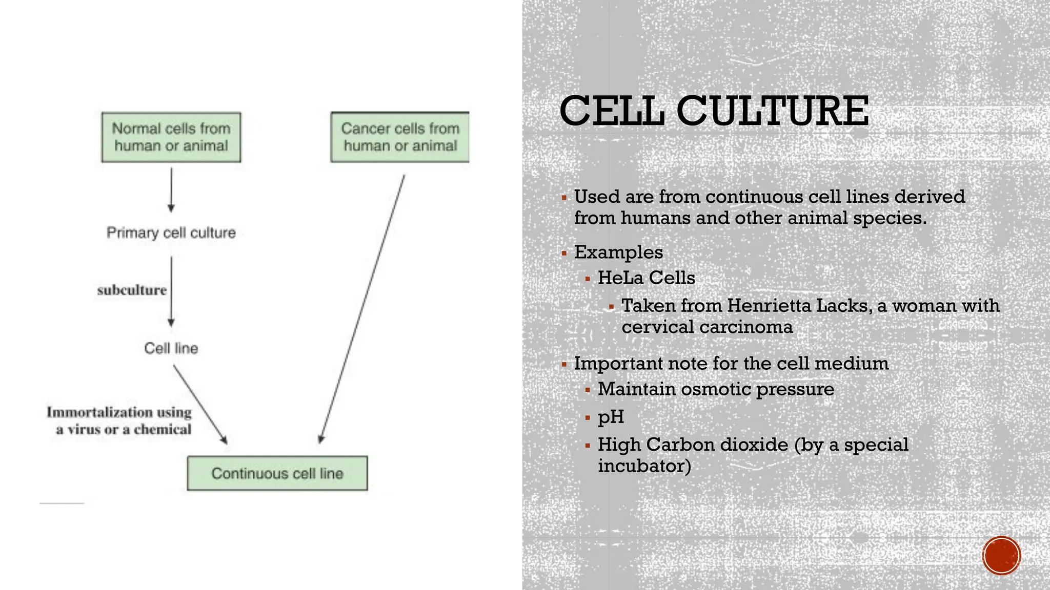
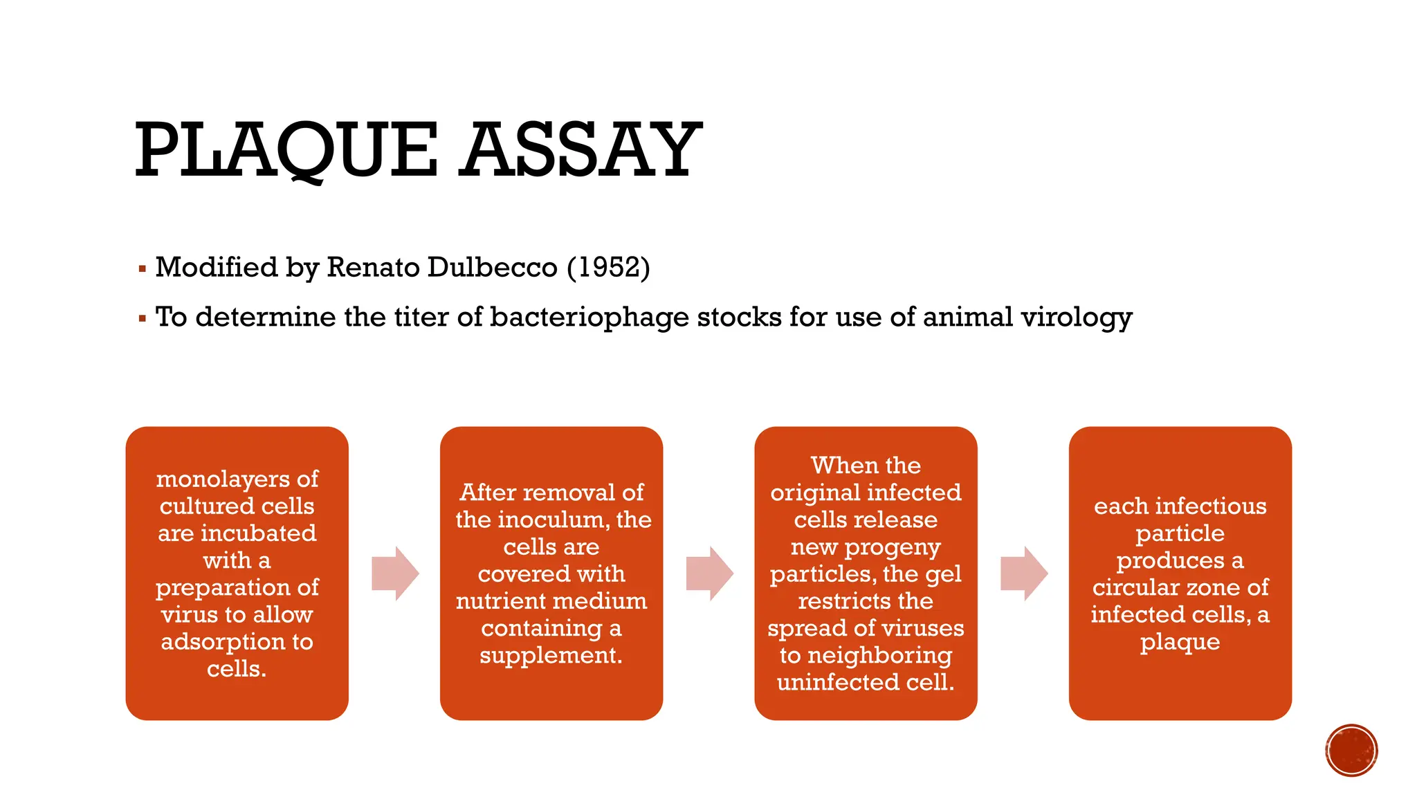
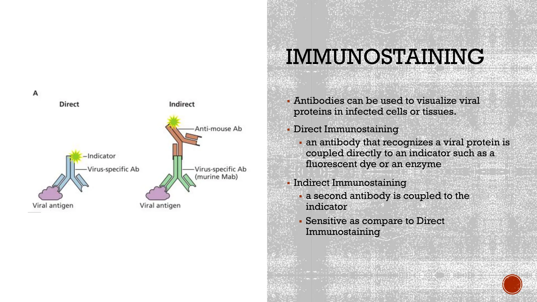
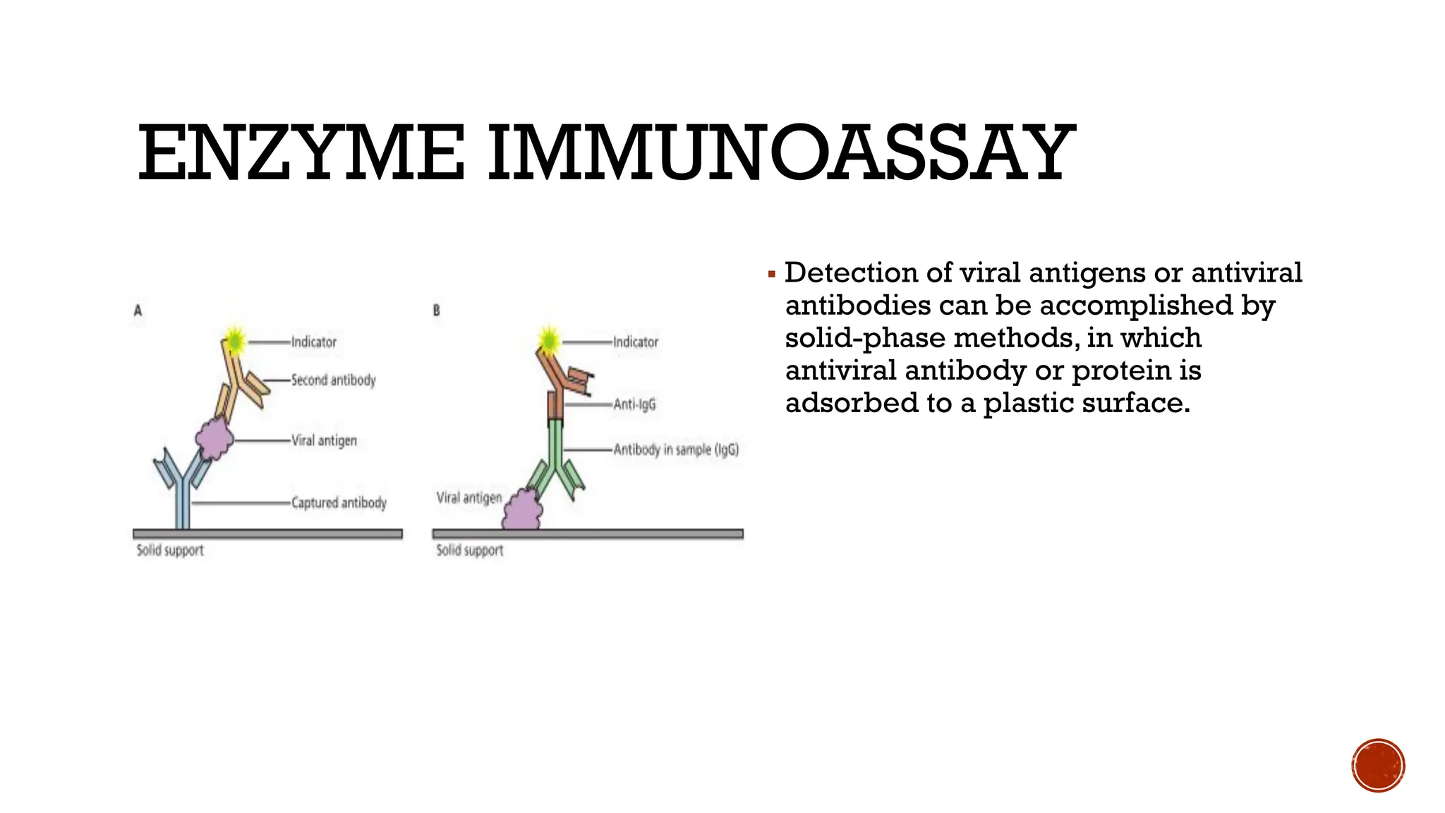
METHOD USED
IN VIROLOGY
VIRUS IDENTIFICATION
▪Cytopathic effects
▪microscopic or macroscopic degenerative changes or
abnormalities in host cells and tissues
▪Serological tests
▪Detect antibodies against viruses in a patient
▪Use antibodies to identify viruses in neutralization tests, viral
hemagglutination, and Western blot
▪Nucleic acids
▪RFLPs
▪PCR
CYTOPATHIC
EFFECT
▪ Observe on a high power
microscopy
▪ Changes in the cell
▪ Used to monitor the progress of
infection
▪ Other viruses do not have the
obvious cytopathic effect
▪ Arenaviridae
▪ Paramyxoviridae
▪ Retroviridae
VIRUS ISOLATION
AND CULTURE
CELL CULTURE
▪ Used are from continuous cell lines derived
from humans and other animal species.
▪ Examples
▪ HeLa Cells
▪ Taken from Henrietta Lacks, a woman with
cervical carcinoma
▪ Important note for the cell medium
▪ Maintain osmotic pressure
▪ pH
▪ High Carbon dioxide (by a special
incubator)
KINDS OF CELL CULTURE
▪ Cell Monolayer
▪ The cells are then suspended in culture medium and placed in plastic flasks or covered
plates. Attached to the plastic to form a monolayer
▪ Commonly used cell lines double in number in 24 to 48 h in such media
▪ E.g Epithelial cell and Fibroblastic cells
▪ Primary culture
▪ They have a limited life span, usually no more than 5 to 20 cell divisions.
▪ Examples are monkey kidneys, human embryonic amnion and kidneys, human
foreskins and respiratory epithelium, and chicken or mouse embryos.
▪ Used for vaccination preparation eg Poliovirus in monkey kidney cells
▪ Continuous Cell line
▪ a single cell type that can be propagated indefinitely in culture.
▪ E.g HeLa Cells
CELL CULTURE
EMBRYONATED
EGGS
▪ At 5 to 14 days after fertilization, a
hole is drilled in the shell and
virus is injected into the site
appropriate for its replication.
▪ Virus propagation for influenza
virus
▪ For vaccine production
LABORATORY
ANIMALS
▪ Use of monkeys in the study of
poliomyelitis
▪ Development of hepatitis B virus
in chimpanzees
ASSAY OF VIRUSES
TWO MAIN TYPES OF ASSAY
FOR DETECTING VIRUSES
▪ BIOLOGICAL
▪ PLAQUE ASSAY
▪ END POINT TITRATION
▪ Do not measure non infectious particles
▪ Viral Titer
▪ the concentration of a virus in a sample.
▪ PHYSICAL
▪ ELECTRON MICROSCOPY
▪ IMMUNOLOGICAL METHODS
▪ Measures both infectious and non infectious particles
BIOLOGICAL
PLAQUE ASSAY
▪ Modified by Renato Dulbecco (1952)
▪ To determine the titer of bacteriophage stocks for use of animal virology
monolayers of
cultured cells
are incubated
with a
preparation of
virus to allow
adsorption to
cells.
After removal of
the inoculum, the
cells are
covered with
nutrient medium
containing a
supplement.
When the
original infected
cells release
new progeny
particles, the gel
restricts the
spread of viruses
to neighboring
uninfected cell.
each infectious
particle
produces a
circular zone of
infected cells, a
plaque
PLAQUE ASSAY
MODIFIED PLAQUE ASSAY
▪ FLUORESCENT FOCUS ASSAY
▪ a modification of the plaque Two hits assay, can be done more rapidly and is useful
in determining the titers of viruses that do not form plaques.
▪ INFECTIOUS CENTER ASSAY
▪ used to determine the fraction of cells in a culture that are infected with a virus.
▪ TRANSFORMATION ASSAY
▪ useful for determining the titers of some retroviruses that do not form plaques.
END POINT DILUTION ASSAY
▪ a measure of virus titer before the
development of the plaque assay.
▪ the result of the assay can be expressed
in terms of 50% lethal dose (LD 50) per
milliliter or 50% paralytic dose (PD 50 )
per milliliter, end points of death and
paralysis,
▪ End point is the dilution of virus that
affects 50% of the test units.
PHYSICAL
ELECTRON MICROSCOPY
▪ Provide detailed views of the smallest bacteria, viruses, internal cellular
structures, and even molecules and large atoms.
▪ Ultrastructure
▪ Two types:
▪ Transmission Electron Microscope
▪ Scanning Electron Microscope
TRANSMISSION
ELECTRON
MICROSCOPE
▪ Generates a beam of electrons that
ultimately produces an image on a
fluorescent screen.
▪ Dense Area: Block electrons (Dark
Area)
▪ Less Dense: Screen fluoresces more
brightly.
▪ Uses Ultramicrotome (there is
sectioning)
HEMMAGGLUTINATI
ON
▪ Members of the Adenoviridae ,
Orthomyxoviridae , and Paramyxoviridae
▪ Viruses bind on the rbc and can form lattice
formation
▪ Example
▪ Influenza virus
▪ Due to glycoprotein called hemagglutinin
SEROLOGICAL
METHODS
VIRUS NEUTRALIZATION
▪ Virus is inoculated to animal and produce antibodies
▪ Antibodies bind to virus particle and can cause neutralization
▪ End point: The highest dilution antibody inhibits the development of cytopathic
effect in cells or virus.
HEMMAGGLUTINATION
INHIBITION
▪ Antibodies against viral proteins with hemagglutination activity can block the
ability of virus to bind red blood cells
▪ it is the method of choice for assaying antibodies to any virus that causes
hemagglutination.
▪ Used in the Avian Influenza A (H7N9) in 2013
IMMUNOSTAINING
▪ Antibodies can be used to visualize viral
proteins in infected cells or tissues.
▪ Direct Immunostaining
▪ an antibody that recognizes a viral protein is
coupled directly to an indicator such as a
fluorescent dye or an enzyme
▪ Indirect Immunostaining
▪ a second antibody is coupled to the
indicator
▪ Sensitive as compare to Direct
Immunostaining
ENZYME IMMUNOASSAY
▪ Detection of viral antigens or antiviral
antibodies can be accomplished by
solid-phase methods, in which
antiviral antibody or protein is
adsorbed to a plastic surface.
DETECTION OF
VIRAL NUCLEIC
ACID
POLYMERASE
CHAIN
REACTION
▪ specific oligonucleotides
are used to amplify viral
DNA sequences from
infected cells or clinical
specimens.
▪ Cycle
▪ Annealing
▪ Extension
▪ Thermal denaturation
DNA MICROARRAYS
▪ Th is approach provides a method for studying the gene expression profi le of a
cell in response to virus infection
▪ in this method, millions of unique viral DNA sequences fixed to glass or silicon
wafers are incubated with complementary sequences amplified from clinical and
environmental samples.
▪ Detected by using fluorescent molecules incorporated into amplified nucleic acids.
VIRUS PURIFICATION
• four commonly used methods
• differential centrifugation and density gradient
centrifugation
• precipitation of viruses
• denaturation of contaminants
• enzymatic digestion of cell constituents
DIFFERENTIAL
CENTRIFUGATION
• separates based
on size
DENSITY GRADIENT
CENTRIFUGATION
involves centrifuging particles (such as virions) or molecules (such
as nucleic acids) in a solution of increasing concentration, and
therefore density.
THANKYOU!!!

METHOD USED IN VIROLOGY.pptx.pdf_Adoximenes

  • 1.
  • 2.
    VIRUS IDENTIFICATION ▪Cytopathic effects ▪microscopicor macroscopic degenerative changes or abnormalities in host cells and tissues ▪Serological tests ▪Detect antibodies against viruses in a patient ▪Use antibodies to identify viruses in neutralization tests, viral hemagglutination, and Western blot ▪Nucleic acids ▪RFLPs ▪PCR
  • 3.
    CYTOPATHIC EFFECT ▪ Observe ona high power microscopy ▪ Changes in the cell ▪ Used to monitor the progress of infection ▪ Other viruses do not have the obvious cytopathic effect ▪ Arenaviridae ▪ Paramyxoviridae ▪ Retroviridae
  • 4.
  • 5.
    CELL CULTURE ▪ Usedare from continuous cell lines derived from humans and other animal species. ▪ Examples ▪ HeLa Cells ▪ Taken from Henrietta Lacks, a woman with cervical carcinoma ▪ Important note for the cell medium ▪ Maintain osmotic pressure ▪ pH ▪ High Carbon dioxide (by a special incubator)
  • 6.
    KINDS OF CELLCULTURE ▪ Cell Monolayer ▪ The cells are then suspended in culture medium and placed in plastic flasks or covered plates. Attached to the plastic to form a monolayer ▪ Commonly used cell lines double in number in 24 to 48 h in such media ▪ E.g Epithelial cell and Fibroblastic cells ▪ Primary culture ▪ They have a limited life span, usually no more than 5 to 20 cell divisions. ▪ Examples are monkey kidneys, human embryonic amnion and kidneys, human foreskins and respiratory epithelium, and chicken or mouse embryos. ▪ Used for vaccination preparation eg Poliovirus in monkey kidney cells ▪ Continuous Cell line ▪ a single cell type that can be propagated indefinitely in culture. ▪ E.g HeLa Cells
  • 7.
  • 8.
    EMBRYONATED EGGS ▪ At 5to 14 days after fertilization, a hole is drilled in the shell and virus is injected into the site appropriate for its replication. ▪ Virus propagation for influenza virus ▪ For vaccine production
  • 9.
    LABORATORY ANIMALS ▪ Use ofmonkeys in the study of poliomyelitis ▪ Development of hepatitis B virus in chimpanzees
  • 10.
  • 11.
    TWO MAIN TYPESOF ASSAY FOR DETECTING VIRUSES ▪ BIOLOGICAL ▪ PLAQUE ASSAY ▪ END POINT TITRATION ▪ Do not measure non infectious particles ▪ Viral Titer ▪ the concentration of a virus in a sample. ▪ PHYSICAL ▪ ELECTRON MICROSCOPY ▪ IMMUNOLOGICAL METHODS ▪ Measures both infectious and non infectious particles
  • 12.
  • 13.
    PLAQUE ASSAY ▪ Modifiedby Renato Dulbecco (1952) ▪ To determine the titer of bacteriophage stocks for use of animal virology monolayers of cultured cells are incubated with a preparation of virus to allow adsorption to cells. After removal of the inoculum, the cells are covered with nutrient medium containing a supplement. When the original infected cells release new progeny particles, the gel restricts the spread of viruses to neighboring uninfected cell. each infectious particle produces a circular zone of infected cells, a plaque
  • 14.
  • 15.
    MODIFIED PLAQUE ASSAY ▪FLUORESCENT FOCUS ASSAY ▪ a modification of the plaque Two hits assay, can be done more rapidly and is useful in determining the titers of viruses that do not form plaques. ▪ INFECTIOUS CENTER ASSAY ▪ used to determine the fraction of cells in a culture that are infected with a virus. ▪ TRANSFORMATION ASSAY ▪ useful for determining the titers of some retroviruses that do not form plaques.
  • 16.
    END POINT DILUTIONASSAY ▪ a measure of virus titer before the development of the plaque assay. ▪ the result of the assay can be expressed in terms of 50% lethal dose (LD 50) per milliliter or 50% paralytic dose (PD 50 ) per milliliter, end points of death and paralysis, ▪ End point is the dilution of virus that affects 50% of the test units.
  • 17.
  • 18.
    ELECTRON MICROSCOPY ▪ Providedetailed views of the smallest bacteria, viruses, internal cellular structures, and even molecules and large atoms. ▪ Ultrastructure ▪ Two types: ▪ Transmission Electron Microscope ▪ Scanning Electron Microscope
  • 19.
    TRANSMISSION ELECTRON MICROSCOPE ▪ Generates abeam of electrons that ultimately produces an image on a fluorescent screen. ▪ Dense Area: Block electrons (Dark Area) ▪ Less Dense: Screen fluoresces more brightly. ▪ Uses Ultramicrotome (there is sectioning)
  • 20.
    HEMMAGGLUTINATI ON ▪ Members ofthe Adenoviridae , Orthomyxoviridae , and Paramyxoviridae ▪ Viruses bind on the rbc and can form lattice formation ▪ Example ▪ Influenza virus ▪ Due to glycoprotein called hemagglutinin
  • 21.
  • 22.
    VIRUS NEUTRALIZATION ▪ Virusis inoculated to animal and produce antibodies ▪ Antibodies bind to virus particle and can cause neutralization ▪ End point: The highest dilution antibody inhibits the development of cytopathic effect in cells or virus.
  • 23.
    HEMMAGGLUTINATION INHIBITION ▪ Antibodies againstviral proteins with hemagglutination activity can block the ability of virus to bind red blood cells ▪ it is the method of choice for assaying antibodies to any virus that causes hemagglutination. ▪ Used in the Avian Influenza A (H7N9) in 2013
  • 24.
    IMMUNOSTAINING ▪ Antibodies canbe used to visualize viral proteins in infected cells or tissues. ▪ Direct Immunostaining ▪ an antibody that recognizes a viral protein is coupled directly to an indicator such as a fluorescent dye or an enzyme ▪ Indirect Immunostaining ▪ a second antibody is coupled to the indicator ▪ Sensitive as compare to Direct Immunostaining
  • 25.
    ENZYME IMMUNOASSAY ▪ Detectionof viral antigens or antiviral antibodies can be accomplished by solid-phase methods, in which antiviral antibody or protein is adsorbed to a plastic surface.
  • 26.
  • 27.
    POLYMERASE CHAIN REACTION ▪ specific oligonucleotides areused to amplify viral DNA sequences from infected cells or clinical specimens. ▪ Cycle ▪ Annealing ▪ Extension ▪ Thermal denaturation
  • 28.
    DNA MICROARRAYS ▪ This approach provides a method for studying the gene expression profi le of a cell in response to virus infection ▪ in this method, millions of unique viral DNA sequences fixed to glass or silicon wafers are incubated with complementary sequences amplified from clinical and environmental samples. ▪ Detected by using fluorescent molecules incorporated into amplified nucleic acids.
  • 29.
    VIRUS PURIFICATION • fourcommonly used methods • differential centrifugation and density gradient centrifugation • precipitation of viruses • denaturation of contaminants • enzymatic digestion of cell constituents
  • 30.
  • 31.
    DENSITY GRADIENT CENTRIFUGATION involves centrifugingparticles (such as virions) or molecules (such as nucleic acids) in a solution of increasing concentration, and therefore density.
  • 32.