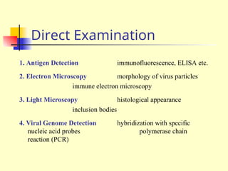
Direct Examination
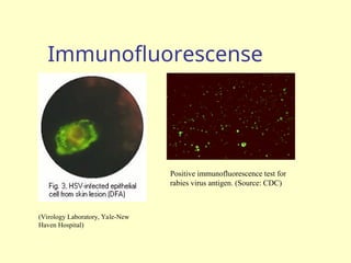
1. AntigenDetection immunofluorescence, ELISA etc.
2. Electron Microscopy morphology of virus particles
immune electron microscopy
3. Light Microscopy histological appearance
inclusion bodies
4. Viral Genome Detection hybridization with specific
nucleic acid probes polymerase chain
reaction (PCR)
4.
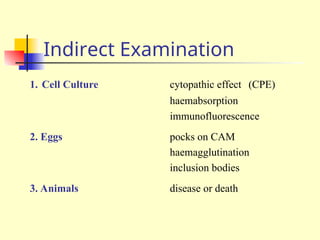
Indirect Examination
1. CellCulture cytopathic effect (CPE)
haemabsorption
immunofluorescence
2. Eggs pocks on CAM
haemagglutination
inclusion bodies
3. Animals disease or death
5.
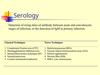
Serology
Detection of risingtitres of antibody between acute and convalescent
stages of infection, or the detection of IgM in primary infection.
Classical Techniques Newer Techniques
1. Complement fixation tests (CFT) 1. Radioimmunoassay (RIA)
2. Haemagglutination inhibition tests 2. Enzyme linked immunosorbent assay (EIA)
3. Immunofluorescence techniques (IF) 3. Particle agglutination
4. Neutralization tests 4. Western Blot (WB)
5. Counter-immunoelectrophoresis 5. RIBA, Line immunoassay
6.
Virus Isolation
Cell Culturesare most widely used for virus isolation, there are 3
types of cell cultures:
1. Primary cells - Monkey Kidney
2. Semi-continuous cells - Human embryonic kidney and skin
fibroblasts
3. Continuous cells - HeLa, Vero, Hep2, LLC-MK2, MDCK
Primary cell culture are widely acknowledged as the best cell culture
systems available since they support the widest range of viruses.
However, they are very expensive and it is often difficult to obtain a
reliable supply. Continuous cells are the most easy to handle but the range
of viruses supported is often limited.
7.
Cell Cultures
Growing virusmay produce
1. Cytopathic Effect (CPE) - such as the ballooning of cells or
syncytia formation, may be specific or non-specific.
2. Haemadsorption - cells acquire the ability to stick to
mammalian red blood cells.
Confirmation of the identity of the virus may be carried out using
neutralization, haemadsorption-inhibition or immunofluorescence
tests.
8.
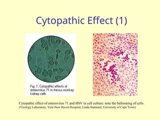
Cytopathic Effect (1)
Cytopathiceffect of enterovirus 71 and HSV in cell culture: note the ballooning of cells.
(Virology Laboratory, Yale-New Haven Hospital, Linda Stannard, University of Cape Town)
9.
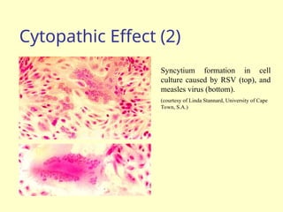
Cytopathic Effect (2)
Syncytiumformation in cell
culture caused by RSV (top), and
measles virus (bottom).
(courtesy of Linda Stannard, University of Cape
Town, S.A.)
10.
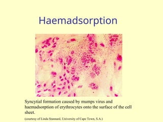
Haemadsorption
Syncytial formation causedby mumps virus and
haemadsorption of erythrocytes onto the surface of the cell
sheet.
(courtesy of Linda Stannard, University of Cape Town, S.A.)
11.
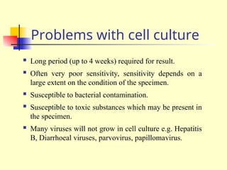
Problems with cellculture
Long period (up to 4 weeks) required for result.
Often very poor sensitivity, sensitivity depends on a
large extent on the condition of the specimen.
Susceptible to bacterial contamination.
Susceptible to toxic substances which may be present in
the specimen.
Many viruses will not grow in cell culture e.g. Hepatitis
B, Diarrhoeal viruses, parvovirus, papillomavirus.
12.
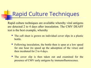
Rapid Culture Techniques
Rapidculture techniques are available whereby viral antigens
are detected 2 to 4 days after inoculation. The CMV DEAFF
test is the best example, whereby
The cell sheet is grown on individual cover slips in a plastic
bottle.
Following inoculation, the bottle then is spun at a low speed
for one hour (to speed up the adsorption of the virus) and
then incubated for 2 to 4 days.
The cover slip is then taken out and examined for the
presence of CMV early antigens by immunofluorescence.
13.
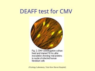
DEAFF test forCMV
(Virology Laboratory, Yale-New Haven Hospital)
14.
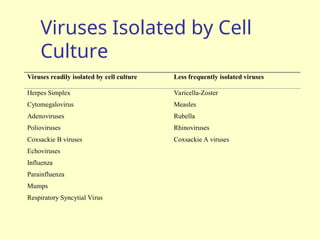
Viruses Isolated byCell
Culture
Viruses readily isolated by cell culture Less frequently isolated viruses
Herpes Simplex Varicella-Zoster
Cytomegalovirus Measles
Adenoviruses Rubella
Polioviruses Rhinoviruses
Coxsackie B viruses Coxsackie A viruses
Echoviruses
Influenza
Parainfluenza
Mumps
Respiratory Syncytial Virus
15.
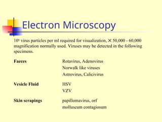
Electron Microscopy
106
virus particlesper ml required for visualization, 50,000 - 60,000
magnification normally used. Viruses may be detected in the following
specimens.
Faeces Rotavirus, Adenovirus
Norwalk like viruses
Astrovirus, Calicivirus
Vesicle Fluid HSV
VZV
Skin scrapings papillomavirus, orf
molluscum contagiosum
Immune Electron
Microscopy
The sensitivityand specificity of EM may be enhanced by
immune electron microscopy. There are two variants:-
Classical Immune electron microscopy (IEM) - the sample is
treated with specific anti-sera before being put up for EM.
Viral particles present will be agglutinated and thus congregate
together by the antibody.
Solid phase immune electron microscopy (SPIEM) - the grid is
coated with specific anti-sera. Virus particles present in the
sample will be absorbed onto the grid by the antibody.
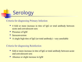
Serology
Criteria for diagnosingPrimary Infection
4 fold or more increase in titre of IgG or total antibody between
acute and convalescent sera
Presence of IgM
Seroconversion
A single high titre of IgG (or total antibody) - very unreliable
Criteria for diagnosing Reinfection
fold or more increase in titre of IgG or total antibody between acute
and convalescent sera
Absence or slight increase in IgM
20.
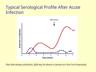
Typical Serological ProfileAfter Acute
Infection
Note that during reinfection, IgM may be absent or present at a low level transiently
21.
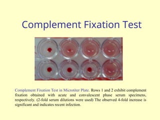
Complement Fixation Test
ComplementFixation Test in Microtiter Plate. Rows 1 and 2 exhibit complement
fixation obtained with acute and convalescent phase serum specimens,
respectively. (2-fold serum dilutions were used) The observed 4-fold increase is
significant and indicates recent infection.
22.
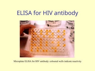
ELISA for HIVantibody
Microplate ELISA for HIV antibody: coloured wells indicate reactivity
23.
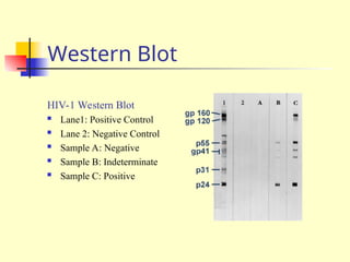
Western Blot
HIV-1 WesternBlot
Lane1: Positive Control
Lane 2: Negative Control
Sample A: Negative
Sample B: Indeterminate
Sample C: Positive
24.
Usefulness of Serological
Results
How useful a serological result is depends on the individual virus.
For example, for viruses such as rubella and hepatitis A, the onset of
clinical symptoms coincide with the development of antibodies. The
detection of IgM or rising titres of IgG in the serum of the patient would
indicate active disease.
However, many viruses often produce clinical disease before the
appearance of antibodies such as respiratory and diarrhoeal viruses. So in
this case, any serological diagnosis would be retrospective and therefore
will not be that useful.
There are also viruses which produce clinical disease months or years
after seroconversion e.g. HIV and rabies. In the case of these viruses, the
mere presence of antibody is sufficient to make a definitive diagnosis.
25.
Problems with Serology
Long period of time required for diagnosis for paired acute and convalescent
sera.
Mild local infections such as HSV genitalis may not produce a detectable
humoral immune response.
Extensive antigenic cross-reactivity between related viruses e.g. HSV and
VZV, Japanese B encephalitis and Dengue, may lead to false positive results.
immunocompromised patients often give a reduced or absent humoral
immune response.
Patients with infectious mononucleosis and those with connective tissue
diseases such as SLE may react non-specifically giving a false positive
result.
Patients given blood or blood products may give a false positive result due to
the transfer of antibody.
26.
CSF antibodies
Usedmainly for the diagnosis of herpes simplex and VZV
encephalitis
CSF normally contain little or no antibodies
presence of antibodies suggest meningitis or
meningoencephalitis
CSF antibody titre > _1_ is indicative of meningitis
Serum antibody titre 100
Diagnosis depends on the presence of an intact blood-brain
barrier
27.
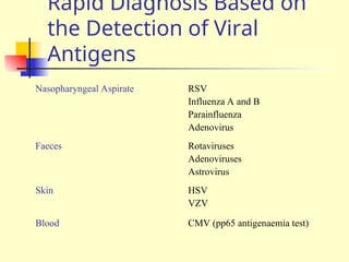
Rapid Diagnosis Basedon
the Detection of Viral
Antigens
Nasopharyngeal Aspirate RSV
Influenza A and B
Parainfluenza
Adenovirus
Faeces Rotaviruses
Adenoviruses
Astrovirus
Skin HSV
VZV
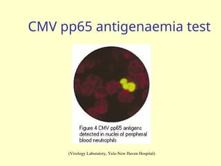
Blood CMV (pp65 antigenaemia test)
Advantages and Disadvantages
Advantages
Result available quickly, usually within a few hours.
Potential Problems
Often very much reduced sensitivity compared to cell culture,
can be as low as 20%. Specificity often poor as well.
Requires good specimens.
The procedures involved are often tedious and time-
consuming and thus expensive in terms of laboratory time.
31.
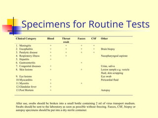
Specimens for RoutineTests
Clinical Category Blood Throat
swab
Faeces CSF Other
1. Meningitis + + + +
2. Encephalitis + + + + Brain biopsy
3. Paralytic disease + + + +
4. Respiratory illness + + Nasopharyngeal aspirate
5. Hepatitis +
6. Gastroenteritis +
7. Congenital diseases + Urine, saliva
8. Skin lesions + + Lesion sample e.g. vesicle
fluid, skin scrapping
9. Eye lesions Eye swab
10.Myocarditis + Pericardial fluid
11.Myositis + +
12.Glandular fever +
13.Post Mortem + Autopsy
After use, swabs should be broken into a small bottle containing 2 ml of virus transport medium.
Swabs should be sent to the laboratory as soon as possible without freezing. Faeces, CSF, biopsy or
autopsy specimens should be put into a dry sterile container.
32.
Molecular Methods
Methodsbased on the detection of viral genome are also
commonly known as molecular methods. It is often said that
molecular methods is the future direction of viral diagnosis.
However in practice, although the use of these methods is
indeed increasing, the role played by molecular methods in a
routine diagnostic virus laboratory is still small compared to
conventional methods.
It is certain though that the role of molecular methods will
increase rapidly in the near future.
33.
Classical Molecular
Techniques
Dot-blot,Southern blot, in-situ hydridization are examples of
classical techniques. They depend on the use of specific
DNA/RNA probes for hybridization.
The specificity of the reaction depends on the conditions used
for hybridization. However, the sensitivity of these techniques
is not better than conventional viral diagnostic methods.
However, since they are usually more tedious and expensive
than conventional techniques, they never found widespread
acceptance.
34.
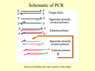
Polymerase Chain Reaction(1)
PCR allows the in vitro amplification of specific target DNA sequences by a
factor of 106
and is thus an extremely sensitive technique.
It is based on an enzymatic reaction involving the use of synthetic
oligonucleotides flanking the target nucleic sequence of interest.
These oligonucleotides act as primers for the thermostable Taq polymerase.
Repeated cycles (usually 25 to 40) of denaturation of the template DNA (at
94o
C), annealing of primers to their complementary sequences (50o
C), and
primer extension (72o
C) result in the exponential production of the specific
target fragment.
Further sensitivity and specificity may be obtained by the nested PCR.
Detection and identification of the PCR product is usually carried out by
agarose gel electrophoresis, hybridization with a specific oligonucleotide
probe, restriction enzyme analysis, or DNA sequencing.
35.
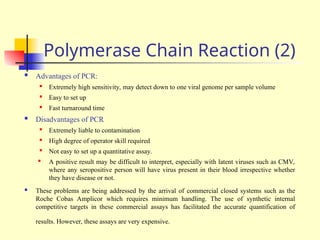
Polymerase Chain Reaction(2)
Advantages of PCR:
Extremely high sensitivity, may detect down to one viral genome per sample volume
Easy to set up
Fast turnaround time
Disadvantages of PCR
Extremely liable to contamination
High degree of operator skill required
Not easy to set up a quantitative assay.
A positive result may be difficult to interpret, especially with latent viruses such as CMV,
where any seropositive person will have virus present in their blood irrespective whether
they have disease or not.
These problems are being addressed by the arrival of commercial closed systems such as the
Roche Cobas Amplicor which requires minimum handling. The use of synthetic internal
competitive targets in these commercial assays has facilitated the accurate quantification of
results. However, these assays are very expensive.
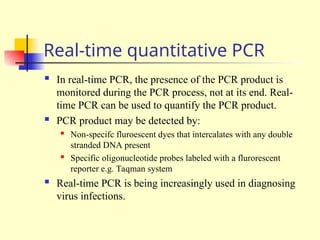
Real-time quantitative PCR
In real-time PCR, the presence of the PCR product is
monitored during the PCR process, not at its end. Real-
time PCR can be used to quantify the PCR product.
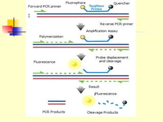
PCR product may be detected by:
Non-specifc fluroescent dyes that intercalates with any double
stranded DNA present
Specific oligonucleotide probes labeled with a flurorescent
reporter e.g. Taqman system
Real-time PCR is being increasingly used in diagnosing
virus infections.
39.
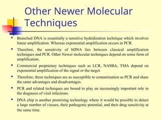
Other Newer Molecular
Techniques
Branched DNA is essentially a sensitive hydridization technique which involves
linear amplification. Whereas exponential amplification occurs in PCR.
Therefore, the sensitivity of bDNA lies between classical amplification
techniques and PCR. Other Newer molecular techniques depend on some form of
amplification.
Commercial proprietary techniques such as LCR, NASBA, TMA depend on
exponential amplification of the signal or the target.
Therefore, these techniques are as susceptible to contamination as PCR and share
the same advantages and disadvantages.
PCR and related techniques are bound to play an increasingly important role in
the diagnosis of viral infections.
DNA chip is another promising technology where it would be possible to detect
a large number of viruses, their pathogenic potential, and their drug sensitivity at
the same time.
40.
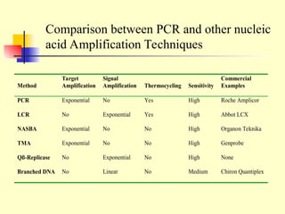
Method
Target
Amplification
Signal
Amplification Thermocycling Sensitivity
Commercial
Examples
PCRExponential No Yes High Roche Amplicor
LCR No Exponential Yes High Abbot LCX
NASBA Exponential No No High Organon Teknika
TMA Exponential No No High Genprobe
Qß-Replicase No Exponential No High None
Branched DNA No Linear No Medium Chiron Quantiplex
Comparison between PCR and other nucleic
acid Amplification Techniques