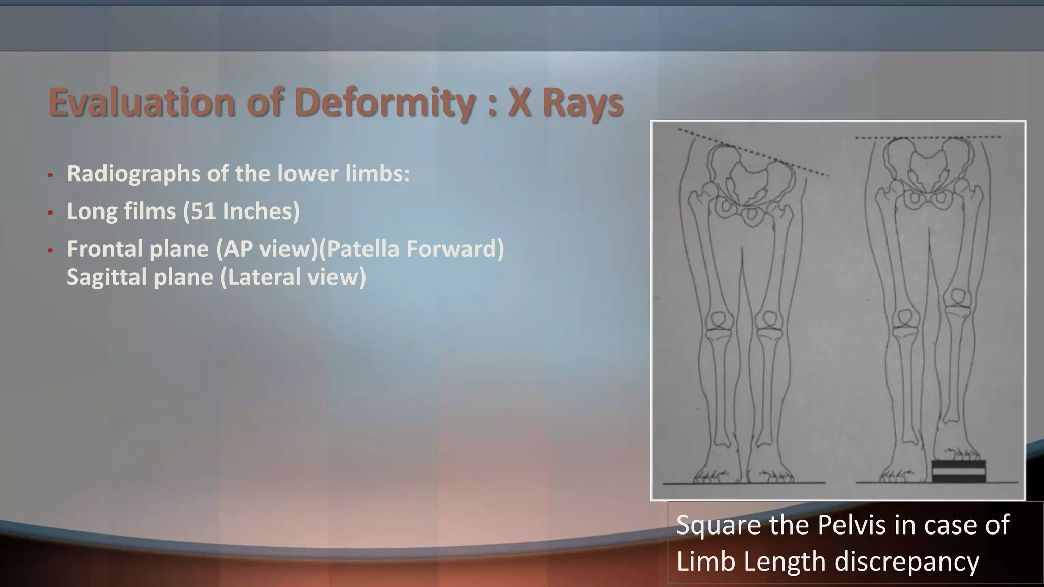
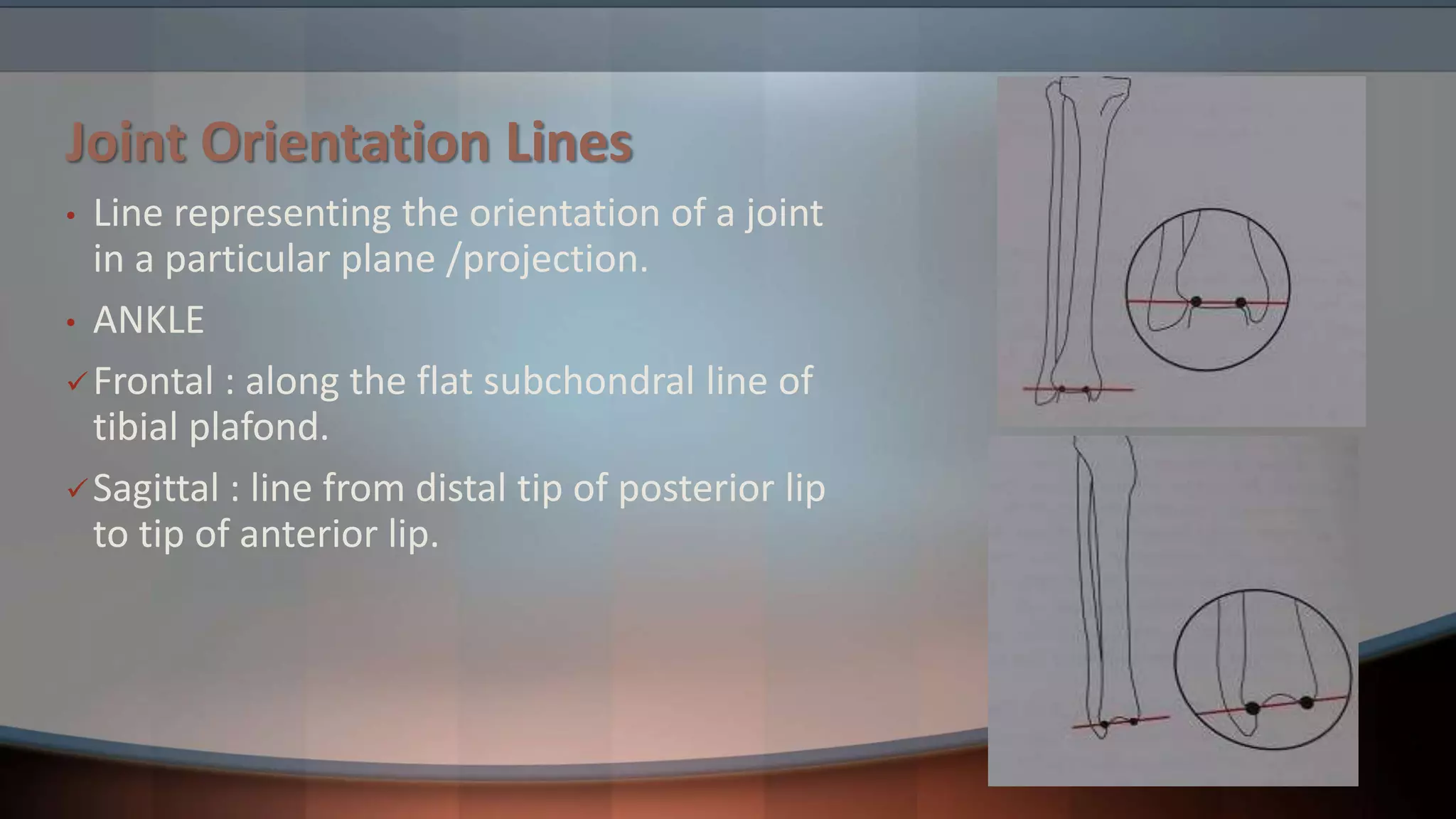
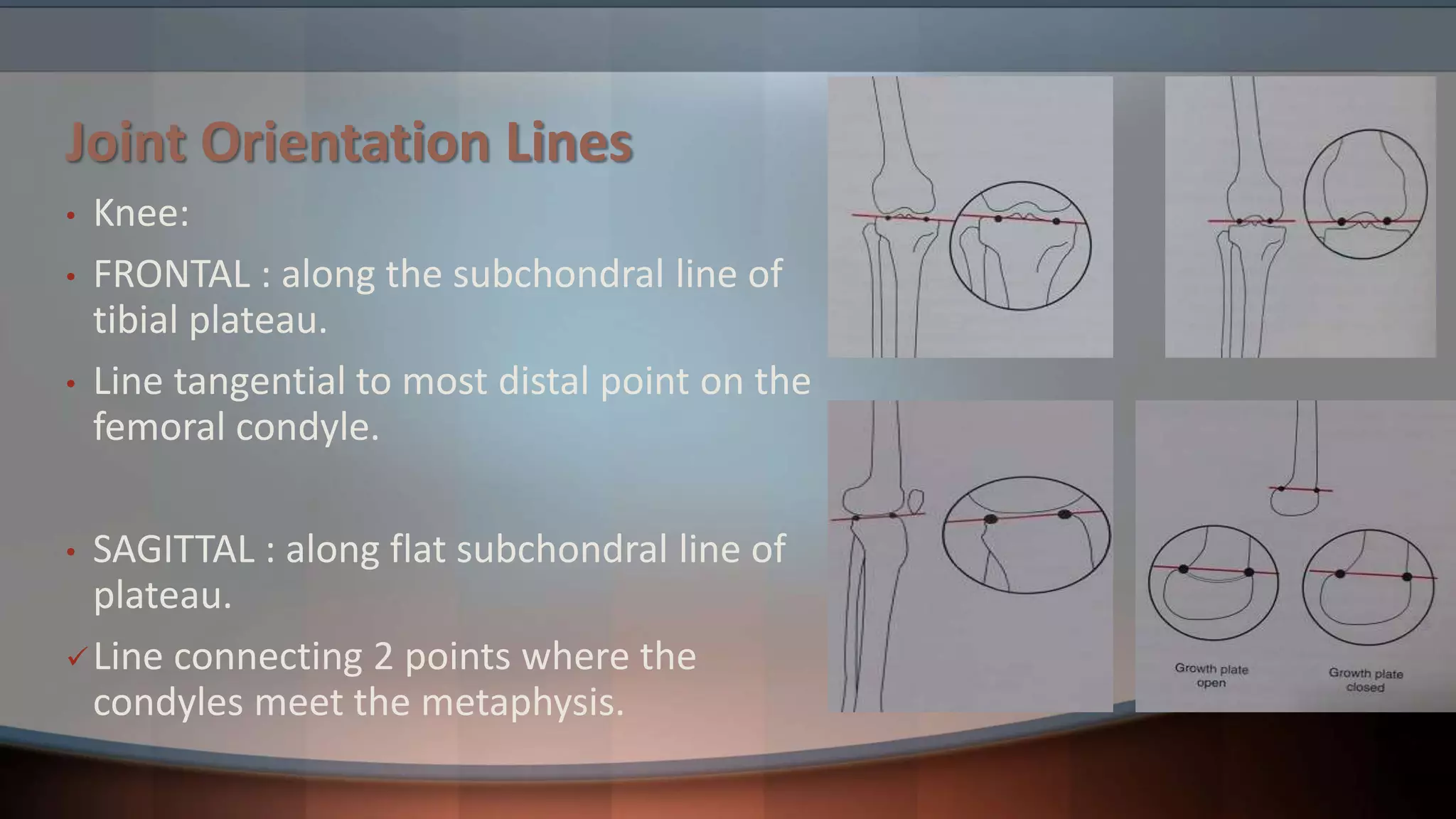
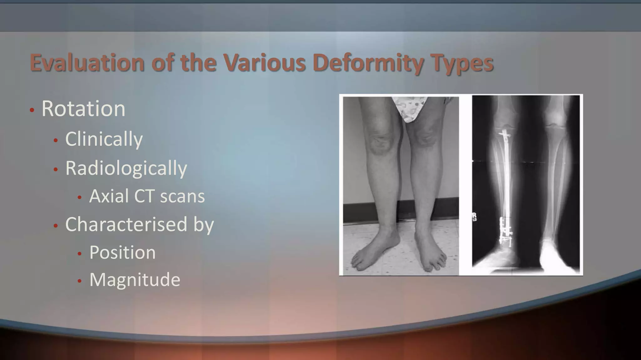
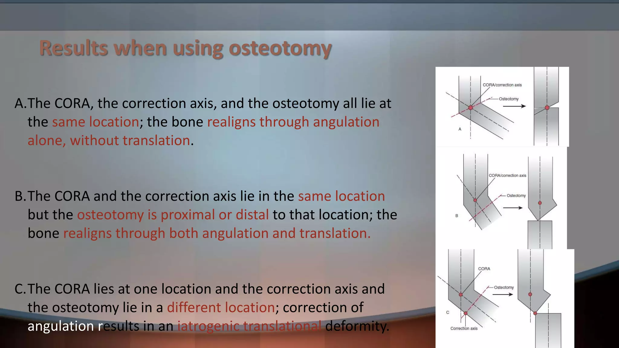
SlideShare a Scribd company logo
Principles of Deformity
Correction
Dr Ankit Madharia
Junior Resident
NKPSIMS & RC
Deformity
• Definition:
• It’s the position of a limb/Joint, from which it cannot be
brought back to its normal anatomical position.
Deformity
• Described as abnormalities of
• Length
• Angulation
• Rotation
• Translation
• The location, magnitude, and direction of the deformity
complete the characterization of a bony deformity
Evaluation of Deformity
• Clinical Examination
• Radiological Examination
• Xrays
• CT Scans
Evaluation of Deformity : X Rays
• Radiographs of the lower limbs:
• Long films (51 Inches)
• Frontal plane (AP view)(Patella Forward)
Sagittal plane (Lateral view)
Evaluation of Deformity : X Rays
• Radiographs of the lower limbs:
• Long films (51 Inches)
• Frontal plane (AP view)(Patella Forward)
Sagittal plane (Lateral view)
Evaluation of Deformity : X Rays
• Radiographs of the lower limbs:
• Long films (51 Inches)
• Frontal plane (AP view)(Patella Forward)
Sagittal plane (Lateral view)
Evaluation of Deformity : X Rays
• Radiographs of the lower limbs:
• Long films (51 Inches)
• Frontal plane (AP view)(Patella Forward)
Sagittal plane (Lateral view)
Square the Pelvis in case of
Limb Length discrepancy
Evaluation of Deformity : CT Scan
• CT Scanogram
AXIS
• Each long bone has 2 axis :-
Mechanical axis
Anatomical axis.
MECHANICAL AXIS
• Straight line connecting the joint
center points of the proximal &
distal joints.
• Its always a straight line whether in
frontal or sagittal plane.
ANATOMICAL AXIS
• Is mid diaphyseal line.
• In a normal bone, the
anatomic axis is a
single straight line.
• In a malunited bone
with angulation, each
bony segment can be
defined by its own
anatomic axis
Limb Alignment
• It involves assessment of the frontal
plane mechanical axis of the entire limb
rather than single bones
Limb Alignment
• Mechanical axis deviation (MAD) is measured
as the distance from the knee joint center to
the line connecting the joint centers of the
hip and ankle.
• Normally, 1 mm to 15 mm medial to the knee
joint center.
Joint Orientation Lines
• Line representing the orientation of a joint
in a particular plane /projection.
• ANKLE
Frontal : along the flat subchondral line of
tibial plafond.
Sagittal : line from distal tip of posterior lip
to tip of anterior lip.
Joint Orientation Lines
• Knee:
• FRONTAL : along the subchondral line of
tibial plateau.
• Line tangential to most distal point on the
femoral condyle.
• SAGITTAL : along flat subchondral line of
plateau.
Line connecting 2 points where the
condyles meet the metaphysis.
Joint Orientation Lines
• Hip:
• FRONTAL : from tip of greater trochanter to
center of femoral head.
• Also from the centre of femoral head along
the anatomical axis of the femoral neck
• The relation between anatomical or
mechanical axes and the joint
orientation lines can be referred to as
joint orientation angles
Joint Orientation Angles
Centre of Rotation of Angulation (CORA)
• The intersection of the proximal axis and
distal axis of a deformed bone is called
the CORA , which is the point about
which a deformity may be rotated to
achieve correction.
• Either Anatomical or Mechanical axis can
be used to identify CORA.
Centre of Rotation of Angulation (CORA)
• Correction of angulation by rotating the bone
around a point on the line that bisects the
angle of the CORA (the “bisector”) ensures
realignment of the anatomic and mechanical
axes without introducing an iatrogenic
translational deformity.
• All points that lie on the bisector can be
considered to be CORAs because angulation
about these points will result in realignment
of the deformed bone
Evaluation of the Various Deformity Types
• Length
• Clinically
• Radiologically
Evaluation of the Various Deformity Types
• Angulation
• Characterized by Magnitude and direction of apex
• Identification of the CORA is key in characterizing
angular deformities and planning their correction
• Angulation can be in Frontal plane or in sagittal
plane or in oblique plane
Evaluation of the Various Deformity Types
• Angulation
Evaluation of the Various Deformity Types
• Rotation
• Clinically
• Radiologically
• Axial CT scans
• Characterised by
• Position
• Magnitude
Evaluation of the Various Deformity Types
• Translation
• Clinically
• Radiologically
• Axial CT scans
• Characterised by
• Plane
• Direction
• Magnitude
• Level
Treatment
• Following evaluation, the deformity is characterized by
its
• type (length, angulation, rotational, translational, or
combined),
• the direction of the apex (anterior, lateral, posterolateral,
etc.),
• the orientation plane,
• It’s magnitude,
• and the level of the CORA
Osteotomies
• An osteotomy is used to separate the deformed bone
segments to allow realignment of the anatomic and
mechanical axes.
• The ability of an osteotomy to restore alignment depends on
• location of the CORA
• Axis about which correction is performed (the correction axis),
• Location of the osteotomy
Results when using osteotomy
A.The CORA, the correction axis, and the osteotomy all lie at
the same location; the bone realigns through angulation
alone, without translation.
B.The CORA and the correction axis lie in the same location
but the osteotomy is proximal or distal to that location; the
bone realigns through both angulation and translation.
C.The CORA lies at one location and the correction axis and
the osteotomy lie in a different location; correction of
angulation results in an iatrogenic translational deformity.
Wedge osteotomy
• The type of wedge osteotomy is determined by the
location of the osteotomy relative to the locations of the
CORA and the correction axis
Wedge osteotomy
A.Opening wedge osteotomy.
• The CORA and correction axis lie on the cortex
on the convex side of the deformity.
• The cortex on the concave side of the deformity
is distracted to restore alignment, opening an
empty wedge that traverses the diameter of
the bone.
• Opening wedge osteotomy increases final bone
length.
Wedge osteotomy
B. Neutral wedge osteotomy.
• The CORA and correction axis lie in the middle of
the bone.
• The concave side cortex is distracted and the
convex side cortex is compressed.
• A bone wedge is removed from the convex side.
• Neutral wedge osteotomy has no effect on final
bone length.
Wedge osteotomies
C. Closing wedge osteotomy.
• The CORA and correction axis lie on the concave
cortex of the deformity.
• The cortex on the convex side of the deformity is
compressed to restore alignment, requiring removal
of a bone wedge across the entire bone diameter.
• A closing wedge osteotomy decreases final bone
length.
Dome Osteotomy
• In a dome osteotomy, the osteotomy site
cannot pass through both the CORA and
the correction axis. Thus, translation will
always occur when using a dome
osteotomy.
Dome Osteotomy
Ideally, the CORA and correction axis are
mutually located with the osteotomy
proximal or distal to that location such
that the angulation and obligatory
translation that occurs at the osteotomy
site results in realignment of the bone
axis.
Dome Osteotomy
When the CORA and correction axis are
not mutually located, a dome osteotomy
through the CORA location results in a
translational deformity.
Dome osteotomy
the CORA and correction axis are mutually located with
the osteotomy distal to that location in all of these
examples.
A.Opening dome osteotomy.
The CORA and correction axis lie on the cortex on the
convex side of the deformity.
Opening dome osteotomy increases final bone length.
Dome osteotomy
B. Neutral dome osteotomy.
The CORA and correction axis lie in the middle of the
bone.
Neutral dome osteotomy has no effect on final bone
length..
Dome osteotomy
C. Closing dome osteotomy.
The CORA and correction axis lie on the concave cortex of
the deformity.
A closing dome osteotomy decreases final bone length
It can result in significant overhang of bone that may
require resection
Treatment By Deformity type : Length
• Acute distraction or compression methods obtain
immediate correction of limb length by acute
lengthening with bone grafting or acute shortening,
respectively
• Gradual correction techniques for length deformities
typically use Ilizarov external fixation/ LRS
Treatment By Deformity type : Angulation
• Correction of angulation deformities involves
making an osteotomy, obtaining realignment of the
bone segments, and securing fixation during healing.
• Alternatively, the correction may be made gradually
using external fixation to both restore alignment and
provide stabilization during healing
Treatment By Deformity type : Rotation
• Correction of a rotational deformity requires an
osteotomy and rotational realignment followed by
stabilization.
Treatment By Deformity type : Translation
• Translational deformities may be
corrected in one of three ways.
Thank you

More Related Content

What's hot

total hip arthroplasty
total hip arthroplastytotal hip arthroplasty
total hip arthroplasty
Sunil Poonia
 
Ilizarov External fixator
Ilizarov External fixatorIlizarov External fixator
Ilizarov External fixatorAbdullah Mamun
 
Proximal humerus fracture Management
Proximal humerus  fracture ManagementProximal humerus  fracture Management
Proximal humerus fracture Managementvaruntandra
 
Osteotomies around the hip
Osteotomies around the hipOsteotomies around the hip
Osteotomies around the hip
Santoshi Tanabuddi
 
Radial head replacement best evidence
Radial head replacement best evidenceRadial head replacement best evidence
Radial head replacement best evidenceorthoprinciples
 
Templating X-rays in THR
Templating X-rays in THR Templating X-rays in THR
Templating X-rays in THR
Dr. Bushu Harna
 
Total knee approaches
Total knee approachesTotal knee approaches
Total knee approaches
jatinder12345
 
Triple arthrodesis seminar by Dr Chirag Patel
Triple arthrodesis seminar by Dr Chirag PatelTriple arthrodesis seminar by Dr Chirag Patel
Triple arthrodesis seminar by Dr Chirag Patel
Chirag Patel
 
Limb deformity
Limb deformityLimb deformity
Limb deformity
ujjalrajbangshi
 
Primary total knee arthroplasty
Primary total knee arthroplastyPrimary total knee arthroplasty
Primary total knee arthroplastyjatinder12345
 
Protrusio acetabuli
Protrusio acetabuliProtrusio acetabuli
Protrusio acetabuli
Ghazwan Bayaty
 
Proximal femur focal def
Proximal femur focal defProximal femur focal def
Proximal femur focal def
Ponnilavan Ponz
 
Acl reconstruction
Acl reconstructionAcl reconstruction
Acl reconstruction
Ponnilavan Ponz
 
Septic arthritis sequelae
Septic arthritis sequelaeSeptic arthritis sequelae
Septic arthritis sequelaeorthoprince
 
spine surgical approaches along with tb spine complications
 spine surgical approaches along with tb spine complications spine surgical approaches along with tb spine complications
spine surgical approaches along with tb spine complications
Pramod Yspam
 
Distal Humerus Fractures.pptx
Distal Humerus Fractures.pptxDistal Humerus Fractures.pptx
Distal Humerus Fractures.pptx
SethiNet presentations
 
Proximal Tibia Surgical approaches
Proximal Tibia Surgical approachesProximal Tibia Surgical approaches
Proximal Tibia Surgical approaches
MOHAMMED ROSHEN
 
osteotomies around hip
osteotomies around hiposteotomies around hip
osteotomies around hip
Prashanth Kumar
 
Humeral shaft
Humeral shaftHumeral shaft
Humeral shaft
Orthosurg2016
 

What's hot (20)

total hip arthroplasty
total hip arthroplastytotal hip arthroplasty
total hip arthroplasty
 
Tkr by dr. saumya agarwal
Tkr by dr. saumya agarwalTkr by dr. saumya agarwal
Tkr by dr. saumya agarwal
 
Ilizarov External fixator
Ilizarov External fixatorIlizarov External fixator
Ilizarov External fixator
 
Proximal humerus fracture Management
Proximal humerus  fracture ManagementProximal humerus  fracture Management
Proximal humerus fracture Management
 
Osteotomies around the hip
Osteotomies around the hipOsteotomies around the hip
Osteotomies around the hip
 
Radial head replacement best evidence
Radial head replacement best evidenceRadial head replacement best evidence
Radial head replacement best evidence
 
Templating X-rays in THR
Templating X-rays in THR Templating X-rays in THR
Templating X-rays in THR
 
Total knee approaches
Total knee approachesTotal knee approaches
Total knee approaches
 
Triple arthrodesis seminar by Dr Chirag Patel
Triple arthrodesis seminar by Dr Chirag PatelTriple arthrodesis seminar by Dr Chirag Patel
Triple arthrodesis seminar by Dr Chirag Patel
 
Limb deformity
Limb deformityLimb deformity
Limb deformity
 
Primary total knee arthroplasty
Primary total knee arthroplastyPrimary total knee arthroplasty
Primary total knee arthroplasty
 
Protrusio acetabuli
Protrusio acetabuliProtrusio acetabuli
Protrusio acetabuli
 
Proximal femur focal def
Proximal femur focal defProximal femur focal def
Proximal femur focal def
 
Acl reconstruction
Acl reconstructionAcl reconstruction
Acl reconstruction
 
Septic arthritis sequelae
Septic arthritis sequelaeSeptic arthritis sequelae
Septic arthritis sequelae
 
spine surgical approaches along with tb spine complications
 spine surgical approaches along with tb spine complications spine surgical approaches along with tb spine complications
spine surgical approaches along with tb spine complications
 
Distal Humerus Fractures.pptx
Distal Humerus Fractures.pptxDistal Humerus Fractures.pptx
Distal Humerus Fractures.pptx
 
Proximal Tibia Surgical approaches
Proximal Tibia Surgical approachesProximal Tibia Surgical approaches
Proximal Tibia Surgical approaches
 
osteotomies around hip
osteotomies around hiposteotomies around hip
osteotomies around hip
 
Humeral shaft
Humeral shaftHumeral shaft
Humeral shaft
 

Viewers also liked

Osteotomy
OsteotomyOsteotomy
Osteotomy
Robins Shah
 
radiological signs of in orthopaedics
radiological signs of in orthopaedicsradiological signs of in orthopaedics
radiological signs of in orthopaedics
sguruprasad311286
 
Orthopaedics Mc Qs
Orthopaedics Mc QsOrthopaedics Mc Qs
Orthopaedics Mc Qsmahmoud beco
 
Introduction to musculoskeletal radiology
Introduction to musculoskeletal radiologyIntroduction to musculoskeletal radiology
Introduction to musculoskeletal radiologySubhanjan Das
 
Bone cement.pptx 2 its science and cementing technique and safe surgical use
Bone cement.pptx 2 its science and cementing technique and safe surgical useBone cement.pptx 2 its science and cementing technique and safe surgical use
Bone cement.pptx 2 its science and cementing technique and safe surgical useMahatma Gandhi Hospital Parel Mumbai
 
Limb length discrepency
Limb length discrepencyLimb length discrepency
Limb length discrepency
Naveed Jumani
 

Viewers also liked (7)

Osteotomy
OsteotomyOsteotomy
Osteotomy
 
radiological signs of in orthopaedics
radiological signs of in orthopaedicsradiological signs of in orthopaedics
radiological signs of in orthopaedics
 
Orthopaedics Mc Qs
Orthopaedics Mc QsOrthopaedics Mc Qs
Orthopaedics Mc Qs
 
Introduction to musculoskeletal radiology
Introduction to musculoskeletal radiologyIntroduction to musculoskeletal radiology
Introduction to musculoskeletal radiology
 
Hip osteotomy
Hip osteotomyHip osteotomy
Hip osteotomy
 
Bone cement.pptx 2 its science and cementing technique and safe surgical use
Bone cement.pptx 2 its science and cementing technique and safe surgical useBone cement.pptx 2 its science and cementing technique and safe surgical use
Bone cement.pptx 2 its science and cementing technique and safe surgical use
 
Limb length discrepency
Limb length discrepencyLimb length discrepency
Limb length discrepency
 

Similar to Principles of deformity correction

MALUNION.pptx
MALUNION.pptxMALUNION.pptx
MALUNION.pptx
Karthik MV
 
malunion.pptx
malunion.pptxmalunion.pptx
malunion.pptx
Pirfa Jo
 
Principle of Deformity Correction in lower Limb
Principle of Deformity Correction in lower Limb Principle of Deformity Correction in lower Limb
Principle of Deformity Correction in lower Limb
Kaushal Kafle
 
Osteotomies around the hip
Osteotomies around the hipOsteotomies around the hip
Osteotomies around the hipSanjay Kumar
 
THR
THRTHR
Fractures around elbow lateral condyle and intercondylar fractures
 Fractures around elbow lateral condyle and intercondylar fractures Fractures around elbow lateral condyle and intercondylar fractures
Fractures around elbow lateral condyle and intercondylar fractures
Siddhartha Sinha
 
Ankle instability
Ankle instabilityAnkle instability
Ankle instability
RziUllah
 
tota knee arthroplasty.pptx
tota knee arthroplasty.pptxtota knee arthroplasty.pptx
tota knee arthroplasty.pptx
ahmedshafik44
 
Fractures around elbow lateral condyle and intercondylar fractures
Fractures around elbow lateral condyle and intercondylar fracturesFractures around elbow lateral condyle and intercondylar fractures
Fractures around elbow lateral condyle and intercondylar fractures
Siddhartha Sinha
 
Osteotomy around elbow
Osteotomy around elbowOsteotomy around elbow
Osteotomy around elbow
Sushil Sharma
 
Distal humeruss.pptx
Distal humeruss.pptxDistal humeruss.pptx
Distal humeruss.pptx
manasil1
 
Proximal tibia fractures(Plateau, spine ,Tubercle and Epiphyseal )
Proximal tibia fractures(Plateau, spine ,Tubercle and Epiphyseal ) Proximal tibia fractures(Plateau, spine ,Tubercle and Epiphyseal )
Proximal tibia fractures(Plateau, spine ,Tubercle and Epiphyseal )
Prasanthmuddada
 
HIgh Tibial Osteotomy: when and how
HIgh Tibial Osteotomy: when and howHIgh Tibial Osteotomy: when and how
HIgh Tibial Osteotomy: when and how
AbhishekKaushik126
 
Mean Value Articulator
Mean Value ArticulatorMean Value Articulator
Mean Value Articulator
kontamuru
 
Acetabulum Fractures - Anatomy, Radiological Evaluation, Classification and M...
Acetabulum Fractures - Anatomy, Radiological Evaluation, Classification and M...Acetabulum Fractures - Anatomy, Radiological Evaluation, Classification and M...
Acetabulum Fractures - Anatomy, Radiological Evaluation, Classification and M...
Arun Upreti
 
Distal humerus revised
Distal humerus revisedDistal humerus revised
Distal humerus revisedAhmed Azab
 
Surgical approach intercondylar/ supracondylar humerus fracture
Surgical approach intercondylar/ supracondylar humerus fractureSurgical approach intercondylar/ supracondylar humerus fracture
Surgical approach intercondylar/ supracondylar humerus fracture
Khadijah Nordin
 
Anatomy of elbow and INTERCONDYLAR FRACTURE OF THE HUMERUS
Anatomy of elbow and INTERCONDYLAR FRACTURE OF THE HUMERUSAnatomy of elbow and INTERCONDYLAR FRACTURE OF THE HUMERUS
Anatomy of elbow and INTERCONDYLAR FRACTURE OF THE HUMERUS
Dr. Vinaykumar S Appannavar
 
Diagnostic aids in implant seminar in detail
Diagnostic aids in implant seminar in detailDiagnostic aids in implant seminar in detail
Diagnostic aids in implant seminar in detail
NikitaChhabariya
 

Similar to Principles of deformity correction (20)

MALUNION.pptx
MALUNION.pptxMALUNION.pptx
MALUNION.pptx
 
malunion.pptx
malunion.pptxmalunion.pptx
malunion.pptx
 
Principle of Deformity Correction in lower Limb
Principle of Deformity Correction in lower Limb Principle of Deformity Correction in lower Limb
Principle of Deformity Correction in lower Limb
 
Osteotomies around the hip
Osteotomies around the hipOsteotomies around the hip
Osteotomies around the hip
 
THR
THRTHR
THR
 
Fractures around elbow lateral condyle and intercondylar fractures
 Fractures around elbow lateral condyle and intercondylar fractures Fractures around elbow lateral condyle and intercondylar fractures
Fractures around elbow lateral condyle and intercondylar fractures
 
Ankle instability
Ankle instabilityAnkle instability
Ankle instability
 
tota knee arthroplasty.pptx
tota knee arthroplasty.pptxtota knee arthroplasty.pptx
tota knee arthroplasty.pptx
 
Fractures around elbow lateral condyle and intercondylar fractures
Fractures around elbow lateral condyle and intercondylar fracturesFractures around elbow lateral condyle and intercondylar fractures
Fractures around elbow lateral condyle and intercondylar fractures
 
Osteotomy around elbow
Osteotomy around elbowOsteotomy around elbow
Osteotomy around elbow
 
Distal humeruss.pptx
Distal humeruss.pptxDistal humeruss.pptx
Distal humeruss.pptx
 
Proximal tibia fractures(Plateau, spine ,Tubercle and Epiphyseal )
Proximal tibia fractures(Plateau, spine ,Tubercle and Epiphyseal ) Proximal tibia fractures(Plateau, spine ,Tubercle and Epiphyseal )
Proximal tibia fractures(Plateau, spine ,Tubercle and Epiphyseal )
 
HIgh Tibial Osteotomy: when and how
HIgh Tibial Osteotomy: when and howHIgh Tibial Osteotomy: when and how
HIgh Tibial Osteotomy: when and how
 
Mean Value Articulator
Mean Value ArticulatorMean Value Articulator
Mean Value Articulator
 
Acetabulum Fractures - Anatomy, Radiological Evaluation, Classification and M...
Acetabulum Fractures - Anatomy, Radiological Evaluation, Classification and M...Acetabulum Fractures - Anatomy, Radiological Evaluation, Classification and M...
Acetabulum Fractures - Anatomy, Radiological Evaluation, Classification and M...
 
Distal humerus revised
Distal humerus revisedDistal humerus revised
Distal humerus revised
 
Surgical approach intercondylar/ supracondylar humerus fracture
Surgical approach intercondylar/ supracondylar humerus fractureSurgical approach intercondylar/ supracondylar humerus fracture
Surgical approach intercondylar/ supracondylar humerus fracture
 
How_to_do_a_TKR.ppt
How_to_do_a_TKR.pptHow_to_do_a_TKR.ppt
How_to_do_a_TKR.ppt
 
Anatomy of elbow and INTERCONDYLAR FRACTURE OF THE HUMERUS
Anatomy of elbow and INTERCONDYLAR FRACTURE OF THE HUMERUSAnatomy of elbow and INTERCONDYLAR FRACTURE OF THE HUMERUS
Anatomy of elbow and INTERCONDYLAR FRACTURE OF THE HUMERUS
 
Diagnostic aids in implant seminar in detail
Diagnostic aids in implant seminar in detailDiagnostic aids in implant seminar in detail
Diagnostic aids in implant seminar in detail
 

Recently uploaded

New Directions in Targeted Therapeutic Approaches for Older Adults With Mantl...
New Directions in Targeted Therapeutic Approaches for Older Adults With Mantl...New Directions in Targeted Therapeutic Approaches for Older Adults With Mantl...
New Directions in Targeted Therapeutic Approaches for Older Adults With Mantl...
i3 Health
 
Are There Any Natural Remedies To Treat Syphilis.pdf
Are There Any Natural Remedies To Treat Syphilis.pdfAre There Any Natural Remedies To Treat Syphilis.pdf
Are There Any Natural Remedies To Treat Syphilis.pdf
Little Cross Family Clinic
 
basicmodesofventilation2022-220313203758.pdf
basicmodesofventilation2022-220313203758.pdfbasicmodesofventilation2022-220313203758.pdf
basicmodesofventilation2022-220313203758.pdf
aljamhori teaching hospital
 
BENIGN PROSTATIC HYPERPLASIA.BPH. BPHpdf
BENIGN PROSTATIC HYPERPLASIA.BPH. BPHpdfBENIGN PROSTATIC HYPERPLASIA.BPH. BPHpdf
BENIGN PROSTATIC HYPERPLASIA.BPH. BPHpdf
DR SETH JOTHAM
 
Tom Selleck Health: A Comprehensive Look at the Iconic Actor’s Wellness Journey
Tom Selleck Health: A Comprehensive Look at the Iconic Actor’s Wellness JourneyTom Selleck Health: A Comprehensive Look at the Iconic Actor’s Wellness Journey
Tom Selleck Health: A Comprehensive Look at the Iconic Actor’s Wellness Journey
greendigital
 
How to Give Better Lectures: Some Tips for Doctors
How to Give Better Lectures: Some Tips for DoctorsHow to Give Better Lectures: Some Tips for Doctors
How to Give Better Lectures: Some Tips for Doctors
LanceCatedral
 
Non-respiratory Functions of the Lungs.pdf
Non-respiratory Functions of the Lungs.pdfNon-respiratory Functions of the Lungs.pdf
Non-respiratory Functions of the Lungs.pdf
MedicoseAcademics
 
Report Back from SGO 2024: What’s the Latest in Cervical Cancer?
Report Back from SGO 2024: What’s the Latest in Cervical Cancer?Report Back from SGO 2024: What’s the Latest in Cervical Cancer?
Report Back from SGO 2024: What’s the Latest in Cervical Cancer?
bkling
 
Couples presenting to the infertility clinic- Do they really have infertility...
Couples presenting to the infertility clinic- Do they really have infertility...Couples presenting to the infertility clinic- Do they really have infertility...
Couples presenting to the infertility clinic- Do they really have infertility...
Sujoy Dasgupta
 
Flu Vaccine Alert in Bangalore Karnataka
Flu Vaccine Alert in Bangalore KarnatakaFlu Vaccine Alert in Bangalore Karnataka
Flu Vaccine Alert in Bangalore Karnataka
addon Scans
 
ANATOMY AND PHYSIOLOGY OF URINARY SYSTEM.pptx
ANATOMY AND PHYSIOLOGY OF URINARY SYSTEM.pptxANATOMY AND PHYSIOLOGY OF URINARY SYSTEM.pptx
ANATOMY AND PHYSIOLOGY OF URINARY SYSTEM.pptx
Swetaba Besh
 
Surgical Site Infections, pathophysiology, and prevention.pptx
Surgical Site Infections, pathophysiology, and prevention.pptxSurgical Site Infections, pathophysiology, and prevention.pptx
Surgical Site Infections, pathophysiology, and prevention.pptx
jval Landero
 
ARTHROLOGY PPT NCISM SYLLABUS AYURVEDA STUDENTS
ARTHROLOGY PPT NCISM SYLLABUS AYURVEDA STUDENTSARTHROLOGY PPT NCISM SYLLABUS AYURVEDA STUDENTS
ARTHROLOGY PPT NCISM SYLLABUS AYURVEDA STUDENTS
Dr. Vinay Pareek
 
Hemodialysis: Chapter 3, Dialysis Water Unit - Dr.Gawad
Hemodialysis: Chapter 3, Dialysis Water Unit - Dr.GawadHemodialysis: Chapter 3, Dialysis Water Unit - Dr.Gawad
Hemodialysis: Chapter 3, Dialysis Water Unit - Dr.Gawad
NephroTube - Dr.Gawad
 
MANAGEMENT OF ATRIOVENTRICULAR CONDUCTION BLOCK.pdf
MANAGEMENT OF ATRIOVENTRICULAR CONDUCTION BLOCK.pdfMANAGEMENT OF ATRIOVENTRICULAR CONDUCTION BLOCK.pdf
MANAGEMENT OF ATRIOVENTRICULAR CONDUCTION BLOCK.pdf
Jim Jacob Roy
 
Ophthalmology Clinical Tests for OSCE exam
Ophthalmology Clinical Tests for OSCE examOphthalmology Clinical Tests for OSCE exam
Ophthalmology Clinical Tests for OSCE exam
KafrELShiekh University
 
heat stroke and heat exhaustion in children
heat stroke and heat exhaustion in childrenheat stroke and heat exhaustion in children
heat stroke and heat exhaustion in children
SumeraAhmad5
 
Cervical & Brachial Plexus By Dr. RIG.pptx
Cervical & Brachial Plexus By Dr. RIG.pptxCervical & Brachial Plexus By Dr. RIG.pptx
Cervical & Brachial Plexus By Dr. RIG.pptx
Dr. Rabia Inam Gandapore
 
POST OPERATIVE OLIGURIA and its management
POST OPERATIVE OLIGURIA and its managementPOST OPERATIVE OLIGURIA and its management
POST OPERATIVE OLIGURIA and its management
touseefaziz1
 
Antiulcer drugs Advance Pharmacology .pptx
Antiulcer drugs Advance Pharmacology .pptxAntiulcer drugs Advance Pharmacology .pptx
Antiulcer drugs Advance Pharmacology .pptx
Rohit chaurpagar
 

Recently uploaded (20)

New Directions in Targeted Therapeutic Approaches for Older Adults With Mantl...
New Directions in Targeted Therapeutic Approaches for Older Adults With Mantl...New Directions in Targeted Therapeutic Approaches for Older Adults With Mantl...
New Directions in Targeted Therapeutic Approaches for Older Adults With Mantl...
 
Are There Any Natural Remedies To Treat Syphilis.pdf
Are There Any Natural Remedies To Treat Syphilis.pdfAre There Any Natural Remedies To Treat Syphilis.pdf
Are There Any Natural Remedies To Treat Syphilis.pdf
 
basicmodesofventilation2022-220313203758.pdf
basicmodesofventilation2022-220313203758.pdfbasicmodesofventilation2022-220313203758.pdf
basicmodesofventilation2022-220313203758.pdf
 
BENIGN PROSTATIC HYPERPLASIA.BPH. BPHpdf
BENIGN PROSTATIC HYPERPLASIA.BPH. BPHpdfBENIGN PROSTATIC HYPERPLASIA.BPH. BPHpdf
BENIGN PROSTATIC HYPERPLASIA.BPH. BPHpdf
 
Tom Selleck Health: A Comprehensive Look at the Iconic Actor’s Wellness Journey
Tom Selleck Health: A Comprehensive Look at the Iconic Actor’s Wellness JourneyTom Selleck Health: A Comprehensive Look at the Iconic Actor’s Wellness Journey
Tom Selleck Health: A Comprehensive Look at the Iconic Actor’s Wellness Journey
 
How to Give Better Lectures: Some Tips for Doctors
How to Give Better Lectures: Some Tips for DoctorsHow to Give Better Lectures: Some Tips for Doctors
How to Give Better Lectures: Some Tips for Doctors
 
Non-respiratory Functions of the Lungs.pdf
Non-respiratory Functions of the Lungs.pdfNon-respiratory Functions of the Lungs.pdf
Non-respiratory Functions of the Lungs.pdf
 
Report Back from SGO 2024: What’s the Latest in Cervical Cancer?
Report Back from SGO 2024: What’s the Latest in Cervical Cancer?Report Back from SGO 2024: What’s the Latest in Cervical Cancer?
Report Back from SGO 2024: What’s the Latest in Cervical Cancer?
 
Couples presenting to the infertility clinic- Do they really have infertility...
Couples presenting to the infertility clinic- Do they really have infertility...Couples presenting to the infertility clinic- Do they really have infertility...
Couples presenting to the infertility clinic- Do they really have infertility...
 
Flu Vaccine Alert in Bangalore Karnataka
Flu Vaccine Alert in Bangalore KarnatakaFlu Vaccine Alert in Bangalore Karnataka
Flu Vaccine Alert in Bangalore Karnataka
 
ANATOMY AND PHYSIOLOGY OF URINARY SYSTEM.pptx
ANATOMY AND PHYSIOLOGY OF URINARY SYSTEM.pptxANATOMY AND PHYSIOLOGY OF URINARY SYSTEM.pptx
ANATOMY AND PHYSIOLOGY OF URINARY SYSTEM.pptx
 
Surgical Site Infections, pathophysiology, and prevention.pptx
Surgical Site Infections, pathophysiology, and prevention.pptxSurgical Site Infections, pathophysiology, and prevention.pptx
Surgical Site Infections, pathophysiology, and prevention.pptx
 
ARTHROLOGY PPT NCISM SYLLABUS AYURVEDA STUDENTS
ARTHROLOGY PPT NCISM SYLLABUS AYURVEDA STUDENTSARTHROLOGY PPT NCISM SYLLABUS AYURVEDA STUDENTS
ARTHROLOGY PPT NCISM SYLLABUS AYURVEDA STUDENTS
 
Hemodialysis: Chapter 3, Dialysis Water Unit - Dr.Gawad
Hemodialysis: Chapter 3, Dialysis Water Unit - Dr.GawadHemodialysis: Chapter 3, Dialysis Water Unit - Dr.Gawad
Hemodialysis: Chapter 3, Dialysis Water Unit - Dr.Gawad
 
MANAGEMENT OF ATRIOVENTRICULAR CONDUCTION BLOCK.pdf
MANAGEMENT OF ATRIOVENTRICULAR CONDUCTION BLOCK.pdfMANAGEMENT OF ATRIOVENTRICULAR CONDUCTION BLOCK.pdf
MANAGEMENT OF ATRIOVENTRICULAR CONDUCTION BLOCK.pdf
 
Ophthalmology Clinical Tests for OSCE exam
Ophthalmology Clinical Tests for OSCE examOphthalmology Clinical Tests for OSCE exam
Ophthalmology Clinical Tests for OSCE exam
 
heat stroke and heat exhaustion in children
heat stroke and heat exhaustion in childrenheat stroke and heat exhaustion in children
heat stroke and heat exhaustion in children
 
Cervical & Brachial Plexus By Dr. RIG.pptx
Cervical & Brachial Plexus By Dr. RIG.pptxCervical & Brachial Plexus By Dr. RIG.pptx
Cervical & Brachial Plexus By Dr. RIG.pptx
 
POST OPERATIVE OLIGURIA and its management
POST OPERATIVE OLIGURIA and its managementPOST OPERATIVE OLIGURIA and its management
POST OPERATIVE OLIGURIA and its management
 
Antiulcer drugs Advance Pharmacology .pptx
Antiulcer drugs Advance Pharmacology .pptxAntiulcer drugs Advance Pharmacology .pptx
Antiulcer drugs Advance Pharmacology .pptx
 

Principles of deformity correction

  • 1. Principles of Deformity Correction Dr Ankit Madharia Junior Resident NKPSIMS & RC
  • 2. Deformity • Definition: • It’s the position of a limb/Joint, from which it cannot be brought back to its normal anatomical position.
  • 3. Deformity • Described as abnormalities of • Length • Angulation • Rotation • Translation • The location, magnitude, and direction of the deformity complete the characterization of a bony deformity
  • 4. Evaluation of Deformity • Clinical Examination • Radiological Examination • Xrays • CT Scans
  • 5. Evaluation of Deformity : X Rays • Radiographs of the lower limbs: • Long films (51 Inches) • Frontal plane (AP view)(Patella Forward) Sagittal plane (Lateral view)
  • 6. Evaluation of Deformity : X Rays • Radiographs of the lower limbs: • Long films (51 Inches) • Frontal plane (AP view)(Patella Forward) Sagittal plane (Lateral view)
  • 7. Evaluation of Deformity : X Rays • Radiographs of the lower limbs: • Long films (51 Inches) • Frontal plane (AP view)(Patella Forward) Sagittal plane (Lateral view)
  • 8. Evaluation of Deformity : X Rays • Radiographs of the lower limbs: • Long films (51 Inches) • Frontal plane (AP view)(Patella Forward) Sagittal plane (Lateral view) Square the Pelvis in case of Limb Length discrepancy
  • 9. Evaluation of Deformity : CT Scan • CT Scanogram
  • 10. AXIS • Each long bone has 2 axis :- Mechanical axis Anatomical axis.
  • 11. MECHANICAL AXIS • Straight line connecting the joint center points of the proximal & distal joints. • Its always a straight line whether in frontal or sagittal plane.
  • 12. ANATOMICAL AXIS • Is mid diaphyseal line. • In a normal bone, the anatomic axis is a single straight line. • In a malunited bone with angulation, each bony segment can be defined by its own anatomic axis
  • 13. Limb Alignment • It involves assessment of the frontal plane mechanical axis of the entire limb rather than single bones
  • 14. Limb Alignment • Mechanical axis deviation (MAD) is measured as the distance from the knee joint center to the line connecting the joint centers of the hip and ankle. • Normally, 1 mm to 15 mm medial to the knee joint center.
  • 15. Joint Orientation Lines • Line representing the orientation of a joint in a particular plane /projection. • ANKLE Frontal : along the flat subchondral line of tibial plafond. Sagittal : line from distal tip of posterior lip to tip of anterior lip.
  • 16. Joint Orientation Lines • Knee: • FRONTAL : along the subchondral line of tibial plateau. • Line tangential to most distal point on the femoral condyle. • SAGITTAL : along flat subchondral line of plateau. Line connecting 2 points where the condyles meet the metaphysis.
  • 17. Joint Orientation Lines • Hip: • FRONTAL : from tip of greater trochanter to center of femoral head. • Also from the centre of femoral head along the anatomical axis of the femoral neck
  • 18. • The relation between anatomical or mechanical axes and the joint orientation lines can be referred to as joint orientation angles Joint Orientation Angles
  • 19. Centre of Rotation of Angulation (CORA) • The intersection of the proximal axis and distal axis of a deformed bone is called the CORA , which is the point about which a deformity may be rotated to achieve correction. • Either Anatomical or Mechanical axis can be used to identify CORA.
  • 20. Centre of Rotation of Angulation (CORA) • Correction of angulation by rotating the bone around a point on the line that bisects the angle of the CORA (the “bisector”) ensures realignment of the anatomic and mechanical axes without introducing an iatrogenic translational deformity. • All points that lie on the bisector can be considered to be CORAs because angulation about these points will result in realignment of the deformed bone
  • 21. Evaluation of the Various Deformity Types • Length • Clinically • Radiologically
  • 22. Evaluation of the Various Deformity Types • Angulation • Characterized by Magnitude and direction of apex • Identification of the CORA is key in characterizing angular deformities and planning their correction • Angulation can be in Frontal plane or in sagittal plane or in oblique plane
  • 23. Evaluation of the Various Deformity Types • Angulation
  • 24. Evaluation of the Various Deformity Types • Rotation • Clinically • Radiologically • Axial CT scans • Characterised by • Position • Magnitude
  • 25. Evaluation of the Various Deformity Types • Translation • Clinically • Radiologically • Axial CT scans • Characterised by • Plane • Direction • Magnitude • Level
  • 26. Treatment • Following evaluation, the deformity is characterized by its • type (length, angulation, rotational, translational, or combined), • the direction of the apex (anterior, lateral, posterolateral, etc.), • the orientation plane, • It’s magnitude, • and the level of the CORA
  • 27. Osteotomies • An osteotomy is used to separate the deformed bone segments to allow realignment of the anatomic and mechanical axes. • The ability of an osteotomy to restore alignment depends on • location of the CORA • Axis about which correction is performed (the correction axis), • Location of the osteotomy
  • 28. Results when using osteotomy A.The CORA, the correction axis, and the osteotomy all lie at the same location; the bone realigns through angulation alone, without translation. B.The CORA and the correction axis lie in the same location but the osteotomy is proximal or distal to that location; the bone realigns through both angulation and translation. C.The CORA lies at one location and the correction axis and the osteotomy lie in a different location; correction of angulation results in an iatrogenic translational deformity.
  • 29. Wedge osteotomy • The type of wedge osteotomy is determined by the location of the osteotomy relative to the locations of the CORA and the correction axis
  • 30. Wedge osteotomy A.Opening wedge osteotomy. • The CORA and correction axis lie on the cortex on the convex side of the deformity. • The cortex on the concave side of the deformity is distracted to restore alignment, opening an empty wedge that traverses the diameter of the bone. • Opening wedge osteotomy increases final bone length.
  • 31. Wedge osteotomy B. Neutral wedge osteotomy. • The CORA and correction axis lie in the middle of the bone. • The concave side cortex is distracted and the convex side cortex is compressed. • A bone wedge is removed from the convex side. • Neutral wedge osteotomy has no effect on final bone length.
  • 32. Wedge osteotomies C. Closing wedge osteotomy. • The CORA and correction axis lie on the concave cortex of the deformity. • The cortex on the convex side of the deformity is compressed to restore alignment, requiring removal of a bone wedge across the entire bone diameter. • A closing wedge osteotomy decreases final bone length.
  • 33. Dome Osteotomy • In a dome osteotomy, the osteotomy site cannot pass through both the CORA and the correction axis. Thus, translation will always occur when using a dome osteotomy.
  • 34. Dome Osteotomy Ideally, the CORA and correction axis are mutually located with the osteotomy proximal or distal to that location such that the angulation and obligatory translation that occurs at the osteotomy site results in realignment of the bone axis.
  • 35. Dome Osteotomy When the CORA and correction axis are not mutually located, a dome osteotomy through the CORA location results in a translational deformity.
  • 36. Dome osteotomy the CORA and correction axis are mutually located with the osteotomy distal to that location in all of these examples. A.Opening dome osteotomy. The CORA and correction axis lie on the cortex on the convex side of the deformity. Opening dome osteotomy increases final bone length.
  • 37. Dome osteotomy B. Neutral dome osteotomy. The CORA and correction axis lie in the middle of the bone. Neutral dome osteotomy has no effect on final bone length..
  • 38. Dome osteotomy C. Closing dome osteotomy. The CORA and correction axis lie on the concave cortex of the deformity. A closing dome osteotomy decreases final bone length It can result in significant overhang of bone that may require resection
  • 39. Treatment By Deformity type : Length • Acute distraction or compression methods obtain immediate correction of limb length by acute lengthening with bone grafting or acute shortening, respectively • Gradual correction techniques for length deformities typically use Ilizarov external fixation/ LRS
  • 40. Treatment By Deformity type : Angulation • Correction of angulation deformities involves making an osteotomy, obtaining realignment of the bone segments, and securing fixation during healing. • Alternatively, the correction may be made gradually using external fixation to both restore alignment and provide stabilization during healing
  • 41. Treatment By Deformity type : Rotation • Correction of a rotational deformity requires an osteotomy and rotational realignment followed by stabilization.
  • 42. Treatment By Deformity type : Translation • Translational deformities may be corrected in one of three ways.

Editor's Notes

  1. The hip joint center is located at the center of the femoral head. The knee joint center is half the distance from the nadir between the tibial spines to the apex of the intercondylar notch on the femur. The ankle joint center is the center of the tibial plafond.
  2. MAD >15 mm medial to the knee midpoint varus malalignment MAD lateral to the knee midpoint valgus malalignment
  3. Magnitude of translation is measured as the horizontal distance from the proximal segment's anatomic axis to the distal segment's anatomic axis at the level of the proximal end of the distal segment