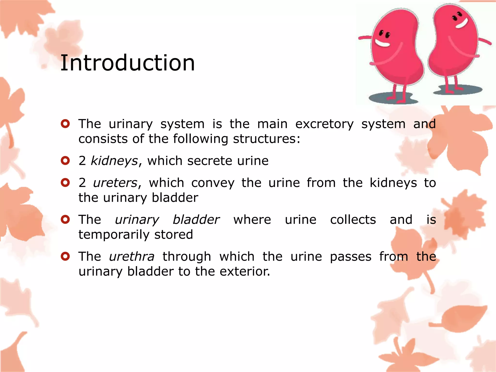
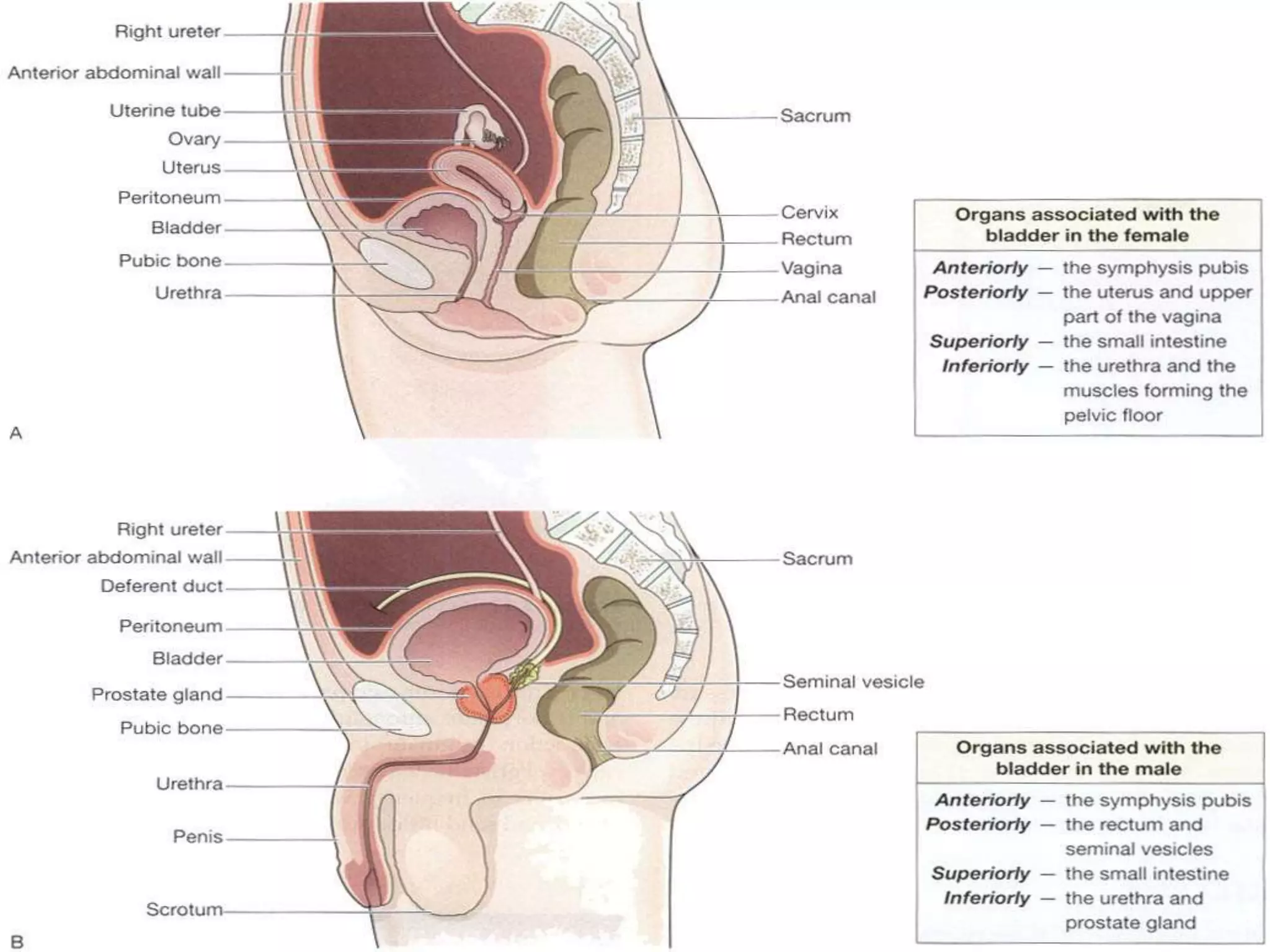
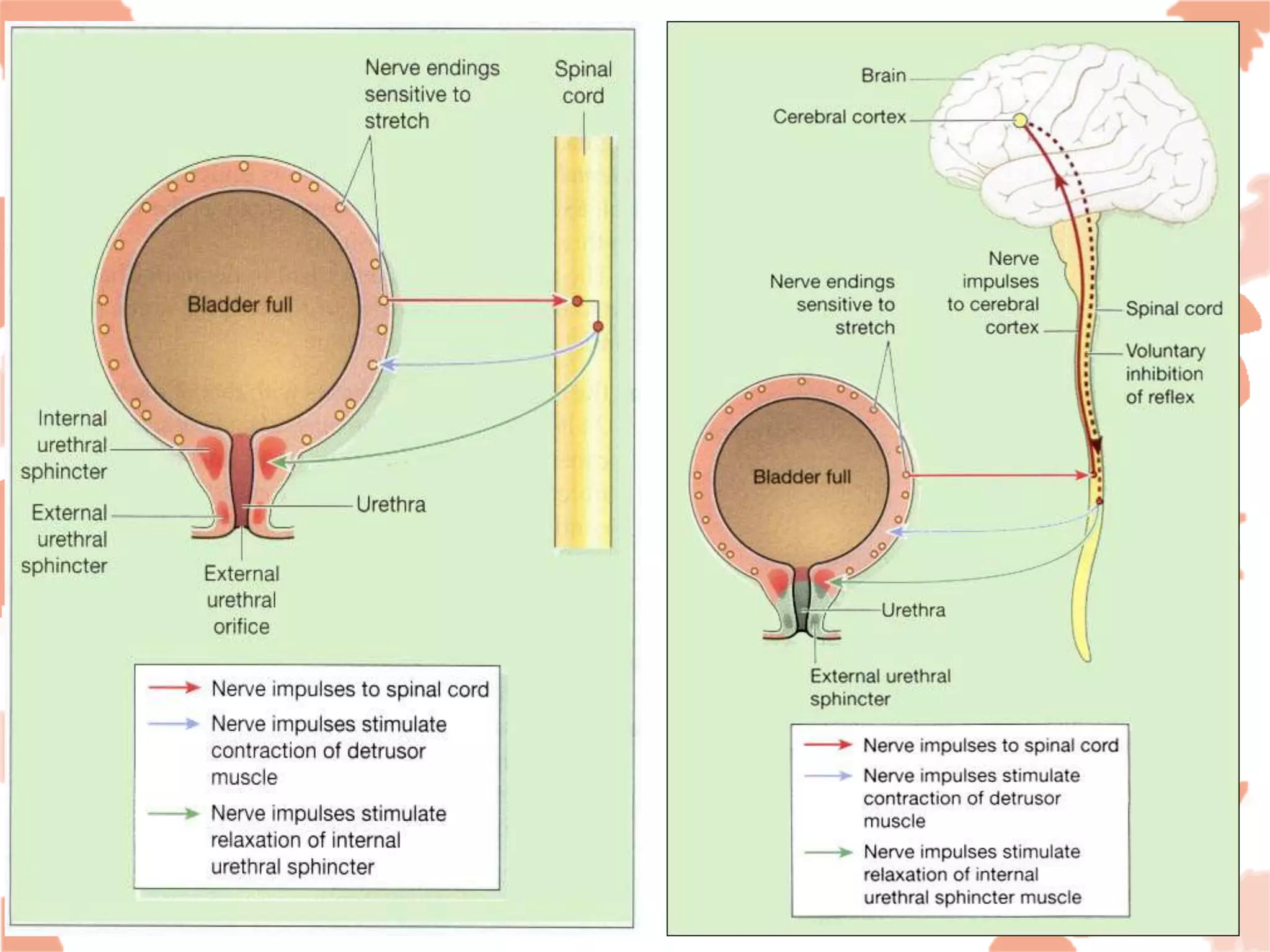
Urinary System
Mo Faishal
Nursing Tutor
Rama College of Nursing
Introduction
 The urinary system is the main excretory system and
consists of the following structures:
 2 kidneys, which secrete urine
 2 ureters, which convey the urine from the kidneys to
the urinary bladder
 The urinary bladder where urine collects and is
temporarily stored
 The urethra through which the urine passes from the
urinary bladder to the exterior.
 The urinary system plays a vital part in maintaining
homeostasis of water and electrolyte concentrations
within the body.
 The kidneys produce urine that contains metabolic waste
products, including the nitrogenous compounds urea and
uric acid, excess ions and some drugs.
Kidneys
 Kidneys are bean-shaped organs, about 11 cm long, 6
cm wide, 3 cm thick and weight about 150 g.
 A sheath of fibrous connective tissue, also known as
the renal fascia, encloses the kidney and the renal fat.
 The kidneys lie on the posterior abdominal wall
 one on each side of the vertebral column, behind the
peritoneum and below the diaphragm.
 They extend from the level of the 12th thoracic vertebra
to the 3rd lumbar vertebra, receiving some protection
from the lower rib cage.
 The right kidney is usually slightly lower than the left,
probably because of the considerable space occupied by
the liver.
Gross structure of the kidney
 There are three areas of tissue
 an outer fibrous capsule, surrounding the kidney
 the cortex, a reddish-brown layer of tissue immediately
below the capsule and outside the pyramids
 the medulla, the innermost layer, consisting of pale
conical-shaped striations, the renal pyramids.
 The hilum is the concave medial border of the kidney
where the renal blood and lymph vessels, the ureter and
nerves enter
 The renal pelvis is the funnel-shaped structure that
collects urine formed by the kidney
 Urine formed in the kidney passes through a
renal papilla at the apex of a pyramid into a minor calyx,
 then into a major calyx before passing through the renal
pelvis into the ureter.
 The walls of the pelvis contain smooth muscle and are
lined with transitional epithelium.
 Peristalsis of the smooth muscle originating in
pacemaker cells in the walls of the calyces propels urine
through the renal pelvis and ureters to the bladder.
 This is an intrinsic property of the smooth muscle, and is
not under nerve control.
Microscopic structure of the
kidney
THE NEPHRON
 The kidney is composed of about 1–2 million functional
units, the nephrons, and a smaller number of collecting
ducts.
 The collecting ducts transport urine through the
pyramids to the calyces and renal pelvis.
 The nephron consists of a tubule closed at one end, the
other end opening into a collecting tubule
 The closed or blind end is indented to form the cup-
shaped glomerular capsule (Bowman’s capsule)
 which almost completely encloses a network of tiny
arterial capillaries, the glomerulus.
 the nephron is about 3 cm long and is described in three
parts:
 the proximal convoluted tubule
 the medullary loop (loop of Henle)
 the distal convoluted tubule, leading into a collecting
duct.
The main functions of the kidneys
are:
 formation and secretion of urine, which regulates
total body water, electrolyte and acid–base balance and
enables excretion of waste products
 production and secretion of erythropoietin, the
hormone that stimulates formation of red blood cells
 production and secretion of renin, an important
enzyme in the control of blood pressure
 Urine is stored in the bladder and excreted by the
process of micturition.
Organs associated with the
kidneys
Right kidney
 Superiorly – the right adrenal gland
 Anteriorly – the right lobe of the liver, the duodenum
and the hepatic flexure of the colon
 Posteriorly – the diaphragm, and muscles of the
posterior abdominal wall
Left kidney
 Superiorly – the left adrenal gland
 Anteriorly – the spleen, stomach, pancreas, jejunum and
splenic flexure of the colon
 Posteriorly – the diaphragm and muscles of the posterior
abdominal wall
Blood & Nerve Supply
Renal Arteries
The renal arteries branch off of the abdominal aorta and
supply the kidneys with blood.
Renal Veins
The renal veins are the veins that drain the kidneys and
connect them to the inferior vena cava.
Renal Plexus
The renal plexus are the source of nervous tissue
innervation within the kidney.
URETERS
 The ureters are the tubes that convey urine from the
kidneys to the urinary bladder
 They are about 25 to 30 cm long with a diameter of
about 3 mm.
 The ureter is continuous with the funnel-shaped renal
pelvis.
 It passes downwards through the abdominal cavity,
behind the peritoneum into the pelvic cavity, and passes
obliquely through the posterior wall of the bladder
 Because of this arrangement, when urine accumulates
and the pressure in the bladder rises,
 the ureters are compressed and the openings occluded.
 This prevents reflux of urine into the ureters (towards
the kidneys) as the bladder fills and during micturition,
 when pressure increases as the muscular bladder wall
contracts.
Structure
 The ureters consist of three layers of tissue:
 An outer covering of fibrous tissue, continuous with
the fibrous capsule of the kidney
 A middle muscular layer consisting of interlacing
smooth muscle fibres that form a syncytium spiralling
round the ureter, some in clockwise and some in
anticlockwise directions and an additional outer
longitudinal layer in the lower third
 An inner layer, the mucosa, lined with transitional
epithelium.
Function
 The ureters propel the urine from the kidneys into the
bladder by peristaltic contraction of the smooth muscle
layer.
 This is an intrinsic property of the smooth muscle
and is not under autonomic nerve control
URINARY BLADDER
 The urinary bladder is a reservoir for urine. It lies in the
pelvic cavity and its size and position vary, depending on
the amount of urine it contains.
 When distended, the bladder rises into the abdominal
cavity.
Structure
 The bladder is roughly pear-shaped, but becomes
more oval as it fills with urine. It has anterior,
superior and posterior surfaces.
 The posterior surface is the base. The bladder opens
into the urethra at its lowest point, the neck.
 When the bladder is empty the inner lining is arranged
in folds, and these gradually disappear as the
bladder fills.
 The bladder is distensible but when it contains 300 to
400 ml the awareness of the desire to urinate is
initiated.
 The total capacity is rarely more than about 600 ml.
 The bladder wall is composed of three layers:
 The outer layer of loose connective tissue, containing
blood and lymphatic vessels and nerves, covered on the
upper surface by the peritoneum
 The middle layer, consisting of a mass of interlacing
smooth muscle fibres and elastic tissue loosely
arranged in three layers.
 This is called the detrusor muscle and it empties the
bladder when it contracts
 The mucosa, lined with transitional epithelium
 The three orifices in the bladder wall form a triangle or
trigone .
 The upper two orifices on the posterior wall are the
openings of the ureters. The lower orifice is the point of
origin of the urethra.
 Where the urethra commences is a thickening of the
smooth muscle layer forming the internal uretharal
sphincter.
 This sphincter is not under voluntary control.
URETHRA
 The urethra is a canal extending from the neck of the
bladder to the exterior, at the external urethral orifice.
 Its length differs in the male and in the female. The male
urethra is associated with the urinary and the reproductive
systems
 The female urethra is approximately 4 cm long.
 It runs downwards and forwards behind the symphysis
pubis and opens at the external urethral orifice just in front
of the vagina.
 The external urethral orifice is guarded by the external
urethral sphincter which is under voluntary control.
 Except during the passage of urine, the walls of the urethra
are in close apposition.
 The male urethra provides a common pathway for the
flow of urine and semen, the combined secretions of the
male reproductive organs.
 It is about 19 to 20 cm long
 but in both sexes the basic structure is the same. Its
walls consist of three layers of tissue.
 The muscle layer, continuous with that of the bladder. At
its origin there is the internal urethral sphincter, consisting
mainly of elastic tissue and smooth muscle fibres,
under autonomic nerve control
 In the middle third there is skeletal muscle surrounding
the urethra, under voluntary nerve control, that forms
the external urethral sphincter
 The sub mucosa, a spongy layer containing blood vessels
and nerves
 The mucosa, which is continuous with that of the bladder
in the upper part.
 In the lower part the lining consists of stratified
squamous epithelium, continuous externally with the skin
of the vulva.
MICTURITION
 The urinary bladder acts as a reservoir for urine. When
300 to 400 ml of urine have accumulated, afferent
autonomic nerve fibres in the bladder wall sensitive to
stretch are stimulated.
 In the infant this initiates a spinal reflex action and
micturition occurs .
 Micturition occurs when autonomic efferent fibres convey
impulses to the bladder causing contraction of the
detrusor muscle and relaxation of the internal urethral
sphincter.
 When the nervous system is fully developed the
micturition reflex is stimulated but sensory impulses
pass upwards to the brain and there is an awareness of
the desire to pass urine.
 By conscious effort, reflex contraction of the bladder wall
and relaxation of the internal sphincter can be inhibited
for a limited period of time
 In adults, micturition occurs when the detrusor muscle
contracts, and there is reflex relaxation of the internal
sphincter and voluntary relaxation of the external
sphincter.
 Over-distension of the bladder is extremely painful, and
when this stage is reached there is a tendency for
involuntary relaxation of the external sphincter to occur
and a small amount of urine to escape, provided there is
no mechanical obstruction
DISEASES OF THE KIDNEYS
 Glomerulonephritis (GN)-This term suggests
inflammatory conditions of the glomerulus, but there are
several types of GN and inflammatory changes are not
always present.
 In many cases immune complexes damage the
glomeruli. These are formed when antigens and
antibodies combine either within the kidney or elsewhere
in the body, and they circulate in the blood.
Effects of glomerulonephritis
 These depend on the type and are listed below.
 Haematuria. This is usually painless and not
accompanied by other symptoms.
 When microscopic, it may be found on routine urinalysis
when red blood cells have passed from the damaged
glomeruli into the filtrate.
 Asymptomatic proteinuria. This may also be found on
routine urinalysis and when it is of a low level does not
cause nephrotic syndrome.
 It occurs as protein passes through the damaged
glomeruli into the filtrate.
 Acute nephritis. This is characterized by the presence
of:
 anuria or oliguria
 hypertension
 haematuria
 fluid retention
Nephrotic syndrome
 This is not a disease in itself but is an important feature
of several kidney diseases.
 The main characteristics are:
 proteinuria
 Hypo-albuminaemia
 generalized edema
 hyperlipidaemia.
Diabetic kidney
 Renal failure is the cause of death in 10% of all diabetics
and up to 50% of cases of the insulin-dependent (type I)
diabetes mellitus.
 There is damage to large and small blood vessels in
many parts of the body
 The effects include:
 Progressive Glomerulosclerosis followed by atrophy of
the tubules
 Acute pyelonephritis with papillary necrosis
 ischemia and hypertension
 Nephritic syndrome.
Renal calculi
 Calculi (stones) form in the kidneys and bladder when
urinary constituents normally in solution are
precipitated.
 The solutes involved are oxalates, phosphates, uriates
and uric acid, and stones usually consist of more than
one substance, deposited in layers
 They are more common in males and after 30 years of
age. Most originate in collecting tubules or in renal
papillae.
 They then pass into the renal pelvis where they may
increase in size.
 Some become too large to pass through the ureter and
may obstruct the outflow of urine causing renal failure.
 Others pass to the bladder and are either excreted or
increase in size and obstruct the urethra.
 Predisposing factors include -
 Dehydration. This leads to increased reabsorption of water from the
tubules but does not change solute reabsorption, resulting in a reduced
volume of highly concentrated filtrate in the collecting tubules.
 pH of urine. When the normally acid filtrate becomes alkaline some
substances may be precipitated, e.g. phosphates.
 Infection. Necrotic material and pus provide foci upon which solutes in
the filtrate may be deposited and the products of infection may alter the
pH of the urine. Infection sometimes leads to alkaline urine .
 Metabolic conditions. These include hyperparathyroidism and gout.
DISEASES OF THE RENAL PELVIS, URETERS, BLADDER
AND URETHRA
Obstruction to the outflow of urine –
 Hydronephrosis-This is dilatation of the renal pelvis
and calyces caused by accumulation of urine. It leads to
destruction of the nephrons, fibrosis and atrophy of the
kidney.
 Spinal lesions - The immediate effect of transverse
spinal cord lesions that damage the nerve supply to the
bladder is that micturition does not occur.
 When the bladder fills the rise in pressure causes
overflow incontinence, back pressure into the ureters
and hydronephrosis.
Complications of urinary tract
obstruction
 Infection. Stasis of urine predisposes to infection and
pyelonephritis. The microbes usually spread upwards in
the urinary tract or are sometimes bloodborne.
 Calculus formation
Infections of the urinary tract
 Infection of any part of the tract may spread upwards
causing pyelonephritis and severe kidney damage.
 Ureteritis - Inflammation of a ureter is usually due to
the upward spread of infection in cystitis.
 Cystitis- This is inflammation of the bladder and may be
due to:
 spread of microbes that are commensals of the bowel
(Escherichia coli and Streptococcus ) from the perineum,
especially in women because of the short wide urethra.
 Tumours of the bladder-
 Urinary incontinence - In this condition there is
involuntary passage of urine due to defective voluntary
control of the external urethral sphincter.
 Stress incontinence - This is leakage of urine when
intra-abdominal pressure is raised, e.g. on coughing,
laughing, sneezing or lifting.
 Retention and overflow incontinence -retention of
urine due to obstruction of urinary outflow, e.g. enlarged
prostate or urethral stricture
 Urge incontinence - Leakage of urine follows a sudden
and intense urge to void and may be due to a urinary
tract infection, calculus, tumor or sudden stress.
Urinary System Anatomy

Urinary System Anatomy

  • 1.
    Urinary System Mo Faishal NursingTutor Rama College of Nursing
  • 2.
    Introduction  The urinarysystem is the main excretory system and consists of the following structures:  2 kidneys, which secrete urine  2 ureters, which convey the urine from the kidneys to the urinary bladder  The urinary bladder where urine collects and is temporarily stored  The urethra through which the urine passes from the urinary bladder to the exterior.
  • 3.
     The urinarysystem plays a vital part in maintaining homeostasis of water and electrolyte concentrations within the body.  The kidneys produce urine that contains metabolic waste products, including the nitrogenous compounds urea and uric acid, excess ions and some drugs.
  • 4.
    Kidneys  Kidneys arebean-shaped organs, about 11 cm long, 6 cm wide, 3 cm thick and weight about 150 g.  A sheath of fibrous connective tissue, also known as the renal fascia, encloses the kidney and the renal fat.  The kidneys lie on the posterior abdominal wall
  • 5.
     one oneach side of the vertebral column, behind the peritoneum and below the diaphragm.  They extend from the level of the 12th thoracic vertebra to the 3rd lumbar vertebra, receiving some protection from the lower rib cage.  The right kidney is usually slightly lower than the left, probably because of the considerable space occupied by the liver.
  • 8.
    Gross structure ofthe kidney  There are three areas of tissue  an outer fibrous capsule, surrounding the kidney  the cortex, a reddish-brown layer of tissue immediately below the capsule and outside the pyramids  the medulla, the innermost layer, consisting of pale conical-shaped striations, the renal pyramids.  The hilum is the concave medial border of the kidney where the renal blood and lymph vessels, the ureter and nerves enter
  • 10.
     The renalpelvis is the funnel-shaped structure that collects urine formed by the kidney  Urine formed in the kidney passes through a renal papilla at the apex of a pyramid into a minor calyx,
  • 11.
     then intoa major calyx before passing through the renal pelvis into the ureter.  The walls of the pelvis contain smooth muscle and are lined with transitional epithelium.  Peristalsis of the smooth muscle originating in pacemaker cells in the walls of the calyces propels urine through the renal pelvis and ureters to the bladder.  This is an intrinsic property of the smooth muscle, and is not under nerve control.
  • 12.
    Microscopic structure ofthe kidney THE NEPHRON  The kidney is composed of about 1–2 million functional units, the nephrons, and a smaller number of collecting ducts.  The collecting ducts transport urine through the pyramids to the calyces and renal pelvis.  The nephron consists of a tubule closed at one end, the other end opening into a collecting tubule
  • 13.
     The closedor blind end is indented to form the cup- shaped glomerular capsule (Bowman’s capsule)  which almost completely encloses a network of tiny arterial capillaries, the glomerulus.
  • 14.
     the nephronis about 3 cm long and is described in three parts:  the proximal convoluted tubule  the medullary loop (loop of Henle)  the distal convoluted tubule, leading into a collecting duct.
  • 16.
    The main functionsof the kidneys are:  formation and secretion of urine, which regulates total body water, electrolyte and acid–base balance and enables excretion of waste products  production and secretion of erythropoietin, the hormone that stimulates formation of red blood cells  production and secretion of renin, an important enzyme in the control of blood pressure  Urine is stored in the bladder and excreted by the process of micturition.
  • 17.
    Organs associated withthe kidneys Right kidney  Superiorly – the right adrenal gland  Anteriorly – the right lobe of the liver, the duodenum and the hepatic flexure of the colon  Posteriorly – the diaphragm, and muscles of the posterior abdominal wall
  • 18.
    Left kidney  Superiorly– the left adrenal gland  Anteriorly – the spleen, stomach, pancreas, jejunum and splenic flexure of the colon  Posteriorly – the diaphragm and muscles of the posterior abdominal wall
  • 19.
    Blood & NerveSupply Renal Arteries The renal arteries branch off of the abdominal aorta and supply the kidneys with blood. Renal Veins The renal veins are the veins that drain the kidneys and connect them to the inferior vena cava. Renal Plexus The renal plexus are the source of nervous tissue innervation within the kidney.
  • 20.
    URETERS  The uretersare the tubes that convey urine from the kidneys to the urinary bladder  They are about 25 to 30 cm long with a diameter of about 3 mm.  The ureter is continuous with the funnel-shaped renal pelvis.  It passes downwards through the abdominal cavity, behind the peritoneum into the pelvic cavity, and passes obliquely through the posterior wall of the bladder
  • 22.
     Because ofthis arrangement, when urine accumulates and the pressure in the bladder rises,  the ureters are compressed and the openings occluded.  This prevents reflux of urine into the ureters (towards the kidneys) as the bladder fills and during micturition,  when pressure increases as the muscular bladder wall contracts.
  • 23.
    Structure  The uretersconsist of three layers of tissue:  An outer covering of fibrous tissue, continuous with the fibrous capsule of the kidney  A middle muscular layer consisting of interlacing smooth muscle fibres that form a syncytium spiralling round the ureter, some in clockwise and some in anticlockwise directions and an additional outer longitudinal layer in the lower third  An inner layer, the mucosa, lined with transitional epithelium.
  • 24.
    Function  The ureterspropel the urine from the kidneys into the bladder by peristaltic contraction of the smooth muscle layer.  This is an intrinsic property of the smooth muscle and is not under autonomic nerve control
  • 25.
    URINARY BLADDER  Theurinary bladder is a reservoir for urine. It lies in the pelvic cavity and its size and position vary, depending on the amount of urine it contains.  When distended, the bladder rises into the abdominal cavity.
  • 26.
    Structure  The bladderis roughly pear-shaped, but becomes more oval as it fills with urine. It has anterior, superior and posterior surfaces.  The posterior surface is the base. The bladder opens into the urethra at its lowest point, the neck.  When the bladder is empty the inner lining is arranged in folds, and these gradually disappear as the bladder fills.  The bladder is distensible but when it contains 300 to 400 ml the awareness of the desire to urinate is initiated.  The total capacity is rarely more than about 600 ml.
  • 27.
     The bladderwall is composed of three layers:  The outer layer of loose connective tissue, containing blood and lymphatic vessels and nerves, covered on the upper surface by the peritoneum  The middle layer, consisting of a mass of interlacing smooth muscle fibres and elastic tissue loosely arranged in three layers.  This is called the detrusor muscle and it empties the bladder when it contracts  The mucosa, lined with transitional epithelium
  • 28.
     The threeorifices in the bladder wall form a triangle or trigone .  The upper two orifices on the posterior wall are the openings of the ureters. The lower orifice is the point of origin of the urethra.  Where the urethra commences is a thickening of the smooth muscle layer forming the internal uretharal sphincter.  This sphincter is not under voluntary control.
  • 31.
    URETHRA  The urethrais a canal extending from the neck of the bladder to the exterior, at the external urethral orifice.  Its length differs in the male and in the female. The male urethra is associated with the urinary and the reproductive systems  The female urethra is approximately 4 cm long.  It runs downwards and forwards behind the symphysis pubis and opens at the external urethral orifice just in front of the vagina.  The external urethral orifice is guarded by the external urethral sphincter which is under voluntary control.  Except during the passage of urine, the walls of the urethra are in close apposition.
  • 33.
     The maleurethra provides a common pathway for the flow of urine and semen, the combined secretions of the male reproductive organs.  It is about 19 to 20 cm long  but in both sexes the basic structure is the same. Its walls consist of three layers of tissue.
  • 35.
     The musclelayer, continuous with that of the bladder. At its origin there is the internal urethral sphincter, consisting mainly of elastic tissue and smooth muscle fibres, under autonomic nerve control  In the middle third there is skeletal muscle surrounding the urethra, under voluntary nerve control, that forms the external urethral sphincter  The sub mucosa, a spongy layer containing blood vessels and nerves  The mucosa, which is continuous with that of the bladder in the upper part.  In the lower part the lining consists of stratified squamous epithelium, continuous externally with the skin of the vulva.
  • 37.
    MICTURITION  The urinarybladder acts as a reservoir for urine. When 300 to 400 ml of urine have accumulated, afferent autonomic nerve fibres in the bladder wall sensitive to stretch are stimulated.  In the infant this initiates a spinal reflex action and micturition occurs .  Micturition occurs when autonomic efferent fibres convey impulses to the bladder causing contraction of the detrusor muscle and relaxation of the internal urethral sphincter.
  • 38.
     When thenervous system is fully developed the micturition reflex is stimulated but sensory impulses pass upwards to the brain and there is an awareness of the desire to pass urine.  By conscious effort, reflex contraction of the bladder wall and relaxation of the internal sphincter can be inhibited for a limited period of time
  • 39.
     In adults,micturition occurs when the detrusor muscle contracts, and there is reflex relaxation of the internal sphincter and voluntary relaxation of the external sphincter.  Over-distension of the bladder is extremely painful, and when this stage is reached there is a tendency for involuntary relaxation of the external sphincter to occur and a small amount of urine to escape, provided there is no mechanical obstruction
  • 41.
    DISEASES OF THEKIDNEYS  Glomerulonephritis (GN)-This term suggests inflammatory conditions of the glomerulus, but there are several types of GN and inflammatory changes are not always present.  In many cases immune complexes damage the glomeruli. These are formed when antigens and antibodies combine either within the kidney or elsewhere in the body, and they circulate in the blood.
  • 42.
    Effects of glomerulonephritis These depend on the type and are listed below.  Haematuria. This is usually painless and not accompanied by other symptoms.  When microscopic, it may be found on routine urinalysis when red blood cells have passed from the damaged glomeruli into the filtrate.  Asymptomatic proteinuria. This may also be found on routine urinalysis and when it is of a low level does not cause nephrotic syndrome.  It occurs as protein passes through the damaged glomeruli into the filtrate.
  • 43.
     Acute nephritis.This is characterized by the presence of:  anuria or oliguria  hypertension  haematuria  fluid retention
  • 44.
    Nephrotic syndrome  Thisis not a disease in itself but is an important feature of several kidney diseases.  The main characteristics are:  proteinuria  Hypo-albuminaemia  generalized edema  hyperlipidaemia.
  • 45.
    Diabetic kidney  Renalfailure is the cause of death in 10% of all diabetics and up to 50% of cases of the insulin-dependent (type I) diabetes mellitus.  There is damage to large and small blood vessels in many parts of the body  The effects include:  Progressive Glomerulosclerosis followed by atrophy of the tubules  Acute pyelonephritis with papillary necrosis  ischemia and hypertension  Nephritic syndrome.
  • 46.
    Renal calculi  Calculi(stones) form in the kidneys and bladder when urinary constituents normally in solution are precipitated.  The solutes involved are oxalates, phosphates, uriates and uric acid, and stones usually consist of more than one substance, deposited in layers  They are more common in males and after 30 years of age. Most originate in collecting tubules or in renal papillae.
  • 47.
     They thenpass into the renal pelvis where they may increase in size.  Some become too large to pass through the ureter and may obstruct the outflow of urine causing renal failure.  Others pass to the bladder and are either excreted or increase in size and obstruct the urethra.  Predisposing factors include -
  • 48.
     Dehydration. Thisleads to increased reabsorption of water from the tubules but does not change solute reabsorption, resulting in a reduced volume of highly concentrated filtrate in the collecting tubules.  pH of urine. When the normally acid filtrate becomes alkaline some substances may be precipitated, e.g. phosphates.  Infection. Necrotic material and pus provide foci upon which solutes in the filtrate may be deposited and the products of infection may alter the pH of the urine. Infection sometimes leads to alkaline urine .  Metabolic conditions. These include hyperparathyroidism and gout.
  • 49.
    DISEASES OF THERENAL PELVIS, URETERS, BLADDER AND URETHRA Obstruction to the outflow of urine –  Hydronephrosis-This is dilatation of the renal pelvis and calyces caused by accumulation of urine. It leads to destruction of the nephrons, fibrosis and atrophy of the kidney.  Spinal lesions - The immediate effect of transverse spinal cord lesions that damage the nerve supply to the bladder is that micturition does not occur.  When the bladder fills the rise in pressure causes overflow incontinence, back pressure into the ureters and hydronephrosis.
  • 50.
    Complications of urinarytract obstruction  Infection. Stasis of urine predisposes to infection and pyelonephritis. The microbes usually spread upwards in the urinary tract or are sometimes bloodborne.  Calculus formation
  • 51.
    Infections of theurinary tract  Infection of any part of the tract may spread upwards causing pyelonephritis and severe kidney damage.  Ureteritis - Inflammation of a ureter is usually due to the upward spread of infection in cystitis.  Cystitis- This is inflammation of the bladder and may be due to:  spread of microbes that are commensals of the bowel (Escherichia coli and Streptococcus ) from the perineum, especially in women because of the short wide urethra.
  • 52.
     Tumours ofthe bladder-  Urinary incontinence - In this condition there is involuntary passage of urine due to defective voluntary control of the external urethral sphincter.  Stress incontinence - This is leakage of urine when intra-abdominal pressure is raised, e.g. on coughing, laughing, sneezing or lifting.  Retention and overflow incontinence -retention of urine due to obstruction of urinary outflow, e.g. enlarged prostate or urethral stricture  Urge incontinence - Leakage of urine follows a sudden and intense urge to void and may be due to a urinary tract infection, calculus, tumor or sudden stress.