IVMS-CV Pharmacology- Antiarrhythmic Agents
•
14 likes•2,357 views

Individualized Webcam facilitated and e-Classroom USMLE Step 1 Tutorials with Dr. Cray. 1 BMS Unit is 4 hr. General Principles and some Organ System require multiple units to complete in preparation for the USMLE Step 1 A HIGH YIELD FOCUS IN Biochemistry / Cell Biology, Microbiology / Immunology and the 4 P’s-Phiso, Pathophys, Path and Pharm. Webcam Facilitated USMLE Step 2 Clinical Knowledge and Clinical Skills diadactic tutorials /1 Unit is 4 hours, individualized one-on-one and group sessions, Including all Internal Medicine sub-sub-specitialities. For questions or more information.. drcray@imhotepvirtualmedsch.com

More Related Content
What's hot
What's hot (20)
Viewers also liked
Viewers also liked (20)
Similar to IVMS-CV Pharmacology- Antiarrhythmic Agents
Similar to IVMS-CV Pharmacology- Antiarrhythmic Agents (20)
More from Imhotep Virtual Medical School
More from Imhotep Virtual Medical School (20)
Recently uploaded
Recently uploaded (20)
IVMS-CV Pharmacology- Antiarrhythmic Agents
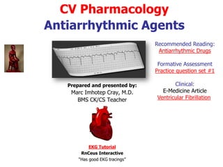
- 1. CV Pharmacology Antiarrhythmic Agents Prepared and presented by: Marc Imhotep Cray, M.D. BMS and CK/CS Teacher Reading: Antiarrhythmic Drugs Related Ppt: Introduction to EKG Interpretation Formative Assessment Practice question set #1 Clinical: e-Medicine articles Ventricular Fibrillation Hypokalemia in Emergency Medicine
- 2. 2 Electrophysiology and Cardiac Arrhythmias Cardiac Rhythm Normal rate: 60-100 beats per minute Impulse Propagation: sinoatrial node atrioventricular (AV node) His-Purkinje distribution throughout the ventricle Normal AV nodal delay (0.15 seconds) -- sufficient to allow atrial ejection of blood into the ventricles See Animated-Interactive Cardiac Cycle Hyper heart by Knowlege Weavers Adobe Shockwave Player
- 3. 3 Electrophysiology and Cardiac Arrhythmias(2) Definition: arrhythmia -- cardiac depolarization different from previous slide sequence -- abnormal origination (not SA nodal) abnormal rate/regularity/rhythm abnormal conduction characteristics See: http://www.rnceus.com/ekg/ekgframe.html
- 4. 4 Cardiac Electrophysiology cardiac action potential is a specialized action potential in heart, with unique properties necessary for function of electrical conduction system of heart cardiac action potential differs significantly in different portions of heart This differentiation of Aps allows different electrical characteristics of different portions of heart For instance, specialized conduction tissue of heart has special property of depolarizing without any external influence known as cardiac muscle automaticity See: Interactive animation illustrating the generation of a cardiac action potential
- 5. 5 Cardiac Electrophysiology(2) In cardiac myocytes, release of Ca2+ from the sarcoplasmic reticulum is induced by Ca2+ influx into cell through voltage-gated calcium channels on sarcolemma This phenomenon is called calcium- induced calcium release and increases myoplasmic free Ca2+ concentration causing muscle contraction
- 7. 7 Cardiac Electrophysiology(4) Note that there are important physiological differences between nodal cells and ventricular cells; the specific differences in ion channels and mechanisms of polarization give rise to unique properties of SA node cells, most importantly the spontaneous depolarizations (cardiac muscle automaticity) necessary for the SA node's pacemaker activity
- 8. 8 Cardiac Electrophysiology(5) Calcium channels Two voltage-dependent calcium channels play critical roles in the physiology of cardiac muscle: 1. L-type calcium channel ('L' for Long-lasting) and 2. T-type calcium channels ('T' for Transient) voltage- gated calcium channels These channels respond differently to voltage changes across the membrane: L-type channels respond to higher membrane potentials, open more slowly, and remain open longer than T-type channels See Notes Page
- 9. 9 Cardiac Electrophysiology(6) resting membrane potential is caused by difference in ionic concentrations and conductance across the membrane of the cell during phase 4 of action potential normal resting membrane potential in ventricular myocardium is about - 85 to -95 mV This potential is determined by the selective permeability of the cell membrane to various ions membrane is most permeable to K+ and relatively impermeable to other ions RMP is therefore dominated by K+ equilibrium potential according to the K+ gradient across the cell membrane The cardiac action potential has five phases
- 10. 10 Cardiac Electrophysiology(7) Maintenance of this electrical gradient is due to various ion pumps and exchange mechanisms, including both Na+-K+ ion exchange pump and Na+-Ca2+ exchanger current Remember: Intracellularly K+ is principal cation, and phosphate and conjugate bases of organic acids are dominant anions Extracellularly Na+ and Cl- predominate
- 11. 11 Cardiac Electrophysiology(8) Transmembrane potential - determined primarily by three ionic gradients: Na+, K+, Ca 2+ water-soluble, -- not free to diffuse through the membrane in response to concentration or electrical gradients: depended upon membrane channels (proteins) Movement through channels depend on controlling "molecular gates" Gate-status controlled by: Ionic conditions Metabolic conditions Transmembrane voltage Maintenance of ionic gradients: Na+/K+ ATPase pump termed "electrogenic" when net current flows as a result of transport (e.g., three Na+ exchange for two K+ ions)
- 12. 12 Cardiac Electrophysiology(9) Initial permeability state - resting membrane potential sodium - relatively impermeable potassium - relatively permeable Cardiac cell permeability and conductance: conductance: determined by characteristics of ion channel protein voltage = (actual membrane potential - membrane potential at which no current would flow, even with channels open) current flow = voltage X conductance
- 13. 13 Cardiac Electrophysiology(10) Sodium Concentration gradient: 140 mmol/L Na+ outside: 10 mmol/L Na+ inside; Electrical gradient: 0 mV outside; -90 mV inside Driving force -- both electrical and concentration -- tending to move Na+ into the cell In the resting state: sodium ion channels are closed therefore no Na+ flow through the membrane In the active state: channels open causing a large influx of sodium which accounts for phase 0 depolarization
- 14. 14 Cardiac Electrophysiology(11) Cardiac Cell Phase 0 and Sodium Current •Note the rapid "upstroke" characteristic of Phase 0 depolarization. •This abrupt change in membrane potential is caused by rapid, synchronous opening of Na+ channels. •Note the relationships between the the ECG tracing and phase 0 Source: http://www.pharmacology2000.com/Cardio/antiarr/antiarrtable.htm
- 15. 15 Cardiac Electrophysiology(12) Potassium: Concentration gradient (140 mmol/L K+ inside; 4 mmol/L K+outside) Concentration gradient -- tends to drive potassium out Electrical gradient tends to hold K+ in Some K+ channels ("inward rectifier") are open in resting state -- however, little K+ current flows because of the balance between K+ concentration and membrane electrical gradients Cardiac resting membrane potential: mainly determined By extracellular potassium concentration and Inward rectifier channel state
- 16. 16 Cardiac Electrophysiology(13) Spontaneous Depolarization (pacemaker cells)- phase 4 depolarization Spontaneous depolarization occurs because: Gradual increase in depolarizing currents (increasing membrane permeability to sodium or calcium) Decrease in repolarizing potassium currents (decreasing membrane potassium permeability) Both Ectopic pacemaker: (not normal SA nodal pacemaker) Facilitated by hypokalemic states Increasing potassium: tends to slow or stop ectopic pacemaker activity
- 17. 17 Cardiac Electrophysiology(14) Ca2+: Channel Activation Sequence similar to sodium; but occurring at more positive membrane potentials (phases 1 and 2) •Following intense inward Na+ current (phase 0), Ca2+currents: •Phases 1 & 2, are slowly inactivated. (Ca2+channel activation occurred later than for Na+) Source: http://www.pharmacology2000.com/Cardio/antiarr/antiarrtable.htm
- 18. 18 Cardiac Electrophysiology(15) Channel Inactivation, Re-establishing Resting Membrane Potential •Final repolarization (phase 3): •complete Na+ and Ca2+ channel inactivation •Increased potassium permeability •Membrane potential approaches K+ equilibrium potential -- which approximates the normal resting membrane potential Source: http://www.pharmacology2000.com/Cardio/antiarr/antiarrtable.htm
- 19. 19 Cardiac Electrophysiology(16) Five Phases: cardiac action potential in associated with HIS-purkinje fibers or ventricular muscle See Notes page for full explanations
- 20. 20 Influence of Membrane Resting Potential on Action Potential Properties: Factors that reduce the membrane resting potential & reduce conduction velocity Hyperkalemia Sodium pump block Ischemic cell damage
- 21. 21 Influence of Membrane Resting Potential on Action Potential Properties(2) Factors that may precipitate or exacerbate arrhythmias Ischemia Hypoxia Acidosis Alkalosis Abnormal electrolytes Excessive catecholamine levels Autonomic nervous system effects (e.g., excess vagal tone) Excessive catecholamine levels Autonomic nervous system effects (e.g., excess vagal tone) Drug effects: e.g., antiarrhythmic drugs may cause arrhythmias) Cardiac fiber stretching (as may occur with ventricular dilatation in congestive heart failure) Presence of scarred/diseased tissue which have altered electrical conduction properties
- 22. 22 Intro to Arrhythmias and Drug Therapy(1) How do Antiarrhythmic Drugs Work? Anti-arrhythmic drugs may work by: (a) Suppressing initiation site (automaticity/after-depolarizations) and/or (b) Preventing early or delayed afterdepolarizations and/or (c) By disrupting a re-entrant pathway Ref. Teaching Cardiac Arrhythmias: A Focus on Pathophysiology and Pharmacology
- 23. Intro to Arrhythmias and Drug Therapy How do Antiarrhythmic Drugs Work? (a) Automaticity: Automaticity may be diminished by: (1) increasing maximum diastolic membrane potential (2) decreasing slope of phase 4 depolarization (3) increasing action potential duration (4) raising threshold potential All of these factors make it take longer or make it more difficult for membrane potential to reach threshold (1) The diastolic membrane potential may be increased by adenosine and acetylcholine. (2) The slope of phase 4 depolarization may be decreased by beta receptor blockers (3) The duration of the action potential may be prolonged by drugs that block cardiac K+ channels (4) The membrane threshold potential may be altered by drugs that block Na+ or Ca2+ channels.
- 24. 24 Intro to Arrhythmias and Drug Therapy How do Antiarrhythmic Drugs Work? (b) Delayed or Early Afterdepolarizations: Delayed or early afterdepolarizations may be blocked by factors that (1) prevent the conditions that lead to afterdepolarizations (2) directly interfere with the inward currents (Na+, Ca2+) that cause afterdepolarizations
- 25. 25 Intro to Arrhythmias and Drug Therapy How do Antiarrhythmic Drugs Work? (c) Reentry For anatomically-determined re-entry such as Wolf- Parkinson-White syndrome (WPW) drugs arrhythmia can be resolved by blocking action potential (AP) propagation In WPW-based arrhythmias, blocking conduction through the AV node may be clinically effective Drugs that prolong nodal refractoriness and slow conduction include: Ca2+ channel blockers, beta- adrenergic blockers, or digitalis glycosides
- 26. 26 Intro to Arrhythmias and Drug Therapy(2) Atrial fibrillation may result in a high ventricular following rate Atrial Fibrillation Accordingly, drugs which may reduce ventricular rate by reducing AV nodal conduction include: 1. calcium channel blockers (verapamil (Isoptin, Calan), diltiazem (Cardiazem)) 2. beta-adrenergic receptor blockers (propranolol (Inderal)), and 3. digitalis glycosides
- 27. 27 Arrhythmias and Drug Therapy(3) calcium channel blockers Treatment of atrial fibrillation(2) Verapamil (Isoptin, Calan) & Diltiazem (Cardiazem) Blocks cardiac calcium channels in slow response tissues, such as the sinus and AV nodes Useful in treating AV reentrant tachyarrhythmias and in management of high ventricular rates secondary to atrial flutter or fibrillation Major adverse effect (i.v. administration) is hypotension Heart block or sinus bradycardia can also occur
- 28. 28 Arrhythmias and Drug Therapy (4) beta-adrenergic receptor blockers Treatment of atrial fibrillation(3): Propranolol (Inderal) Antiarrhythmic effects are due mainly to beta-adrenergic receptor blockade Normally, sympathetic drive results in increased in Ca2+ ,K+ ,and Cl- currents
- 29. 29 Arrhythmias and Drug Therapy (5) beta-adrenergic receptor blockers Increased sympathetic tone also increases phase 4 depolarization (heart rate goes up), and increases DAD (delayed afterdepolarizations) and EAD (early afterdepolarization) mediated arrhythmias These effects are blocked by beta-adrenergic receptor blockers
- 30. 30 Arrhythmias and Drug Therapy (6) beta-adrenergic receptor blockers Beta-adrenergic receptor blockers increase AV conduction time (takes longer) and increase AV nodal refractoriness, thereby helping to terminate nodal reentrant arrhythmias
- 31. 31 Arrhythmias and Drug Therapy (7) beta-adrenergic receptor blockers Beta-adrenergic receptor blockade can also help reduce ventricular following rates in atrial flutter and fibrillation, again by acting at the AV node
- 32. 32 Arrhythmias and Drug Therapy (8) beta-adrenergic receptor blockers Adverse effects of beta blocker therapy can lead to 1. fatigue, 2. bronchospasm, 3. depression, 4. impotence, 5. attenuation of hypoglycemic symptoms in diabetic patients 6. worsening of congestive heart failure
- 33. 33 Class I Antiarrhythmic Drugs Class I: Sodium Channel Blockers Sodium channel blocking antiarrhythmic drugs are classified as use-dependent in that they bind to open sodium channels Their effectiveness is therefore dependent upon the frequency of channel opening.
- 34. 34 Class I Antiarrhythmic Drugs Type Ia quinidine There are three classes or types of sodium channel blockers: Type Ia: prototype: quinidine gluconate (Quinaglute, Quinalan Type Ia drugs slow the rate of AP rise and prolong ventricular effective refractory period
- 35. 35 Quinidine Overview dextroisomer of quinine; quinidine gluconate (Quinaglute, Quinalan) also has antimalarial and antipyretic effects Pharmacokinetics: 80%-90%: bound to plasma albumin Rapid oral absorption; rapid attainment of peak blood levels (60-90 minutes) Elimination half-life: 5-12 hours IM injection, possible but not recommended due to injection site discomfort IV administration: limited due to myocardial depression & peripheral vasodilation
- 36. 36 Quinidine Metabolism: Hepatic: hydroxylation to inactive metabolites; followed by renal excretion 20% excreted unchanged in urine Impaired hepatic/renal function: accumulation of quinidine and metabolites Sensitive to enzyme induction by other agents-- decreased quinidine blood levels with phenytoin, phenobarbital, rifampin
- 37. 37 Quinidine Mechanism of antiarrhythmic action-- primarily activated sodium channel blockade which results in: Depression of ectopic pacemaker activity Depression of conduction velocity may convert a one-way conduction blockade to a two-way (bidirectional) block -- terminating reentry arrhythmias Depression of excitability (particularly in partially depolarized tissue) Also see notes page
- 38. 38 Quinidine Effect on the ECG: QT interval lengthening Basis: quinidine-mediated reduction in repolarizing outward potassium current Result: Longer action potential duration Increased effective refractory period Reduces reentry frequency; reduced rate in tachyarrhythmias Sodium channel blockade results in an increased threshold decreased automaticity
- 39. 39 Quinidine Uses Used to manage nearly every form of arrhythmia especially acute and chronic supraventricular dysrhythmias Ventricular tachycardia (VT) Frequent indications: Prevent recurrence of supraventricular tachyarrhythmias (SVT) Suppression ventricular premature contractions Approximately 20% of patients with atrial fibrillation will convert to normal sinus rhythm following quinidine treatment Supraventricular tachyarrhythmia due to Wolff- Parkinson-White syndrome (WPW) effective suppression by quinidine Also see notes page
- 40. 40 Quinidine Side Effects Cardiovascular--at (high) plasma concentrations (> 2ug/ml) Prolongation (ECG) of PR interval, QRS complex, QT interval Heart block likely with 50% increase in QRS complex duration (reduced dosage) Quinidine syncope: may be caused by delayed intraventricular conduction, resulting in ventricular dysrhythmia Patients with preexisting QT interval prolongation or evidence of existing A-V block (ECG): probably should not be treated with quinidine
- 41. 41 Quinidine Side Effects (cont.) Quinidine is associated with Torsades de pointes, a ventricular arrhythmias associated with marked QT prolongation Torsades de pointes: Electrophysiological Features ventricular origin wide QRS complexes with multiple morphologies changing R - R intervals axis seems to twist about isoelectric line This potentially serious arrhythmia occurs in 2% - 8% if patients, even if they have a therapeutic or subtherapeutic quinidine blood level
- 42. 42 Quinidine Side Effects (cont.) Other quinidine adverse effects include: cinchonism blurred vision, decreased hearing acuity, gastrointestinal upset,headaches and tinnitus. Nausea, vomiting, diarrhea (30% frequency) Drug-drug interaction:quinidine gluconate (Quinaglute, Quinalan)-digoxin (Lanoxin, Lanoxicaps) Quinidine increases digoxin plasma concentration; may cause digitalis toxicity in patients taking digoxin or digitoxin
- 43. 43 Quinidine Side Effects (cont.) Effects on neuromuscular transmission: Quinidine gluconate (Quinaglute, Quinalan) interferes with normal neuromuscular transmission; enhancing effect of neuromuscular-blocking drugs Recurrence of skeletal muscle paralysis postoperatively may be associated with quinidine administration
- 44. 44 Class I Antiarrhythmic Drugs Type Ia Procainamide Overview: Local anesthetic (procaine) analog Long-term use avoided because of lupus-related side effect
- 45. 45 Procainamide Metabolism: Elimination: renal excretion & hepatic metabolism; procainamide is highly resistant to hydrolysis by plasma esterases 40%-60% excreted unchanged (renal) Renal dysfunction requires procainamide dosage reduction Hepatic metabolism -- acetylation cardioactive metabolite: N-acetylprocainamide (NAPA); NAPA accumulation may lead to Torsades de pointes
- 46. 46 Procainamide Quinidine and Procainamide similar: electrophysiological properties Possibly somewhat less effective in suppressing automaticity; possibly more effective in sodium channel blockade in depolarized cells Useful in acute management of supraventricular and ventricular arrhythmias. Drug of second choice for management of sustained ventricular arrhythmias (in the acute myocardial infarction setting) Effective in suppression of premature ventricular contractions & paroxysmal ventricular tachycardia rapidly following IV administration
- 47. 47 Procainamide Most important difference compared quinidine: procainamide does not exhibit vagolytic (antimuscarinic) activity Procainamide is less likely to produce hypotension, unless following rapid IV infusion Ganglionic-Blocking Activity
- 48. 48 Procainamide Side Effects & Toxicities Long term use can be associated with drug-induced, reversible lupus erythematosus-like syndrome which occurs at a frequency of 25% to 50% Consists of serositis, arthralgia & arthritis Occasionally: pleuritis, pericarditis, parenchymal pulmonary disease Rare: renal lupus Vasculitis not typically present (unlike systemic lupus erythematosus) Positive antinuclear antibody test is common; symptoms disappear upon drug discontinuation In slow acetylators the procainamide-induced lupus syndrome occurs more frequently and earlier in therapy than in rapid acetylators Nausea, Vomiting - most common early, noncardiac complication
- 49. 49 Class I Antiarrhythmic Drugs Type Ia Disopyramide (Norpace) Overview: Very similar to quinidine gluconate (Quinaglute, Quinalan) Greater antimuscarinic effects (in management of atrial flutter & fibrillation, pre- treatment with a drug that reduces AV conduction velocity is required) Approved use (USA): ventricular arrhythmias
- 50. 50 Disopyramide (Norpace) Metabolism: Dealkylated metabolite (hepatic); less anticholinergic, less antiarrhythmic effect compared to parent compound 50% -- excreted unchanged, renal Electrophysiological effects similar to quinidine gluconate (Quinaglute, Quinalan) Similar to quinidine gluconate in effective ventricular and atrial tachyarrhythmia suppression Uses: prescribed to maintain normal sinus rhythm in patients prone to atrial fibrillation and flutter also used to prevent ventricular fibrillation or tachycardia
- 51. 51 Disopyramide (Norpace) Side Effects & Toxicity Adverse side-effect profile: different from qunidine's in that disopyramide (Norpace) is not an alpha-adrenergic receptor blocker but is anti-vagal Most common side effects: (anticholinergic) dry mouth urinary hesitancy Other side effects: blurred vision, nausea
- 52. 52 Disopyramide Side Effects &Toxicity (cont.) Cardiovascular: QT interval prolongation (ECG) paradoxical ventricular tachycardia (quinidine-like) Negative inotropism (significant myocardial depressive effects)- undesirable with preexisting left ventricular dysfunction (may promote congestive heart failure, even in patients with no prior evidence of myocardial dysfunction) Disopyramide is not a first-line antiarrhythmic agent because of its negative inotropic effects If used, great caution must be exercised in patients with congestive heart failure Can cause torsades de pointes, a ventricular arrhythmia
- 53. 53 Class I Antiarrhythmic Drugs Type Ib Class Ib agents are often effective in treating ventricular arrhythmias Example:lidocaine Type Ib agents exhibit rapid association and dissociation from the channel
- 54. 54 Class I Antiarrhythmic Drugs Type Ib (Class IB, Sodium Channel Blocker) Mexiletine (Mexitil) Overview Amine analog of lidocaine (Xylocaine), but with reduced first-pass metabolism Suitable for oral administration Similar electrophysiologically to lidocaine
- 55. 55 Class I Antiarrhythmic Drugs Type Ib Mexiletine Clinical Use: Chronic suppression of ventricular tachyarrhythmias Combination with a beta adrenergic receptor blocker or another antiarrhythmic drug (e.g. quinidine gluconate (Quinaglute, Quinalan) or procainamide (Procan SR, Pronestyl-SR)): synergistic effects allow: reduced mexiletine dosage decreased side effect incidence
- 56. 56 Class I Antiarrhythmic Drugs Type Ib Mexiletine (Cont.) Possibly effective: decreasing neuropathic pain when alternative medications have proven ineffective-- applications (on-label use): diabetic neuropathy nerve injury Side effects: Epigastric burning: usually relieved by a taking drug with food nausea (common) Neurologic side effects: diplopia, vertigo, slurred speech (occasionally), tremor
- 57. 57 Class I Antiarrhythmic Drugs Type Ib (Class IB, Sodium Channel Blocker) Lidocaine (Xylocaine) Overview and Pharmacokinetics: Local anesthetic administered by i.v. for therapy of ventricular arrhythmias Extensive first-pass effect requires IV administration Half-life: two hours Infusion rate: should be adjusted based on lidocaine plasma levels Metabolism Hepatic;some active metabolites
- 58. 58 Lidocaine (Xylocaine) (Class Ib, Sodium Channel Blocker) Factors influencing loading and maintenance doses: Congestive heart failure (decreasing volume of distribution and total body clearance) Liver disease: plasma clearance -- reduced; volume of distribution -- increased; elimination half-life substantially increased (3 X or more) Drugs that decrease liver blood flow (e.g. cimetadine, propranolol), decreased lidocaine clearance (increased possible toxicity) Pharmacokinetics cont. :
- 59. 59 Lidocaine (Xylocaine) (Class Ib, Sodium Channel Blocker) (Cont.) Cardiovascular Effects: Site of Action: Sodium Channels Blocks activated and inactivated sodium channels (quinidine blocks sodium channels only in the activated state) No significant effect on QRS or QT interval or on AV conduction (normal doses) Lidocaine (Xylocaine) decreases automaticity by reducing the phase 4 slope and by increasing threshold Pharmacodynamics:
- 60. 60 Lidocaine (Xylocaine) (Cont.) lidocaine is more effective in suppressing activity in depolarized, arrhythmogenic cardiac tissue but little effect on normal cardiac tissue -basis for this drug's selectivity Very effective antiarrhythmic agent for arrhythmia suppression associated with depolarization (e.g., digitalis toxicity or ischemia) Comparatively ineffective in treating arrhythmias occurring in normally polarized issue (e.g., atrial fibrillation or atrial flutter)
- 61. 61 Lidocaine (Xylocaine) (Cont.) Clinical Uses: Suppression of ventricular arrhythmias (limited effect on supraventricular tachyarrhythmias) May reduce incidence of ventricular fibrillation during initial time frame following acute myocardial infarction Suppression of reentry-type rhythm disorders: premature ventricular contractions (PVCs) ventricular tachycardia
- 62. 62 Lidocaine (Xylocaine) (Cont.) Side Effect/Toxicities Overdosage: vasodilation direct cardiac depression decreased cardiac conduction - bradycardia; prolonged PR interval; widening QRS on ECG Major side effect - neurological Large doses, rapidly administered can result in seizure Factors that reduce seizure threshold for lidocaine: hypoxemia, hyperkalemia, acidosis Otherwise: CNS depression, apnea
- 63. 63 Tocainide (Class I, Sodium Channel Blocker) Tocainide Amine analog of lidocaine, similar to mexiletine, orally active --but with reduced first-pass metabolism Used for chronic suppression of ventricular tachyarrhythmias refractory to less toxic agents Electrophysiologically similar to lidocaine Similar to mexiletine: tocainide + beta-adrenergic receptor blocker or another antiarrhythmic drug: synergism e.g.-Combination with quinidine may increase efficacy and diminish adverse effects
- 64. 64 Tocainide (Class I, Sodium Channel Blocker) (cont.) Side Effects: Profile similar to mexiletine suitable for oral administration, but RARELY USED due to possibly fatal bone marrow aplasia and pulmonary fibrosis tremor and nausea are major dose-related adverse side effects Excreted by kidney, accordingly dose should be reduced in patients with renal disease
- 65. 65 Cardiac Electrophysiology online animations and interactive tutorials Electro Cardio Gram by Knowlege Weavers Interpreting an EKG EKG Tutorial RnCeus Interactive Electrocardiogram -ECG Technician Nobel eMuseum Hyper heart by Knowlege Weavers The Arrhythma Center HeartCenterOnline
- 66. 66 Reference Resource (Textbooks) Principles of Pharmacology: The Pathophysiologic Basis of Drug Therapy Cairo CW, Simon JB, Golan DE. (Eds.); LLW 2012 (Google Books Online). Goodman and Gilman’s The Pharmacological Basis of Therapeutics. Brunton LL, Chabner BA , Knollmann BC (Eds.); M-H 12th ed. 2011. Basic and Clinical Pharmacology, Katzung, Masters, Trevor; M-H 12th ed.