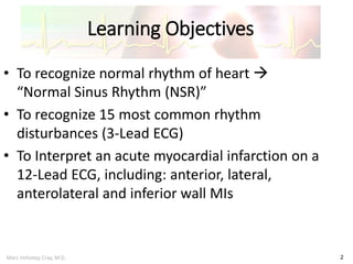
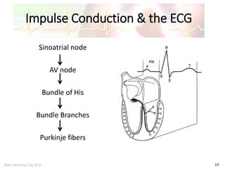
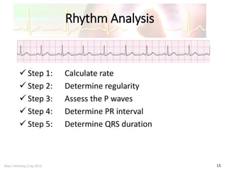
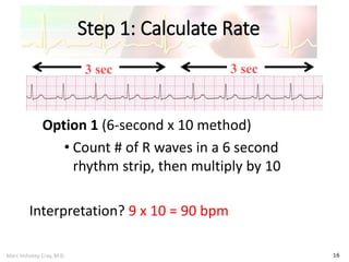
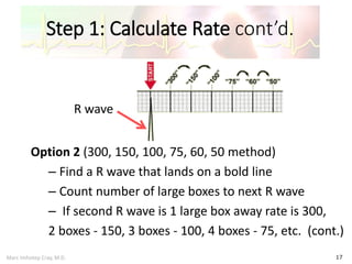
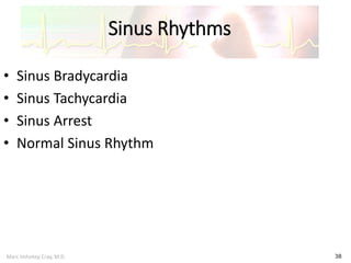
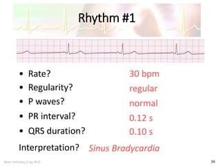
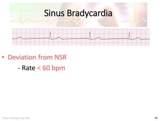
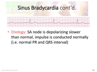
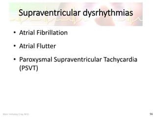
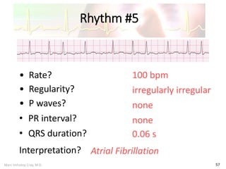
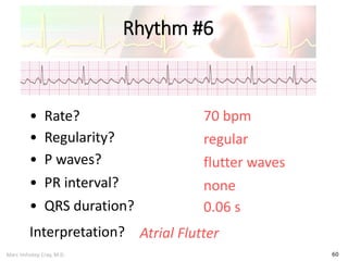
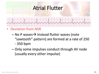
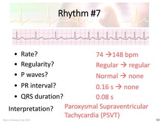
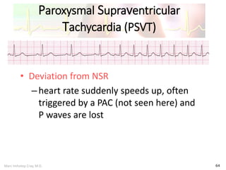
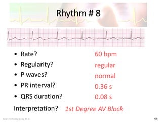
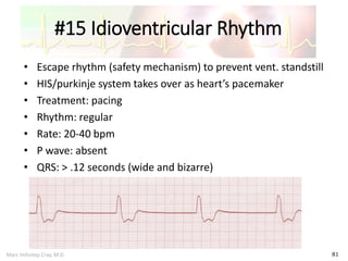
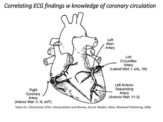
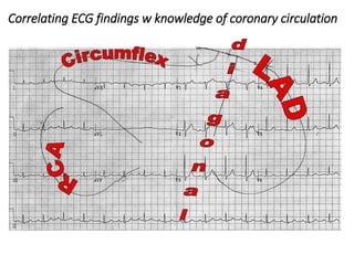
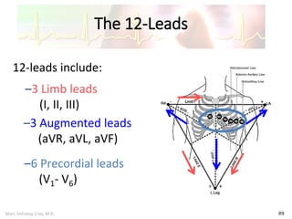
SlideShare a Scribd company logo
Electrocardiogram (ECG)
Interpretation
Marc Imhotep Cray, M.D.
Module 1 of 2
Marc Imhotep Cray, M.D.
Learning Objectives
2
• To recognize normal rhythm of heart 
“Normal Sinus Rhythm (NSR)”
• To recognize 15 most common rhythm
disturbances (3-Lead ECG)
• To Interpret an acute myocardial infarction on a
12-Lead ECG, including: anterior, lateral,
anterolateral and inferior wall MIs
Marc Imhotep Cray, M.D.
Topics Outline
3
• ECG Basics
• How to Analyze a Rhythm
• Normal Sinus Rhythm
• Cardiac Dysrhythmias
• Diagnosing a Myocardial Infarction
Marc Imhotep Cray, M.D.
Features include:
 Regular rhythm at 60-100 bpm
 Normal P wave morphology and axis
(upright in I and II, inverted in aVR)
 Narrow QRS complexes (< 100 ms
wide)
 Each P wave is followed by a QRS
complex
 The PR interval is constant
Review of ECG Basics
Normal ECG Morphology
Marc Imhotep Cray, M.D.
EKG Paper
 Horizontal axis of EKG paper
records time, w black marks at
top indicating 3 second intervals
o Each second is marked by 5
large grid blocks thus each
large block equals 0.2 second
 Vertical axis records EKG
amplitude (voltage)
o Two large blocks equal 1
millivolt (mV)
o Each small block equals 0.1 Mv
 Within large blocks are 5 small
blocks each representing 0.04
seconds
ECG tracings are recorded on grid paper
Marc Imhotep Cray, M.D.
ECG Paper cont’d.
6
• Horizontally
– One small box - 0.04 s
– One large box - 0.20 s
• Vertically
– One large box - 0.5 mV
Marc Imhotep Cray, M.D.
ECG Paper cont’d.
7
• Every 3 seconds (15 large boxes) is marked by a
vertical line
• This Helps When Calculating Heart Rate
Note: Strip above and those that follow are not
marked but all are 6 seconds long
3 sec 3 sec
Marc Imhotep Cray, M.D.
Autorhythmicity
• Some heart cells (SA, AV node and Purkinje) show
automaticity ability to generate a heart beat
– These cells have an intrinsic rhythmicity which generates a
pacemaker potential
• Heart does not require nerve or hormonal input to
beat
– Heart transplant patients nerves are severed but heart
beats on
Marc Imhotep Cray, M.D.
Normal Impulse Conduction
9
Sinoatrial node
AV node
Bundle of His
Bundle Branches
Purkinje fibers
Marc Imhotep Cray, M.D.
Impulse Conduction & the ECG
10
Sinoatrial node
AV node
Bundle of His
Bundle Branches
Purkinje fibers
Marc Imhotep Cray, M.D.
“PQRST”
11
Marc Imhotep Cray, M.D.
PR Interval
12
Atrial depolarization + delay in AV
junction (AV node/Bundle of His)
delay allows time for atria to
contract before ventricles contract
Marc Imhotep Cray, M.D.
Pacemakers of Heart
13
• SA Node - Dominant pacemaker with an intrinsic
rate of 60 - 100 beats/ minute
• AV Node - Back-up pacemaker with an intrinsic
rate of 40 - 60 beats/minute
• Ventricular cells - Back-up pacemaker with an
intrinsic rate of 20 - 45 bpm
How to Analyze a Rhythm
Marc Imhotep Cray, M.D.
Rhythm Analysis
15
 Step 1: Calculate rate
 Step 2: Determine regularity
 Step 3: Assess the P waves
 Step 4: Determine PR interval
 Step 5: Determine QRS duration
Marc Imhotep Cray, M.D.
Step 1: Calculate Rate
16
Option 1 (6-second x 10 method)
• Count # of R waves in a 6 second
rhythm strip, then multiply by 10
Interpretation? 9 x 10 = 90 bpm
3 sec 3 sec
Marc Imhotep Cray, M.D.
Step 1: Calculate Rate cont’d.
17
Option 2 (300, 150, 100, 75, 60, 50 method)
– Find a R wave that lands on a bold line
– Count number of large boxes to next R wave
– If second R wave is 1 large box away rate is 300,
2 boxes - 150, 3 boxes - 100, 4 boxes - 75, etc. (cont.)
R wave
Marc Imhotep Cray, M.D.
Step 1: Calculate Rate
18
• Option 2 (cont.)
– Memorize the sequence:
300 - 150 - 100 - 75 - 60 - 50
Interpretation? Approx. 1 box less than 100 = 95 bpm
3
0
0
1
5
0
1
0
0
7
5
6
0
5
0
Marc Imhotep Cray, M.D.
Step 2: Determine regularity
19
• Look at R-R distances (using a caliper or markings on
a pen or paper)
• Regular (are they equidistant apart)? Occasionally
irregular? Regularly irregular? Irregularly irregular?
Interpretation? Regular
R R
Marc Imhotep Cray, M.D.
Step 3: Assess the P waves
20
• Are there P waves?
• Do the P waves all look alike?
• Do the P waves occur at a regular rate?
• Is there one P wave before each QRS?
Interpretation? Normal P waves w 1 P wave for every QRS
Marc Imhotep Cray, M.D.
Step 4: Determine PR interval
21
• Normal: 0.12 - 0.20 seconds (3 - 5 sm boxes)
Interpretation? 0.12 seconds
Marc Imhotep Cray, M.D.
Step 5: QRS duration
22
• Normal: 0.04 - 0.12 seconds. (1 - 3 sm boxes)
Interpretation? 0.08 seconds
Marc Imhotep Cray, M.D.
NSR Summary
23
• Rate 90-95 bpm
• Regularity regular
• P waves normal
• PR interval 0.12 s
• QRS duration 0.08 s
Interpretation? Normal Sinus Rhythm
Normal Sinus Rhythm
Marc Imhotep Cray, M.D.
Basic Rhythm Analysis
• Rate – too fast or too slow?
• Rhythm – regular or irregular?
• Is there a normal looking QRS? Is it wide
or narrow?
• Are P waves present?
• What is relationship of P waves to QRS
complex?
Marc Imhotep Cray, M.D.
NSR Parameters
• Rate 60 - 100 bpm
• Regularity regular
• P waves normal
• PR interval 0.12 - 0.20 s
• QRS duration 0.04 - 0.12 s
Any deviation from above is
• Sinus tachycardia
• Sinus bradycardia or
• A dysrhythmia
Marc Imhotep Cray, M.D.
Normal Sinus Rhythm
27
12 lead ECG in sinus rhythm
Marc Imhotep Cray, M.D.
Arrhythmia Formation
28
Arrhythmias can arise from problems in:
• Sinus node
• Atrial cells
• AV junction
• Ventricular cells
Marc Imhotep Cray, M.D.
SA Node Problems
29
SA Node can:
• fire too slow
• fire too fast
Sinus Bradycardia
Sinus Tachycardia*
*Sinus Tachycardia may be an appropriate response
to stress.
Marc Imhotep Cray, M.D.
Atrial Cell Problems
30
Atrial cells can:
• fire occasionally from a focus Premature
Atrial Contractions (PACs)
• fire continuously due to a looping re-
entrant circuit Atrial Flutter
Marc Imhotep Cray, M.D.
Atrial Cell Problems
25
Atrial cells can also:
• fire continuously from multiple foci Atrial Fibrillation
or
• fire continuously due to multiple micro re-entrant
“wavelets” Atrial Fibrillation
Marc Imhotep Cray, M.D.
Key Scientific Point
32
Atrial tissue
 Multiple micro re-entrant
“wavelets” refers to wandering
small areas of activation which
generate fine chaotic impulses
 Colliding wavelets can, in turn,
generate new foci of activation
Marc Imhotep Cray, M.D.
AV Junctional Problems
33
AV junction can:
• fire continuously due to a looping re-entrant circuit
Paroxysmal Supraventricular Tachycardia (PSVT)
• block impulses coming from SA Node AV Junctional
Blocks
Marc Imhotep Cray, M.D.
Ventricular Cell Problems
34
Ventricular cells can:
• fire occasionally from 1 or more foci Premature
Ventricular Contractions (PVCs)
• fire continuously from multiple foci Ventricular
Fibrillation (VF)
• fire continuously due to a looping re-entrant circuit 
Ventricular Tachycardia (VT)
Marc Imhotep Cray, M.D.
Analyzing a Rhythm Table
Component Characteristics
Rate bpm is commonly ventricular rate. If atrial and ventricular rates
differ, as in a 3rd-degree block, measure both rates. Normal: 60–100
bpm. Slow (bradycardia): <60 bpm. Fast (tachycardia): >100 bpm
Regularity Measure R-R intervals and P-P intervals. Regular: Intervals consistent.
Regularly irregular: Repeating pattern. Irregular: No pattern
P Waves If present: Same in size, shape, position? Does each QRS have a P
wave? Normal: Upright (positive) and uniform
PR Interval Constant: Intervals are same. Variable: Intervals differ.
Normal: 0.12–0.20 sec and constant
QRS Interval Normal: 0.06–0.10 sec. Wide: >0.10 sec. None: Absent
QT Interval Beginning of R wave to end of T wave Varies with HR. Normal: Less
than half the R-R interval
Dropped
beats
Occur in AV blocks. Occur in sinus arrest.
Cardiac Dysrhythmias
Marc Imhotep Cray, M.D.
Dysrhythmias
37
• Sinus Rhythms
• Premature Beats
• Supraventricular Arrhythmias
• Ventricular Arrhythmias
• AV Junctional Blocks
Marc Imhotep Cray, M.D.
Sinus Rhythms
38
• Sinus Bradycardia
• Sinus Tachycardia
• Sinus Arrest
• Normal Sinus Rhythm
Marc Imhotep Cray, M.D.
Rhythm #1
39
30 bpm• Rate?
• Regularity? regular
normal
0.10 s
• P waves?
• PR interval? 0.12 s
• QRS duration?
Interpretation? Sinus Bradycardia
Marc Imhotep Cray, M.D.
Sinus Bradycardia
40
• Deviation from NSR
- Rate < 60 bpm
Marc Imhotep Cray, M.D.
Sinus Bradycardia cont’d.
41
• Etiology: SA node is depolarizing slower
than normal, impulse is conducted normally
(i.e. normal PR and QRS interval)
Marc Imhotep Cray, M.D.
Rhythm #2
42
130 bpm• Rate?
• Regularity? regular
normal
0.08 s
• P waves?
• PR interval? 0.16 s
• QRS duration?
Interpretation? Sinus Tachycardia
Marc Imhotep Cray, M.D.
Sinus Tachycardia
43
• Deviation from NSR
- Rate > 100 bpm
Marc Imhotep Cray, M.D.
Sinus Tachycardia cont’d.
44
• Etiology: SA node is depolarizing faster than
normal, impulse is conducted normally
• Remember: sinus tachycardia is a response
to physical or psychological stress, not a
primary arrhythmia.
Marc Imhotep Cray, M.D.
Sinus Arrest
45
• Etiology: SA node fails to depolarize and no
compensatory mechanisms take over
• Sinus arrest is usually a transient pause in sinus
node activity
Marc Imhotep Cray, M.D.
Premature Beats
46
• Premature Atrial Contractions
(PACs)
• Premature Ventricular Contractions
(PVCs)
Marc Imhotep Cray, M.D.
Rhythm #3
47
70 bpm• Rate?
• Regularity? occasionally irreg.
2/7 different contour
0.08 s
• P waves?
• PR interval? 0.14 s (except 2/7)
• QRS duration?
Interpretation? NSR w PAC
Marc Imhotep Cray, M.D.
Premature Atrial Contractions
(PACs)
48
• Deviation from NSR
– These ectopic beats originate in atria (but not
in SA node) therefore, contour of P wave, PR
interval, and timing are different than a
normally generated pulse from SA node
Marc Imhotep Cray, M.D.
PAC cont’d.
49
• Etiology: Excitation of an atrial cell forms an
impulse that is then conducted normally
through AV node and ventricles
Marc Imhotep Cray, M.D.
Key Scientific Point
50
• When an impulse originates anywhere in atria
(SA node, atrial cells, AV node, Bundle of His)
and then is conducted normally through
ventricles QRS will be narrow (0.04 - 0.12 s)
Marc Imhotep Cray, M.D.
Rhythm #4
51
60 bpm• Rate?
• Regularity? occasionally irreg.
none for 7th QRS
0.08 s (7th wide)
• P waves?
• PR interval? 0.14 s
• QRS duration?
Interpretation? Sinus Rhythm with 1 PVC
Marc Imhotep Cray, M.D.
Premature ventricular
contractions (PVCs)
52
• Deviation from NSR
– Ectopic beats originate in ventricles resulting
in wide and bizarre QRS complexes
– When there are more than 1 premature beats
and they look alike, called “uniform”
– When they look different, called “multiform”
Marc Imhotep Cray, M.D.
PVCs cont’d.
53
• Etiology: One or more ventricular cells are
depolarizing and impulses are abnormally
conducting through ventricles
Marc Imhotep Cray, M.D.
Key Scientific Point
54
• When an impulse originates in a ventricle,
conduction through ventricles will be
inefficient and QRS will be wide and bizarre
Marc Imhotep Cray, M.D.
Ventricular Conduction
55
Normal
Signal moves rapidly
through ventricles
Abnormal
Signal moves slowly
through ventricles
Marc Imhotep Cray, M.D.
Supraventricular dysrhythmias
56
• Atrial Fibrillation
• Atrial Flutter
• Paroxysmal Supraventricular Tachycardia
(PSVT)
Marc Imhotep Cray, M.D.
Rhythm #5
57
100 bpm• Rate?
• Regularity? irregularly irregular
none
0.06 s
• P waves?
• PR interval? none
• QRS duration?
Interpretation? Atrial Fibrillation
Marc Imhotep Cray, M.D.
Atrial Fibrillation cont’d.
58
• Deviation from NSR
– No organized atrial depolarization so no normal P
waves (impulses are not originating from SA node)
– Atrial activity is chaotic (resulting in an irregularly
irregular rate)
– Common, affects 2-4%, up to 5-10% if > 80 years old
Marc Imhotep Cray, M.D.
Atrial Fibrillation cont’d.
59
• Etiology: due to multiple re-entrant wavelets
conducted betw R & L atria and impulses are formed
in a totally unpredictable fashion
• AV node allows some of impulses to pass through at
variable intervals (so rhythm is irregularly irregular)
Marc Imhotep Cray, M.D.
Rhythm #6
60
70 bpm• Rate?
• Regularity? regular
flutter waves
0.06 s
• P waves?
• PR interval? none
• QRS duration?
Interpretation? Atrial Flutter
Marc Imhotep Cray, M.D.
Atrial Flutter
61
• Deviation from NSR
– No P waves Instead flutter waves (note
“sawtooth” pattern) are formed at a rate of 250
- 350 bpm
– Only some impulses conduct through AV node
(usually every other impulse)
Marc Imhotep Cray, M.D.
Atrial Flutter cont’d.
62
• Etiology: Reentrant pathway in right atrium with
every 2nd, 3rd or 4th impulse generating a QRS
(others are blocked in AV node as node repolarizes)
Marc Imhotep Cray, M.D.
Rhythm #7
63
74 148 bpm• Rate?
• Regularity? Regular  regular
Normal  none
0.08 s
• P waves?
• PR interval? 0.16 s  none
• QRS duration?
Interpretation? Paroxysmal Supraventricular
Tachycardia (PSVT)
Marc Imhotep Cray, M.D.
Paroxysmal Supraventricular
Tachycardia (PSVT)
64
• Deviation from NSR
–heart rate suddenly speeds up, often
triggered by a PAC (not seen here) and
P waves are lost
Marc Imhotep Cray, M.D.
AV Nodal Blocks
65
• 1st Degree AV Block
• 2nd Degree AV Block, Type I
• 2nd Degree AV Block, Type II
• 3rd Degree AV Block
Marc Imhotep Cray, M.D.
Rhythm # 8
66
60 bpm• Rate?
• Regularity? regular
normal
0.08 s
• P waves?
• PR interval? 0.36 s
• QRS duration?
Interpretation? 1st Degree AV Block
Marc Imhotep Cray, M.D.
1st Degree AV Block
67
• Deviation from NSR
–PR Interval > 0.20 s
Marc Imhotep Cray, M.D.
1st Degree AV Block cont’d.
68
• Etiology: Prolonged conduction delay in AV
node or Bundle of His
Marc Imhotep Cray, M.D.
Rhythm # 9
69
50 bpm• Rate?
• Regularity? regularly irregular
nml, but 4th no QRS
0.08 s
• P waves?
• PR interval? lengthens
• QRS duration?
Interpretation? 2nd Degree AV Block, Type I
Marc Imhotep Cray, M.D.
2nd Degree AV Block, Type I
Mobitz type I (Wenckebach)
70
• Deviation from NSR
– PR interval progressively lengthens, then impulse
is completely blocked (P wave not followed by
QRS)
Marc Imhotep Cray, M.D.
2nd Degree AV Block, Type I cont’d.
71
• Etiology: Each successive atrial impulse
encounters a longer and longer delay in AV
node until one impulse (usually 3rd or 4th)
fails to make it through AV node
Marc Imhotep Cray, M.D.
Rhythm # 10
72
40 bpm• Rate?
• Regularity? regular
nml, 2 of 3 no QRS
0.08 s
• P waves?
• PR interval? 0.14 s
• QRS duration?
Interpretation? 2nd Degree AV Block, Type II
Marc Imhotep Cray, M.D.
2nd Degree AV Block, Type II
Mobitz type II
73
• Deviation from NSR
– Occasional P waves are completely blocked (P
wave not followed by QRS)
– Progressive lengthening of PR interval until a QRS
is dropped
– Consistent ratio of conducted to dropped QRS
complexes
NB: Requires a pacemaker
Marc Imhotep Cray, M.D.
Rhythm #11
74
40 bpm• Rate?
• Regularity? regular
no relation to QRS
wide (> 0.12 s)
• P waves?
• PR interval? none
• QRS duration?
Interpretation? 3rd Degree AV Block
Marc Imhotep Cray, M.D.
3rd Degree AV Block
75
• Deviation from NSR
– P waves are completely blocked in AV junction
QRS complexes originate independently from
below junction
– Complete dissociation between atrial and
ventricular rates
NB: Requires a pacemaker
Marc Imhotep Cray, M.D.
3rd Degree AV Block cont’d.
76
• Etiology: There is complete block of conduction in
AV junction atria and ventricles form impulses
independently of each other
• Without impulses from atria, ventricles own
intrinsic pacemaker kicks in at around 30 - 45 bpm
Marc Imhotep Cray, M.D.
Remember
77
• When an impulse originates in a ventricle, conduction
through ventricles will be inefficient and QRS will be
wide and bizarre
Marc Imhotep Cray, M.D.
#12 Ventricular Tachycardia
78
• Ventricular cells fire continuously due to a looping re-entrant circuit
• Rate usually regular, 100 - 250 bpm
• P wave: may be absent, inverted or retrograde
• QRS: complexes bizarre, > .12
• Rhythm: usually regular
Marc Imhotep Cray, M.D.
#13 Ventricular Fibrillation
79
• Rhythm: irregular-coarse or fine, wave form varies in size & shape
• Fires continuously from multiple foci
• No organized electrical activity
• No cardiac output
• Causes: MI, ischemia, untreated VT, underlying CAD, acid base
imbalance, electrolyte imbalance, hypothermia etc.
Marc Imhotep Cray, M.D.
#14 Asystole
80
• Ventricular standstill, no electrical activity, no cardiac output
– no pulse!
• Cardiac arrest, may follow VF or PEA (Pulseless electrical
activity)
• Remember! No defibrillation with Asystole
• Rate: absent due to absence of ventricular activity
Occasional P wave may be identified
Marc Imhotep Cray, M.D.
#15 Idioventricular Rhythm
81
• Escape rhythm (safety mechanism) to prevent vent. standstill
• HIS/purkinje system takes over as heart’s pacemaker
• Treatment: pacing
• Rhythm: regular
• Rate: 20-40 bpm
• P wave: absent
• QRS: > .12 seconds (wide and bizarre)
Diagnosing a Myocardial Infarction
Marc Imhotep Cray, M.D.
Diagnosing a myocardial infarction
83
To Dx a MI you need to go beyond looking at a rhythm
strip and obtain a 12-Lead ECG
Rhythm Strip
Marc Imhotep Cray, M.D.
The 12-Lead ECG
84
• 12-Lead ECG sees heart from 12 different views helps you
see what is happening in different portions of heart
– rhythm strip is only 1 of these 12 views
12 lead ECG in sinus rhythm
Marc Imhotep Cray, M.D.
Which coronary artery?
 Site of infarct can be determined by correlating ECG
findings w knowledge of coronary circulation
o However, nml coronary vasculature varies widely from
individual to individual so only possible to make
generalizations
 Idea is you should be able to look at a 12 lead ECG
pattern of ischemia or infarction and predict
coronary lesion location that will show at cardiac
catheterization
Two main coronary arteries are right coronary artery
and left anterior descending coronary artery
Tao Le T and Bhushan V, Cardiovascular, In: First Aid for the USMLE Step 1 2017.
New York, NY: McGraw-Hill ,2017; 271.
 SA and AV nodes are supplied
by branches of RCA
• Infarct may cause nodal
dysfunction (bradycardia or
heart block)
 Right-dominant circulation
(85%) = PDA arises from RCA
 Left-dominant circulation
(8%) = PDA arises from LCX
 Codominant circulation (7%)
= PDA arises from both LCX
and RCA
 Coronary artery occlusion
most commonly in LAD
 Coronary blood flow peaks in
early diastole
Taylor GJ. 150 practice ECGs: Interpretation and Review, 3rd ed. Malden, Mass: Blackwell Publishing, 2006.
Correlating ECG findings w knowledge of coronary circulation
Marc Imhotep Cray, M.D.
The 12-Leads
89
12-leads include:
–3 Limb leads
(I, II, III)
–3 Augmented leads
(aVR, aVL, aVF)
–6 Precordial leads
(V1- V6)
Marc Imhotep Cray, M.D.
Views of the Heart
90
Some leads get a good view of:
Anterior portion of heart
Lateral portion of heart
Inferior portion of heart
Marc Imhotep Cray, M.D.
Where do you see ST-T wave changes for
following areas of ischemia or infarction?
Diagram showing the contiguous leads in same color
Marc Imhotep Cray, M.D.
ST Elevation
92
• One way to diagnose an
acute MI is to look for
elevation of ST segment
Marc Imhotep Cray, M.D.
ST Elevation cont’d.
93
• Elevation of ST segment
(greater than 1 small box)
in 2 leads is consistent w a
myocardial infarction
Marc Imhotep Cray, M.D.
Anterior View of Heart
94
• Anterior portion of heart is best viewed
using leads V1- V4
Marc Imhotep Cray, M.D.
Anterior Myocardial Infarction
95
• If you see changes in leads V1 - V4 that are
consistent w a MI you can conclude
that it is an anterior wall myocardial
infarction
Marc Imhotep Cray, M.D.
Putting it all Together
96
Do you think this person is having a MI If so, where?
Marc Imhotep Cray, M.D.
Interpretation
97
Yes, this person in previous slide is having an
acute anterior wall myocardial infarction
Marc Imhotep Cray, M.D.
Other MI Locations
98
Now that you know where to look for an anterior
wall MI let’s look at how you would determine if
MI involves lateral wall or inferior wall of heart
Marc Imhotep Cray, M.D.
Other MI Locations
99
 Remember 12-leads of ECG look at different portions of heart
– limb and augmented leads “see” electrical activity moving inferiorly (II, III
and aVF), to left (I, aVL) and to right (aVR)
– Whereas, precordial leads “see” electrical activity in posterior to anterior
direction
Limb Leads Augmented Leads Precordial Leads
Marc Imhotep Cray, M.D.
Other MI Locations
100
• Now, using these 3 diagrams let’s figure where to
look for a lateral wall and inferior wall MI
Limb Leads Augmented Leads Precordial Leads
Marc Imhotep Cray, M.D.
Anterior MI
101
• Remember anterior portion of the heart is best
viewed using leads V1- V4
Limb Leads Augmented Leads Precordial Leads
Marc Imhotep Cray, M.D.
Lateral MI
102
• So what leads do you think lateral portion of
heart is best viewed?
Limb Leads Augmented Leads Precordial Leads
Leads I, aVL, and V5- V6
Marc Imhotep Cray, M.D.
Inferior MI
103
Now how about inferior portion of heart?
Limb Leads Augmented Leads Precordial Leads
Leads II, III and aVF
Marc Imhotep Cray, M.D.
Putting it all Together
104
Now, where do you think this person is having a MI?
Marc Imhotep Cray, M.D.
Inferior Wall MI
105
This is an inferior MI. Note ST elevation in leads II, III
and aVF
Marc Imhotep Cray, M.D.
Putting it all Together
106
How about now?
Marc Imhotep Cray, M.D.
Anterolateral MI
107
This person’s MI involves both the anterior wall (V2-V4) and
lateral wall (V5-V6, I, and aVL)!
Marc Imhotep Cray, M.D.
Next Module:
Reading 12-Lead ECGs
108
 Best way to read 12-lead ECGs is to develop a step-by-
step approach (just as we did for analyzing a rhythm
strip)
 In next Module we present a 6-step and 9-step approaches:
1. Calculate Rate
2. Determine Rhythm
3. Determine QRS axis
4. Calculate Intervals
5. Assess for Hypertrophy
6. Look for evidence of Infarction
See next slide for links to tools and resources for further study.
THE END
Marc Imhotep Cray, M.D.
Companion study tools
110
Video Playlist:
ECG Interpretation
Strong Medicine ECG Video Edu. , Dr. Eric Strong
Reading:
Olivieri , B et al. Ch. 28 ECG (Pgs. 820-28). In: USMLE Step 1
Secrets 3rd. Ed. (Eds. Brown TA and Shah SJ) Saunders, 2013.
ECG eBook:
Shade B. Pocket ECGs: A Quick Information Guide. New York, NY:
McGraw-Hill, 2008. (A good one for beginners.)

More Related Content

What's hot

Normal ecg interpretation
Normal ecg interpretationNormal ecg interpretation
Normal ecg interpretation
Subhashini N
 
Arrhythmias general
Arrhythmias generalArrhythmias general
Arrhythmias general
Adarsh
 
ECG BASICS AND ARRTHYMIAS
ECG BASICS AND ARRTHYMIASECG BASICS AND ARRTHYMIAS
ECG BASICS AND ARRTHYMIAS
Prudhvi Krishna
 
Electrophysiology study basics
Electrophysiology study basicsElectrophysiology study basics
Electrophysiology study basics
Satyam Rajvanshi
 
ECG BASICS IN DETAIL
ECG BASICS IN DETAILECG BASICS IN DETAIL
Basic ecg1
Basic ecg1Basic ecg1
Basic ecg1
ebrahim jalili
 
Ecg made easy
Ecg made easyEcg made easy
Ecg made easy
Ramachandra Barik
 
Ecg quiz
Ecg quizEcg quiz
Ecg quiz
shafei lashin
 
Basic ecg
Basic ecgBasic ecg
Basic ecg
A.Salam Sharif
 
Cardiac arrhythmias
Cardiac arrhythmiasCardiac arrhythmias
Cardiac arrhythmias
Farhan Ali
 
Differentiation between AVNRT and AVRT_advanced lecture
Differentiation between AVNRT and AVRT_advanced lectureDifferentiation between AVNRT and AVRT_advanced lecture
Differentiation between AVNRT and AVRT_advanced lectureTaiwan Heart Rhythm Society
 
An ECG workshop
An ECG workshopAn ECG workshop
An ECG workshop
Dr Shibu Chacko MBE
 
Wide complex tachycardia
Wide complex tachycardiaWide complex tachycardia
Wide complex tachycardia
Amir Mahmoud
 
SOME INTRESTING ECG
SOME INTRESTING ECGSOME INTRESTING ECG
SOME INTRESTING ECG
Pradip Katwal
 
ECG PART 5
ECG PART 5ECG PART 5
ECG PART 5
Ramzan Ali
 
The Basics of ECG Interpretation
The Basics of ECG InterpretationThe Basics of ECG Interpretation
The Basics of ECG Interpretationmeducationdotnet
 

What's hot (20)

Normal ecg interpretation
Normal ecg interpretationNormal ecg interpretation
Normal ecg interpretation
 
Arrhythmias general
Arrhythmias generalArrhythmias general
Arrhythmias general
 
Intro to ECG
Intro to ECGIntro to ECG
Intro to ECG
 
ECG BASICS AND ARRTHYMIAS
ECG BASICS AND ARRTHYMIASECG BASICS AND ARRTHYMIAS
ECG BASICS AND ARRTHYMIAS
 
Electrophysiology study basics
Electrophysiology study basicsElectrophysiology study basics
Electrophysiology study basics
 
ECG BASICS IN DETAIL
ECG BASICS IN DETAILECG BASICS IN DETAIL
ECG BASICS IN DETAIL
 
Basic ecg1
Basic ecg1Basic ecg1
Basic ecg1
 
Ecg made easy
Ecg made easyEcg made easy
Ecg made easy
 
Ecg quiz
Ecg quizEcg quiz
Ecg quiz
 
Basic ecg
Basic ecgBasic ecg
Basic ecg
 
Cardiac arrhythmias
Cardiac arrhythmiasCardiac arrhythmias
Cardiac arrhythmias
 
Differentiation between AVNRT and AVRT_advanced lecture
Differentiation between AVNRT and AVRT_advanced lectureDifferentiation between AVNRT and AVRT_advanced lecture
Differentiation between AVNRT and AVRT_advanced lecture
 
An ECG workshop
An ECG workshopAn ECG workshop
An ECG workshop
 
Wide complex tachycardia
Wide complex tachycardiaWide complex tachycardia
Wide complex tachycardia
 
SOME INTRESTING ECG
SOME INTRESTING ECGSOME INTRESTING ECG
SOME INTRESTING ECG
 
Basics of ECG
Basics of ECGBasics of ECG
Basics of ECG
 
ECG PART 5
ECG PART 5ECG PART 5
ECG PART 5
 
electrocardiography
electrocardiographyelectrocardiography
electrocardiography
 
The Basics of ECG Interpretation
The Basics of ECG InterpretationThe Basics of ECG Interpretation
The Basics of ECG Interpretation
 
Ecg !
Ecg !Ecg !
Ecg !
 

Viewers also liked

Ecg interpretation
Ecg interpretationEcg interpretation
Ecg interpretationPk Doctors
 
Presentation e acls aremt
Presentation   e acls aremtPresentation   e acls aremt
Presentation e acls aremt
aremt
 
Ecg interpretation
Ecg interpretationEcg interpretation
Ecg interpretation
Nurhijrani Said
 
Ecg interpretation
Ecg interpretationEcg interpretation
Ecg interpretationSudhir Dev
 
Systematic ECG Interpretation
Systematic ECG InterpretationSystematic ECG Interpretation
Systematic ECG Interpretation
SCGH ED CME
 
Essentials of ecg interpretation aphrs
Essentials of ecg interpretation aphrsEssentials of ecg interpretation aphrs
Essentials of ecg interpretation aphrs
Benjamin Jacob
 
Principles of ECG Interpretation
Principles of ECG InterpretationPrinciples of ECG Interpretation
Principles of ECG Interpretation
Henry Tran
 
ECG Interpretation
ECG InterpretationECG Interpretation
ECG Interpretation
Mark Hall
 
ECG interpretation: NSTEMI
ECG interpretation: NSTEMIECG interpretation: NSTEMI
ECG interpretation: NSTEMI
Martin Jack
 
ECG interpretation
ECG interpretationECG interpretation
ECG interpretation
SCGH ED CME
 
ECG Interpretation by USAMA ELSAYED
ECG Interpretation by USAMA ELSAYEDECG Interpretation by USAMA ELSAYED
ECG Interpretation by USAMA ELSAYED
usama elsayed
 
ECG interpretation: the basics
ECG interpretation: the basicsECG interpretation: the basics
ECG interpretation: the basics
Jamie Ranse
 

Viewers also liked (12)

Ecg interpretation
Ecg interpretationEcg interpretation
Ecg interpretation
 
Presentation e acls aremt
Presentation   e acls aremtPresentation   e acls aremt
Presentation e acls aremt
 
Ecg interpretation
Ecg interpretationEcg interpretation
Ecg interpretation
 
Ecg interpretation
Ecg interpretationEcg interpretation
Ecg interpretation
 
Systematic ECG Interpretation
Systematic ECG InterpretationSystematic ECG Interpretation
Systematic ECG Interpretation
 
Essentials of ecg interpretation aphrs
Essentials of ecg interpretation aphrsEssentials of ecg interpretation aphrs
Essentials of ecg interpretation aphrs
 
Principles of ECG Interpretation
Principles of ECG InterpretationPrinciples of ECG Interpretation
Principles of ECG Interpretation
 
ECG Interpretation
ECG InterpretationECG Interpretation
ECG Interpretation
 
ECG interpretation: NSTEMI
ECG interpretation: NSTEMIECG interpretation: NSTEMI
ECG interpretation: NSTEMI
 
ECG interpretation
ECG interpretationECG interpretation
ECG interpretation
 
ECG Interpretation by USAMA ELSAYED
ECG Interpretation by USAMA ELSAYEDECG Interpretation by USAMA ELSAYED
ECG Interpretation by USAMA ELSAYED
 
ECG interpretation: the basics
ECG interpretation: the basicsECG interpretation: the basics
ECG interpretation: the basics
 

Similar to Electrocardiogram (ECG) Interpretation_Module 1 of 2

4- ECG.ppt
4- ECG.ppt4- ECG.ppt
4- ECG.ppt
mostafasayed645609
 
Ecg well
Ecg wellEcg well
Ecg well
saadfaysal
 
Electro Cardio Gram - Basics of ECG
Electro Cardio Gram - Basics of ECG Electro Cardio Gram - Basics of ECG
Electro Cardio Gram - Basics of ECG
Dr. Dinesh C. Sharma
 
ECG from Alpha to Omega
ECG from Alpha to OmegaECG from Alpha to Omega
ECG from Alpha to Omega
Kerolus Shehata
 
ECG Rhythm Interpretation (ECG Rhythm Analysis)
ECG Rhythm Interpretation (ECG Rhythm Analysis)ECG Rhythm Interpretation (ECG Rhythm Analysis)
ECG Rhythm Interpretation (ECG Rhythm Analysis)
shohel rana
 
Basic EKG and Rhythm Interpretation Symposia - The CRUDEM Foundation
Basic EKG and Rhythm Interpretation Symposia - The CRUDEM FoundationBasic EKG and Rhythm Interpretation Symposia - The CRUDEM Foundation
Basic EKG and Rhythm Interpretation Symposia - The CRUDEM Foundation
The CRUDEM Foundation
 
1 CARDIAC DYSARHYTHMIAS - INTERPRETATION.pptx
1 CARDIAC DYSARHYTHMIAS - INTERPRETATION.pptx1 CARDIAC DYSARHYTHMIAS - INTERPRETATION.pptx
1 CARDIAC DYSARHYTHMIAS - INTERPRETATION.pptx
PeruguMuniPrathiba
 
Introduction to Electrocardiogram (ECG) Basics
Introduction to Electrocardiogram (ECG) BasicsIntroduction to Electrocardiogram (ECG) Basics
Introduction to Electrocardiogram (ECG) Basics
Jason Foster
 
Skill ECG (2) arrhythmia for students.pptx
Skill ECG (2) arrhythmia for students.pptxSkill ECG (2) arrhythmia for students.pptx
Skill ECG (2) arrhythmia for students.pptx
Jane390174
 
3. common cardiac arrythemias
3. common cardiac arrythemias3. common cardiac arrythemias
3. common cardiac arrythemias
Debre Markos University
 
Anesthesia related presentation very helpful
Anesthesia related presentation very helpfulAnesthesia related presentation very helpful
Anesthesia related presentation very helpful
MalikArifUllah
 
Interpretation of ECG lab session.ppt
Interpretation of ECG  lab session.pptInterpretation of ECG  lab session.ppt
Interpretation of ECG lab session.ppt
Dr Rajesh Chandra Sharma
 
ECG
ECGECG
ECG
aanmol
 
Ecg presentation1
Ecg presentation1Ecg presentation1
Ecg presentation1
University of Gondar
 
Basic of ECG Referesher Course
Basic of ECG Referesher CourseBasic of ECG Referesher Course
Basic of ECG Referesher Course
Ame Mehadi
 
Ecg step y step 2015 hashem
Ecg step  y step 2015 hashemEcg step  y step 2015 hashem
Ecg step y step 2015 hashem
Hashem Yaseen
 
Interpretation of arrhythmias
Interpretation of arrhythmiasInterpretation of arrhythmias
Interpretation of arrhythmiasJeya Rajathurai
 
Arrythmia 411.ppt
Arrythmia 411.pptArrythmia 411.ppt
Arrythmia 411.ppt
ssuser196c47
 

Similar to Electrocardiogram (ECG) Interpretation_Module 1 of 2 (20)

4- ECG.ppt
4- ECG.ppt4- ECG.ppt
4- ECG.ppt
 
Ecg well
Ecg wellEcg well
Ecg well
 
Electro Cardio Gram - Basics of ECG
Electro Cardio Gram - Basics of ECG Electro Cardio Gram - Basics of ECG
Electro Cardio Gram - Basics of ECG
 
ECG from Alpha to Omega
ECG from Alpha to OmegaECG from Alpha to Omega
ECG from Alpha to Omega
 
ECG Rhythm Interpretation (ECG Rhythm Analysis)
ECG Rhythm Interpretation (ECG Rhythm Analysis)ECG Rhythm Interpretation (ECG Rhythm Analysis)
ECG Rhythm Interpretation (ECG Rhythm Analysis)
 
Basic EKG and Rhythm Interpretation Symposia - The CRUDEM Foundation
Basic EKG and Rhythm Interpretation Symposia - The CRUDEM FoundationBasic EKG and Rhythm Interpretation Symposia - The CRUDEM Foundation
Basic EKG and Rhythm Interpretation Symposia - The CRUDEM Foundation
 
Ecg
EcgEcg
Ecg
 
1 CARDIAC DYSARHYTHMIAS - INTERPRETATION.pptx
1 CARDIAC DYSARHYTHMIAS - INTERPRETATION.pptx1 CARDIAC DYSARHYTHMIAS - INTERPRETATION.pptx
1 CARDIAC DYSARHYTHMIAS - INTERPRETATION.pptx
 
Introduction to Electrocardiogram (ECG) Basics
Introduction to Electrocardiogram (ECG) BasicsIntroduction to Electrocardiogram (ECG) Basics
Introduction to Electrocardiogram (ECG) Basics
 
Skill ECG (2) arrhythmia for students.pptx
Skill ECG (2) arrhythmia for students.pptxSkill ECG (2) arrhythmia for students.pptx
Skill ECG (2) arrhythmia for students.pptx
 
3. common cardiac arrythemias
3. common cardiac arrythemias3. common cardiac arrythemias
3. common cardiac arrythemias
 
Anesthesia related presentation very helpful
Anesthesia related presentation very helpfulAnesthesia related presentation very helpful
Anesthesia related presentation very helpful
 
Interpretation of ECG lab session.ppt
Interpretation of ECG  lab session.pptInterpretation of ECG  lab session.ppt
Interpretation of ECG lab session.ppt
 
ECG
ECGECG
ECG
 
Ecg presentation1
Ecg presentation1Ecg presentation1
Ecg presentation1
 
Ecg
EcgEcg
Ecg
 
Basic of ECG Referesher Course
Basic of ECG Referesher CourseBasic of ECG Referesher Course
Basic of ECG Referesher Course
 
Ecg step y step 2015 hashem
Ecg step  y step 2015 hashemEcg step  y step 2015 hashem
Ecg step y step 2015 hashem
 
Interpretation of arrhythmias
Interpretation of arrhythmiasInterpretation of arrhythmias
Interpretation of arrhythmias
 
Arrythmia 411.ppt
Arrythmia 411.pptArrythmia 411.ppt
Arrythmia 411.ppt
 

More from Imhotep Virtual Medical School

Oncologic Pathology_A Case-based Organ Systems Review (USMLE Step 1)
Oncologic Pathology_A Case-based Organ Systems Review (USMLE Step 1)Oncologic Pathology_A Case-based Organ Systems Review (USMLE Step 1)
Oncologic Pathology_A Case-based Organ Systems Review (USMLE Step 1)
Imhotep Virtual Medical School
 
Pathology and Pathophysiology of Shock
Pathology and Pathophysiology of ShockPathology and Pathophysiology of Shock
Pathology and Pathophysiology of Shock
Imhotep Virtual Medical School
 
Drugs Used In Disorders of the Reproductive System
Drugs Used In Disorders of the Reproductive SystemDrugs Used In Disorders of the Reproductive System
Drugs Used In Disorders of the Reproductive System
Imhotep Virtual Medical School
 
Reproductive System Pathology_FM Breast and FM Reproductive Systems
Reproductive System Pathology_FM Breast and FM Reproductive SystemsReproductive System Pathology_FM Breast and FM Reproductive Systems
Reproductive System Pathology_FM Breast and FM Reproductive Systems
Imhotep Virtual Medical School
 
Reproductive System Pathology_Male Reproductive Systems
Reproductive System Pathology_Male Reproductive SystemsReproductive System Pathology_Male Reproductive Systems
Reproductive System Pathology_Male Reproductive Systems
Imhotep Virtual Medical School
 
Nervous System Pathology_A Case-based Learning Approach
Nervous System Pathology_A Case-based Learning ApproachNervous System Pathology_A Case-based Learning Approach
Nervous System Pathology_A Case-based Learning Approach
Imhotep Virtual Medical School
 
CVS Function, Regulation of the Heart and Overview of Therapeutic Goals in CV...
CVS Function, Regulation of the Heart and Overview of Therapeutic Goals in CV...CVS Function, Regulation of the Heart and Overview of Therapeutic Goals in CV...
CVS Function, Regulation of the Heart and Overview of Therapeutic Goals in CV...
Imhotep Virtual Medical School
 
Cardiovascular Pathology Case-based_Gross and Microscopic
Cardiovascular Pathology Case-based_Gross and MicroscopicCardiovascular Pathology Case-based_Gross and Microscopic
Cardiovascular Pathology Case-based_Gross and Microscopic
Imhotep Virtual Medical School
 
HIV / AIDS Pathology
HIV / AIDS PathologyHIV / AIDS Pathology
HIV / AIDS Pathology
Imhotep Virtual Medical School
 
Sepsis & Septic Shock
Sepsis & Septic ShockSepsis & Septic Shock
Sepsis & Septic Shock
Imhotep Virtual Medical School
 
Drugs Used in infectious Disease_Antibiotics
Drugs Used in infectious Disease_AntibioticsDrugs Used in infectious Disease_Antibiotics
Drugs Used in infectious Disease_Antibiotics
Imhotep Virtual Medical School
 
Hematopoietic and Lymphoid Systems Pathology
Hematopoietic and Lymphoid Systems  PathologyHematopoietic and Lymphoid Systems  Pathology
Hematopoietic and Lymphoid Systems Pathology
Imhotep Virtual Medical School
 
Drugs Used in Neoplastic Disorders
Drugs Used in Neoplastic DisordersDrugs Used in Neoplastic Disorders
Drugs Used in Neoplastic Disorders
Imhotep Virtual Medical School
 
Neoplasia & Oncologic Pathology
Neoplasia & Oncologic PathologyNeoplasia & Oncologic Pathology
Neoplasia & Oncologic Pathology
Imhotep Virtual Medical School
 
Clinical Pharmacology for Medical Students_USMLE Step 1 & 2 Review
Clinical Pharmacology for Medical Students_USMLE Step 1 & 2 ReviewClinical Pharmacology for Medical Students_USMLE Step 1 & 2 Review
Clinical Pharmacology for Medical Students_USMLE Step 1 & 2 Review
Imhotep Virtual Medical School
 
Make the Dx_ A Case-based Intro to Select Cardiovascular and Respiratory Dise...
Make the Dx_ A Case-based Intro to Select Cardiovascular and Respiratory Dise...Make the Dx_ A Case-based Intro to Select Cardiovascular and Respiratory Dise...
Make the Dx_ A Case-based Intro to Select Cardiovascular and Respiratory Dise...
Imhotep Virtual Medical School
 
Myocardial infarction_ Causes, Symptoms, Diagnosis, Treatment, and Pathology
Myocardial infarction_ Causes, Symptoms, Diagnosis, Treatment, and PathologyMyocardial infarction_ Causes, Symptoms, Diagnosis, Treatment, and Pathology
Myocardial infarction_ Causes, Symptoms, Diagnosis, Treatment, and Pathology
Imhotep Virtual Medical School
 
Basic CXR Interpretation_Diagnostic Radiographs
Basic CXR Interpretation_Diagnostic RadiographsBasic CXR Interpretation_Diagnostic Radiographs
Basic CXR Interpretation_Diagnostic Radiographs
Imhotep Virtual Medical School
 
Autonomic Nervous System Physiology and Pharmacology_Overview| Review of ANS
Autonomic Nervous System Physiology and Pharmacology_Overview| Review of ANSAutonomic Nervous System Physiology and Pharmacology_Overview| Review of ANS
Autonomic Nervous System Physiology and Pharmacology_Overview| Review of ANS
Imhotep Virtual Medical School
 
Evidence-Based Physical Diagnosis_Lect. 1_ What is Evidence-Based Physical Di...
Evidence-Based Physical Diagnosis_Lect. 1_ What is Evidence-Based Physical Di...Evidence-Based Physical Diagnosis_Lect. 1_ What is Evidence-Based Physical Di...
Evidence-Based Physical Diagnosis_Lect. 1_ What is Evidence-Based Physical Di...
Imhotep Virtual Medical School
 

More from Imhotep Virtual Medical School (20)

Oncologic Pathology_A Case-based Organ Systems Review (USMLE Step 1)
Oncologic Pathology_A Case-based Organ Systems Review (USMLE Step 1)Oncologic Pathology_A Case-based Organ Systems Review (USMLE Step 1)
Oncologic Pathology_A Case-based Organ Systems Review (USMLE Step 1)
 
Pathology and Pathophysiology of Shock
Pathology and Pathophysiology of ShockPathology and Pathophysiology of Shock
Pathology and Pathophysiology of Shock
 
Drugs Used In Disorders of the Reproductive System
Drugs Used In Disorders of the Reproductive SystemDrugs Used In Disorders of the Reproductive System
Drugs Used In Disorders of the Reproductive System
 
Reproductive System Pathology_FM Breast and FM Reproductive Systems
Reproductive System Pathology_FM Breast and FM Reproductive SystemsReproductive System Pathology_FM Breast and FM Reproductive Systems
Reproductive System Pathology_FM Breast and FM Reproductive Systems
 
Reproductive System Pathology_Male Reproductive Systems
Reproductive System Pathology_Male Reproductive SystemsReproductive System Pathology_Male Reproductive Systems
Reproductive System Pathology_Male Reproductive Systems
 
Nervous System Pathology_A Case-based Learning Approach
Nervous System Pathology_A Case-based Learning ApproachNervous System Pathology_A Case-based Learning Approach
Nervous System Pathology_A Case-based Learning Approach
 
CVS Function, Regulation of the Heart and Overview of Therapeutic Goals in CV...
CVS Function, Regulation of the Heart and Overview of Therapeutic Goals in CV...CVS Function, Regulation of the Heart and Overview of Therapeutic Goals in CV...
CVS Function, Regulation of the Heart and Overview of Therapeutic Goals in CV...
 
Cardiovascular Pathology Case-based_Gross and Microscopic
Cardiovascular Pathology Case-based_Gross and MicroscopicCardiovascular Pathology Case-based_Gross and Microscopic
Cardiovascular Pathology Case-based_Gross and Microscopic
 
HIV / AIDS Pathology
HIV / AIDS PathologyHIV / AIDS Pathology
HIV / AIDS Pathology
 
Sepsis & Septic Shock
Sepsis & Septic ShockSepsis & Septic Shock
Sepsis & Septic Shock
 
Drugs Used in infectious Disease_Antibiotics
Drugs Used in infectious Disease_AntibioticsDrugs Used in infectious Disease_Antibiotics
Drugs Used in infectious Disease_Antibiotics
 
Hematopoietic and Lymphoid Systems Pathology
Hematopoietic and Lymphoid Systems  PathologyHematopoietic and Lymphoid Systems  Pathology
Hematopoietic and Lymphoid Systems Pathology
 
Drugs Used in Neoplastic Disorders
Drugs Used in Neoplastic DisordersDrugs Used in Neoplastic Disorders
Drugs Used in Neoplastic Disorders
 
Neoplasia & Oncologic Pathology
Neoplasia & Oncologic PathologyNeoplasia & Oncologic Pathology
Neoplasia & Oncologic Pathology
 
Clinical Pharmacology for Medical Students_USMLE Step 1 & 2 Review
Clinical Pharmacology for Medical Students_USMLE Step 1 & 2 ReviewClinical Pharmacology for Medical Students_USMLE Step 1 & 2 Review
Clinical Pharmacology for Medical Students_USMLE Step 1 & 2 Review
 
Make the Dx_ A Case-based Intro to Select Cardiovascular and Respiratory Dise...
Make the Dx_ A Case-based Intro to Select Cardiovascular and Respiratory Dise...Make the Dx_ A Case-based Intro to Select Cardiovascular and Respiratory Dise...
Make the Dx_ A Case-based Intro to Select Cardiovascular and Respiratory Dise...
 
Myocardial infarction_ Causes, Symptoms, Diagnosis, Treatment, and Pathology
Myocardial infarction_ Causes, Symptoms, Diagnosis, Treatment, and PathologyMyocardial infarction_ Causes, Symptoms, Diagnosis, Treatment, and Pathology
Myocardial infarction_ Causes, Symptoms, Diagnosis, Treatment, and Pathology
 
Basic CXR Interpretation_Diagnostic Radiographs
Basic CXR Interpretation_Diagnostic RadiographsBasic CXR Interpretation_Diagnostic Radiographs
Basic CXR Interpretation_Diagnostic Radiographs
 
Autonomic Nervous System Physiology and Pharmacology_Overview| Review of ANS
Autonomic Nervous System Physiology and Pharmacology_Overview| Review of ANSAutonomic Nervous System Physiology and Pharmacology_Overview| Review of ANS
Autonomic Nervous System Physiology and Pharmacology_Overview| Review of ANS
 
Evidence-Based Physical Diagnosis_Lect. 1_ What is Evidence-Based Physical Di...
Evidence-Based Physical Diagnosis_Lect. 1_ What is Evidence-Based Physical Di...Evidence-Based Physical Diagnosis_Lect. 1_ What is Evidence-Based Physical Di...
Evidence-Based Physical Diagnosis_Lect. 1_ What is Evidence-Based Physical Di...
 

Recently uploaded

Embracing GenAI - A Strategic Imperative
Embracing GenAI - A Strategic ImperativeEmbracing GenAI - A Strategic Imperative
Embracing GenAI - A Strategic Imperative
Peter Windle
 
Honest Reviews of Tim Han LMA Course Program.pptx
Honest Reviews of Tim Han LMA Course Program.pptxHonest Reviews of Tim Han LMA Course Program.pptx
Honest Reviews of Tim Han LMA Course Program.pptx
timhan337
 
Instructions for Submissions thorugh G- Classroom.pptx
Instructions for Submissions thorugh G- Classroom.pptxInstructions for Submissions thorugh G- Classroom.pptx
Instructions for Submissions thorugh G- Classroom.pptx
Jheel Barad
 
Adversarial Attention Modeling for Multi-dimensional Emotion Regression.pdf
Adversarial Attention Modeling for Multi-dimensional Emotion Regression.pdfAdversarial Attention Modeling for Multi-dimensional Emotion Regression.pdf
Adversarial Attention Modeling for Multi-dimensional Emotion Regression.pdf
Po-Chuan Chen
 
Guidance_and_Counselling.pdf B.Ed. 4th Semester
Guidance_and_Counselling.pdf B.Ed. 4th SemesterGuidance_and_Counselling.pdf B.Ed. 4th Semester
Guidance_and_Counselling.pdf B.Ed. 4th Semester
Atul Kumar Singh
 
Mule 4.6 & Java 17 Upgrade | MuleSoft Mysore Meetup #46
Mule 4.6 & Java 17 Upgrade | MuleSoft Mysore Meetup #46Mule 4.6 & Java 17 Upgrade | MuleSoft Mysore Meetup #46
Mule 4.6 & Java 17 Upgrade | MuleSoft Mysore Meetup #46
MysoreMuleSoftMeetup
 
678020731-Sumas-y-Restas-Para-Colorear.pdf
678020731-Sumas-y-Restas-Para-Colorear.pdf678020731-Sumas-y-Restas-Para-Colorear.pdf
678020731-Sumas-y-Restas-Para-Colorear.pdf
CarlosHernanMontoyab2
 
1.4 modern child centered education - mahatma gandhi-2.pptx
1.4 modern child centered education - mahatma gandhi-2.pptx1.4 modern child centered education - mahatma gandhi-2.pptx
1.4 modern child centered education - mahatma gandhi-2.pptx
JosvitaDsouza2
 
TESDA TM1 REVIEWER FOR NATIONAL ASSESSMENT WRITTEN AND ORAL QUESTIONS WITH A...
TESDA TM1 REVIEWER  FOR NATIONAL ASSESSMENT WRITTEN AND ORAL QUESTIONS WITH A...TESDA TM1 REVIEWER  FOR NATIONAL ASSESSMENT WRITTEN AND ORAL QUESTIONS WITH A...
TESDA TM1 REVIEWER FOR NATIONAL ASSESSMENT WRITTEN AND ORAL QUESTIONS WITH A...
EugeneSaldivar
 
Chapter 3 - Islamic Banking Products and Services.pptx
Chapter 3 - Islamic Banking Products and Services.pptxChapter 3 - Islamic Banking Products and Services.pptx
Chapter 3 - Islamic Banking Products and Services.pptx
Mohd Adib Abd Muin, Senior Lecturer at Universiti Utara Malaysia
 
How to Make a Field invisible in Odoo 17
How to Make a Field invisible in Odoo 17How to Make a Field invisible in Odoo 17
How to Make a Field invisible in Odoo 17
Celine George
 
Palestine last event orientationfvgnh .pptx
Palestine last event orientationfvgnh .pptxPalestine last event orientationfvgnh .pptx
Palestine last event orientationfvgnh .pptx
RaedMohamed3
 
Unit 2- Research Aptitude (UGC NET Paper I).pdf
Unit 2- Research Aptitude (UGC NET Paper I).pdfUnit 2- Research Aptitude (UGC NET Paper I).pdf
Unit 2- Research Aptitude (UGC NET Paper I).pdf
Thiyagu K
 
2024.06.01 Introducing a competency framework for languag learning materials ...
2024.06.01 Introducing a competency framework for languag learning materials ...2024.06.01 Introducing a competency framework for languag learning materials ...
2024.06.01 Introducing a competency framework for languag learning materials ...
Sandy Millin
 
The Challenger.pdf DNHS Official Publication
The Challenger.pdf DNHS Official PublicationThe Challenger.pdf DNHS Official Publication
The Challenger.pdf DNHS Official Publication
Delapenabediema
 
CACJapan - GROUP Presentation 1- Wk 4.pdf
CACJapan - GROUP Presentation 1- Wk 4.pdfCACJapan - GROUP Presentation 1- Wk 4.pdf
CACJapan - GROUP Presentation 1- Wk 4.pdf
camakaiclarkmusic
 
Biological Screening of Herbal Drugs in detailed.
Biological Screening of Herbal Drugs in detailed.Biological Screening of Herbal Drugs in detailed.
Biological Screening of Herbal Drugs in detailed.
Ashokrao Mane college of Pharmacy Peth-Vadgaon
 
Language Across the Curriculm LAC B.Ed.
Language Across the  Curriculm LAC B.Ed.Language Across the  Curriculm LAC B.Ed.
Language Across the Curriculm LAC B.Ed.
Atul Kumar Singh
 
Phrasal Verbs.XXXXXXXXXXXXXXXXXXXXXXXXXX
Phrasal Verbs.XXXXXXXXXXXXXXXXXXXXXXXXXXPhrasal Verbs.XXXXXXXXXXXXXXXXXXXXXXXXXX
Phrasal Verbs.XXXXXXXXXXXXXXXXXXXXXXXXXX
MIRIAMSALINAS13
 
Francesca Gottschalk - How can education support child empowerment.pptx
Francesca Gottschalk - How can education support child empowerment.pptxFrancesca Gottschalk - How can education support child empowerment.pptx
Francesca Gottschalk - How can education support child empowerment.pptx
EduSkills OECD
 

Recently uploaded (20)

Embracing GenAI - A Strategic Imperative
Embracing GenAI - A Strategic ImperativeEmbracing GenAI - A Strategic Imperative
Embracing GenAI - A Strategic Imperative
 
Honest Reviews of Tim Han LMA Course Program.pptx
Honest Reviews of Tim Han LMA Course Program.pptxHonest Reviews of Tim Han LMA Course Program.pptx
Honest Reviews of Tim Han LMA Course Program.pptx
 
Instructions for Submissions thorugh G- Classroom.pptx
Instructions for Submissions thorugh G- Classroom.pptxInstructions for Submissions thorugh G- Classroom.pptx
Instructions for Submissions thorugh G- Classroom.pptx
 
Adversarial Attention Modeling for Multi-dimensional Emotion Regression.pdf
Adversarial Attention Modeling for Multi-dimensional Emotion Regression.pdfAdversarial Attention Modeling for Multi-dimensional Emotion Regression.pdf
Adversarial Attention Modeling for Multi-dimensional Emotion Regression.pdf
 
Guidance_and_Counselling.pdf B.Ed. 4th Semester
Guidance_and_Counselling.pdf B.Ed. 4th SemesterGuidance_and_Counselling.pdf B.Ed. 4th Semester
Guidance_and_Counselling.pdf B.Ed. 4th Semester
 
Mule 4.6 & Java 17 Upgrade | MuleSoft Mysore Meetup #46
Mule 4.6 & Java 17 Upgrade | MuleSoft Mysore Meetup #46Mule 4.6 & Java 17 Upgrade | MuleSoft Mysore Meetup #46
Mule 4.6 & Java 17 Upgrade | MuleSoft Mysore Meetup #46
 
678020731-Sumas-y-Restas-Para-Colorear.pdf
678020731-Sumas-y-Restas-Para-Colorear.pdf678020731-Sumas-y-Restas-Para-Colorear.pdf
678020731-Sumas-y-Restas-Para-Colorear.pdf
 
1.4 modern child centered education - mahatma gandhi-2.pptx
1.4 modern child centered education - mahatma gandhi-2.pptx1.4 modern child centered education - mahatma gandhi-2.pptx
1.4 modern child centered education - mahatma gandhi-2.pptx
 
TESDA TM1 REVIEWER FOR NATIONAL ASSESSMENT WRITTEN AND ORAL QUESTIONS WITH A...
TESDA TM1 REVIEWER  FOR NATIONAL ASSESSMENT WRITTEN AND ORAL QUESTIONS WITH A...TESDA TM1 REVIEWER  FOR NATIONAL ASSESSMENT WRITTEN AND ORAL QUESTIONS WITH A...
TESDA TM1 REVIEWER FOR NATIONAL ASSESSMENT WRITTEN AND ORAL QUESTIONS WITH A...
 
Chapter 3 - Islamic Banking Products and Services.pptx
Chapter 3 - Islamic Banking Products and Services.pptxChapter 3 - Islamic Banking Products and Services.pptx
Chapter 3 - Islamic Banking Products and Services.pptx
 
How to Make a Field invisible in Odoo 17
How to Make a Field invisible in Odoo 17How to Make a Field invisible in Odoo 17
How to Make a Field invisible in Odoo 17
 
Palestine last event orientationfvgnh .pptx
Palestine last event orientationfvgnh .pptxPalestine last event orientationfvgnh .pptx
Palestine last event orientationfvgnh .pptx
 
Unit 2- Research Aptitude (UGC NET Paper I).pdf
Unit 2- Research Aptitude (UGC NET Paper I).pdfUnit 2- Research Aptitude (UGC NET Paper I).pdf
Unit 2- Research Aptitude (UGC NET Paper I).pdf
 
2024.06.01 Introducing a competency framework for languag learning materials ...
2024.06.01 Introducing a competency framework for languag learning materials ...2024.06.01 Introducing a competency framework for languag learning materials ...
2024.06.01 Introducing a competency framework for languag learning materials ...
 
The Challenger.pdf DNHS Official Publication
The Challenger.pdf DNHS Official PublicationThe Challenger.pdf DNHS Official Publication
The Challenger.pdf DNHS Official Publication
 
CACJapan - GROUP Presentation 1- Wk 4.pdf
CACJapan - GROUP Presentation 1- Wk 4.pdfCACJapan - GROUP Presentation 1- Wk 4.pdf
CACJapan - GROUP Presentation 1- Wk 4.pdf
 
Biological Screening of Herbal Drugs in detailed.
Biological Screening of Herbal Drugs in detailed.Biological Screening of Herbal Drugs in detailed.
Biological Screening of Herbal Drugs in detailed.
 
Language Across the Curriculm LAC B.Ed.
Language Across the  Curriculm LAC B.Ed.Language Across the  Curriculm LAC B.Ed.
Language Across the Curriculm LAC B.Ed.
 
Phrasal Verbs.XXXXXXXXXXXXXXXXXXXXXXXXXX
Phrasal Verbs.XXXXXXXXXXXXXXXXXXXXXXXXXXPhrasal Verbs.XXXXXXXXXXXXXXXXXXXXXXXXXX
Phrasal Verbs.XXXXXXXXXXXXXXXXXXXXXXXXXX
 
Francesca Gottschalk - How can education support child empowerment.pptx
Francesca Gottschalk - How can education support child empowerment.pptxFrancesca Gottschalk - How can education support child empowerment.pptx
Francesca Gottschalk - How can education support child empowerment.pptx
 

Electrocardiogram (ECG) Interpretation_Module 1 of 2

  • 2. Marc Imhotep Cray, M.D. Learning Objectives 2 • To recognize normal rhythm of heart  “Normal Sinus Rhythm (NSR)” • To recognize 15 most common rhythm disturbances (3-Lead ECG) • To Interpret an acute myocardial infarction on a 12-Lead ECG, including: anterior, lateral, anterolateral and inferior wall MIs
  • 3. Marc Imhotep Cray, M.D. Topics Outline 3 • ECG Basics • How to Analyze a Rhythm • Normal Sinus Rhythm • Cardiac Dysrhythmias • Diagnosing a Myocardial Infarction
  • 4. Marc Imhotep Cray, M.D. Features include:  Regular rhythm at 60-100 bpm  Normal P wave morphology and axis (upright in I and II, inverted in aVR)  Narrow QRS complexes (< 100 ms wide)  Each P wave is followed by a QRS complex  The PR interval is constant Review of ECG Basics Normal ECG Morphology
  • 5. Marc Imhotep Cray, M.D. EKG Paper  Horizontal axis of EKG paper records time, w black marks at top indicating 3 second intervals o Each second is marked by 5 large grid blocks thus each large block equals 0.2 second  Vertical axis records EKG amplitude (voltage) o Two large blocks equal 1 millivolt (mV) o Each small block equals 0.1 Mv  Within large blocks are 5 small blocks each representing 0.04 seconds ECG tracings are recorded on grid paper
  • 6. Marc Imhotep Cray, M.D. ECG Paper cont’d. 6 • Horizontally – One small box - 0.04 s – One large box - 0.20 s • Vertically – One large box - 0.5 mV
  • 7. Marc Imhotep Cray, M.D. ECG Paper cont’d. 7 • Every 3 seconds (15 large boxes) is marked by a vertical line • This Helps When Calculating Heart Rate Note: Strip above and those that follow are not marked but all are 6 seconds long 3 sec 3 sec
  • 8. Marc Imhotep Cray, M.D. Autorhythmicity • Some heart cells (SA, AV node and Purkinje) show automaticity ability to generate a heart beat – These cells have an intrinsic rhythmicity which generates a pacemaker potential • Heart does not require nerve or hormonal input to beat – Heart transplant patients nerves are severed but heart beats on
  • 9. Marc Imhotep Cray, M.D. Normal Impulse Conduction 9 Sinoatrial node AV node Bundle of His Bundle Branches Purkinje fibers
  • 10. Marc Imhotep Cray, M.D. Impulse Conduction & the ECG 10 Sinoatrial node AV node Bundle of His Bundle Branches Purkinje fibers
  • 11. Marc Imhotep Cray, M.D. “PQRST” 11
  • 12. Marc Imhotep Cray, M.D. PR Interval 12 Atrial depolarization + delay in AV junction (AV node/Bundle of His) delay allows time for atria to contract before ventricles contract
  • 13. Marc Imhotep Cray, M.D. Pacemakers of Heart 13 • SA Node - Dominant pacemaker with an intrinsic rate of 60 - 100 beats/ minute • AV Node - Back-up pacemaker with an intrinsic rate of 40 - 60 beats/minute • Ventricular cells - Back-up pacemaker with an intrinsic rate of 20 - 45 bpm
  • 14. How to Analyze a Rhythm
  • 15. Marc Imhotep Cray, M.D. Rhythm Analysis 15  Step 1: Calculate rate  Step 2: Determine regularity  Step 3: Assess the P waves  Step 4: Determine PR interval  Step 5: Determine QRS duration
  • 16. Marc Imhotep Cray, M.D. Step 1: Calculate Rate 16 Option 1 (6-second x 10 method) • Count # of R waves in a 6 second rhythm strip, then multiply by 10 Interpretation? 9 x 10 = 90 bpm 3 sec 3 sec
  • 17. Marc Imhotep Cray, M.D. Step 1: Calculate Rate cont’d. 17 Option 2 (300, 150, 100, 75, 60, 50 method) – Find a R wave that lands on a bold line – Count number of large boxes to next R wave – If second R wave is 1 large box away rate is 300, 2 boxes - 150, 3 boxes - 100, 4 boxes - 75, etc. (cont.) R wave
  • 18. Marc Imhotep Cray, M.D. Step 1: Calculate Rate 18 • Option 2 (cont.) – Memorize the sequence: 300 - 150 - 100 - 75 - 60 - 50 Interpretation? Approx. 1 box less than 100 = 95 bpm 3 0 0 1 5 0 1 0 0 7 5 6 0 5 0
  • 19. Marc Imhotep Cray, M.D. Step 2: Determine regularity 19 • Look at R-R distances (using a caliper or markings on a pen or paper) • Regular (are they equidistant apart)? Occasionally irregular? Regularly irregular? Irregularly irregular? Interpretation? Regular R R
  • 20. Marc Imhotep Cray, M.D. Step 3: Assess the P waves 20 • Are there P waves? • Do the P waves all look alike? • Do the P waves occur at a regular rate? • Is there one P wave before each QRS? Interpretation? Normal P waves w 1 P wave for every QRS
  • 21. Marc Imhotep Cray, M.D. Step 4: Determine PR interval 21 • Normal: 0.12 - 0.20 seconds (3 - 5 sm boxes) Interpretation? 0.12 seconds
  • 22. Marc Imhotep Cray, M.D. Step 5: QRS duration 22 • Normal: 0.04 - 0.12 seconds. (1 - 3 sm boxes) Interpretation? 0.08 seconds
  • 23. Marc Imhotep Cray, M.D. NSR Summary 23 • Rate 90-95 bpm • Regularity regular • P waves normal • PR interval 0.12 s • QRS duration 0.08 s Interpretation? Normal Sinus Rhythm
  • 25. Marc Imhotep Cray, M.D. Basic Rhythm Analysis • Rate – too fast or too slow? • Rhythm – regular or irregular? • Is there a normal looking QRS? Is it wide or narrow? • Are P waves present? • What is relationship of P waves to QRS complex?
  • 26. Marc Imhotep Cray, M.D. NSR Parameters • Rate 60 - 100 bpm • Regularity regular • P waves normal • PR interval 0.12 - 0.20 s • QRS duration 0.04 - 0.12 s Any deviation from above is • Sinus tachycardia • Sinus bradycardia or • A dysrhythmia
  • 27. Marc Imhotep Cray, M.D. Normal Sinus Rhythm 27 12 lead ECG in sinus rhythm
  • 28. Marc Imhotep Cray, M.D. Arrhythmia Formation 28 Arrhythmias can arise from problems in: • Sinus node • Atrial cells • AV junction • Ventricular cells
  • 29. Marc Imhotep Cray, M.D. SA Node Problems 29 SA Node can: • fire too slow • fire too fast Sinus Bradycardia Sinus Tachycardia* *Sinus Tachycardia may be an appropriate response to stress.
  • 30. Marc Imhotep Cray, M.D. Atrial Cell Problems 30 Atrial cells can: • fire occasionally from a focus Premature Atrial Contractions (PACs) • fire continuously due to a looping re- entrant circuit Atrial Flutter
  • 31. Marc Imhotep Cray, M.D. Atrial Cell Problems 25 Atrial cells can also: • fire continuously from multiple foci Atrial Fibrillation or • fire continuously due to multiple micro re-entrant “wavelets” Atrial Fibrillation
  • 32. Marc Imhotep Cray, M.D. Key Scientific Point 32 Atrial tissue  Multiple micro re-entrant “wavelets” refers to wandering small areas of activation which generate fine chaotic impulses  Colliding wavelets can, in turn, generate new foci of activation
  • 33. Marc Imhotep Cray, M.D. AV Junctional Problems 33 AV junction can: • fire continuously due to a looping re-entrant circuit Paroxysmal Supraventricular Tachycardia (PSVT) • block impulses coming from SA Node AV Junctional Blocks
  • 34. Marc Imhotep Cray, M.D. Ventricular Cell Problems 34 Ventricular cells can: • fire occasionally from 1 or more foci Premature Ventricular Contractions (PVCs) • fire continuously from multiple foci Ventricular Fibrillation (VF) • fire continuously due to a looping re-entrant circuit  Ventricular Tachycardia (VT)
  • 35. Marc Imhotep Cray, M.D. Analyzing a Rhythm Table Component Characteristics Rate bpm is commonly ventricular rate. If atrial and ventricular rates differ, as in a 3rd-degree block, measure both rates. Normal: 60–100 bpm. Slow (bradycardia): <60 bpm. Fast (tachycardia): >100 bpm Regularity Measure R-R intervals and P-P intervals. Regular: Intervals consistent. Regularly irregular: Repeating pattern. Irregular: No pattern P Waves If present: Same in size, shape, position? Does each QRS have a P wave? Normal: Upright (positive) and uniform PR Interval Constant: Intervals are same. Variable: Intervals differ. Normal: 0.12–0.20 sec and constant QRS Interval Normal: 0.06–0.10 sec. Wide: >0.10 sec. None: Absent QT Interval Beginning of R wave to end of T wave Varies with HR. Normal: Less than half the R-R interval Dropped beats Occur in AV blocks. Occur in sinus arrest.
  • 37. Marc Imhotep Cray, M.D. Dysrhythmias 37 • Sinus Rhythms • Premature Beats • Supraventricular Arrhythmias • Ventricular Arrhythmias • AV Junctional Blocks
  • 38. Marc Imhotep Cray, M.D. Sinus Rhythms 38 • Sinus Bradycardia • Sinus Tachycardia • Sinus Arrest • Normal Sinus Rhythm
  • 39. Marc Imhotep Cray, M.D. Rhythm #1 39 30 bpm• Rate? • Regularity? regular normal 0.10 s • P waves? • PR interval? 0.12 s • QRS duration? Interpretation? Sinus Bradycardia
  • 40. Marc Imhotep Cray, M.D. Sinus Bradycardia 40 • Deviation from NSR - Rate < 60 bpm
  • 41. Marc Imhotep Cray, M.D. Sinus Bradycardia cont’d. 41 • Etiology: SA node is depolarizing slower than normal, impulse is conducted normally (i.e. normal PR and QRS interval)
  • 42. Marc Imhotep Cray, M.D. Rhythm #2 42 130 bpm• Rate? • Regularity? regular normal 0.08 s • P waves? • PR interval? 0.16 s • QRS duration? Interpretation? Sinus Tachycardia
  • 43. Marc Imhotep Cray, M.D. Sinus Tachycardia 43 • Deviation from NSR - Rate > 100 bpm
  • 44. Marc Imhotep Cray, M.D. Sinus Tachycardia cont’d. 44 • Etiology: SA node is depolarizing faster than normal, impulse is conducted normally • Remember: sinus tachycardia is a response to physical or psychological stress, not a primary arrhythmia.
  • 45. Marc Imhotep Cray, M.D. Sinus Arrest 45 • Etiology: SA node fails to depolarize and no compensatory mechanisms take over • Sinus arrest is usually a transient pause in sinus node activity
  • 46. Marc Imhotep Cray, M.D. Premature Beats 46 • Premature Atrial Contractions (PACs) • Premature Ventricular Contractions (PVCs)
  • 47. Marc Imhotep Cray, M.D. Rhythm #3 47 70 bpm• Rate? • Regularity? occasionally irreg. 2/7 different contour 0.08 s • P waves? • PR interval? 0.14 s (except 2/7) • QRS duration? Interpretation? NSR w PAC
  • 48. Marc Imhotep Cray, M.D. Premature Atrial Contractions (PACs) 48 • Deviation from NSR – These ectopic beats originate in atria (but not in SA node) therefore, contour of P wave, PR interval, and timing are different than a normally generated pulse from SA node
  • 49. Marc Imhotep Cray, M.D. PAC cont’d. 49 • Etiology: Excitation of an atrial cell forms an impulse that is then conducted normally through AV node and ventricles
  • 50. Marc Imhotep Cray, M.D. Key Scientific Point 50 • When an impulse originates anywhere in atria (SA node, atrial cells, AV node, Bundle of His) and then is conducted normally through ventricles QRS will be narrow (0.04 - 0.12 s)
  • 51. Marc Imhotep Cray, M.D. Rhythm #4 51 60 bpm• Rate? • Regularity? occasionally irreg. none for 7th QRS 0.08 s (7th wide) • P waves? • PR interval? 0.14 s • QRS duration? Interpretation? Sinus Rhythm with 1 PVC
  • 52. Marc Imhotep Cray, M.D. Premature ventricular contractions (PVCs) 52 • Deviation from NSR – Ectopic beats originate in ventricles resulting in wide and bizarre QRS complexes – When there are more than 1 premature beats and they look alike, called “uniform” – When they look different, called “multiform”
  • 53. Marc Imhotep Cray, M.D. PVCs cont’d. 53 • Etiology: One or more ventricular cells are depolarizing and impulses are abnormally conducting through ventricles
  • 54. Marc Imhotep Cray, M.D. Key Scientific Point 54 • When an impulse originates in a ventricle, conduction through ventricles will be inefficient and QRS will be wide and bizarre
  • 55. Marc Imhotep Cray, M.D. Ventricular Conduction 55 Normal Signal moves rapidly through ventricles Abnormal Signal moves slowly through ventricles
  • 56. Marc Imhotep Cray, M.D. Supraventricular dysrhythmias 56 • Atrial Fibrillation • Atrial Flutter • Paroxysmal Supraventricular Tachycardia (PSVT)
  • 57. Marc Imhotep Cray, M.D. Rhythm #5 57 100 bpm• Rate? • Regularity? irregularly irregular none 0.06 s • P waves? • PR interval? none • QRS duration? Interpretation? Atrial Fibrillation
  • 58. Marc Imhotep Cray, M.D. Atrial Fibrillation cont’d. 58 • Deviation from NSR – No organized atrial depolarization so no normal P waves (impulses are not originating from SA node) – Atrial activity is chaotic (resulting in an irregularly irregular rate) – Common, affects 2-4%, up to 5-10% if > 80 years old
  • 59. Marc Imhotep Cray, M.D. Atrial Fibrillation cont’d. 59 • Etiology: due to multiple re-entrant wavelets conducted betw R & L atria and impulses are formed in a totally unpredictable fashion • AV node allows some of impulses to pass through at variable intervals (so rhythm is irregularly irregular)
  • 60. Marc Imhotep Cray, M.D. Rhythm #6 60 70 bpm• Rate? • Regularity? regular flutter waves 0.06 s • P waves? • PR interval? none • QRS duration? Interpretation? Atrial Flutter
  • 61. Marc Imhotep Cray, M.D. Atrial Flutter 61 • Deviation from NSR – No P waves Instead flutter waves (note “sawtooth” pattern) are formed at a rate of 250 - 350 bpm – Only some impulses conduct through AV node (usually every other impulse)
  • 62. Marc Imhotep Cray, M.D. Atrial Flutter cont’d. 62 • Etiology: Reentrant pathway in right atrium with every 2nd, 3rd or 4th impulse generating a QRS (others are blocked in AV node as node repolarizes)
  • 63. Marc Imhotep Cray, M.D. Rhythm #7 63 74 148 bpm• Rate? • Regularity? Regular  regular Normal  none 0.08 s • P waves? • PR interval? 0.16 s  none • QRS duration? Interpretation? Paroxysmal Supraventricular Tachycardia (PSVT)
  • 64. Marc Imhotep Cray, M.D. Paroxysmal Supraventricular Tachycardia (PSVT) 64 • Deviation from NSR –heart rate suddenly speeds up, often triggered by a PAC (not seen here) and P waves are lost
  • 65. Marc Imhotep Cray, M.D. AV Nodal Blocks 65 • 1st Degree AV Block • 2nd Degree AV Block, Type I • 2nd Degree AV Block, Type II • 3rd Degree AV Block
  • 66. Marc Imhotep Cray, M.D. Rhythm # 8 66 60 bpm• Rate? • Regularity? regular normal 0.08 s • P waves? • PR interval? 0.36 s • QRS duration? Interpretation? 1st Degree AV Block
  • 67. Marc Imhotep Cray, M.D. 1st Degree AV Block 67 • Deviation from NSR –PR Interval > 0.20 s
  • 68. Marc Imhotep Cray, M.D. 1st Degree AV Block cont’d. 68 • Etiology: Prolonged conduction delay in AV node or Bundle of His
  • 69. Marc Imhotep Cray, M.D. Rhythm # 9 69 50 bpm• Rate? • Regularity? regularly irregular nml, but 4th no QRS 0.08 s • P waves? • PR interval? lengthens • QRS duration? Interpretation? 2nd Degree AV Block, Type I
  • 70. Marc Imhotep Cray, M.D. 2nd Degree AV Block, Type I Mobitz type I (Wenckebach) 70 • Deviation from NSR – PR interval progressively lengthens, then impulse is completely blocked (P wave not followed by QRS)
  • 71. Marc Imhotep Cray, M.D. 2nd Degree AV Block, Type I cont’d. 71 • Etiology: Each successive atrial impulse encounters a longer and longer delay in AV node until one impulse (usually 3rd or 4th) fails to make it through AV node
  • 72. Marc Imhotep Cray, M.D. Rhythm # 10 72 40 bpm• Rate? • Regularity? regular nml, 2 of 3 no QRS 0.08 s • P waves? • PR interval? 0.14 s • QRS duration? Interpretation? 2nd Degree AV Block, Type II
  • 73. Marc Imhotep Cray, M.D. 2nd Degree AV Block, Type II Mobitz type II 73 • Deviation from NSR – Occasional P waves are completely blocked (P wave not followed by QRS) – Progressive lengthening of PR interval until a QRS is dropped – Consistent ratio of conducted to dropped QRS complexes NB: Requires a pacemaker
  • 74. Marc Imhotep Cray, M.D. Rhythm #11 74 40 bpm• Rate? • Regularity? regular no relation to QRS wide (> 0.12 s) • P waves? • PR interval? none • QRS duration? Interpretation? 3rd Degree AV Block
  • 75. Marc Imhotep Cray, M.D. 3rd Degree AV Block 75 • Deviation from NSR – P waves are completely blocked in AV junction QRS complexes originate independently from below junction – Complete dissociation between atrial and ventricular rates NB: Requires a pacemaker
  • 76. Marc Imhotep Cray, M.D. 3rd Degree AV Block cont’d. 76 • Etiology: There is complete block of conduction in AV junction atria and ventricles form impulses independently of each other • Without impulses from atria, ventricles own intrinsic pacemaker kicks in at around 30 - 45 bpm
  • 77. Marc Imhotep Cray, M.D. Remember 77 • When an impulse originates in a ventricle, conduction through ventricles will be inefficient and QRS will be wide and bizarre
  • 78. Marc Imhotep Cray, M.D. #12 Ventricular Tachycardia 78 • Ventricular cells fire continuously due to a looping re-entrant circuit • Rate usually regular, 100 - 250 bpm • P wave: may be absent, inverted or retrograde • QRS: complexes bizarre, > .12 • Rhythm: usually regular
  • 79. Marc Imhotep Cray, M.D. #13 Ventricular Fibrillation 79 • Rhythm: irregular-coarse or fine, wave form varies in size & shape • Fires continuously from multiple foci • No organized electrical activity • No cardiac output • Causes: MI, ischemia, untreated VT, underlying CAD, acid base imbalance, electrolyte imbalance, hypothermia etc.
  • 80. Marc Imhotep Cray, M.D. #14 Asystole 80 • Ventricular standstill, no electrical activity, no cardiac output – no pulse! • Cardiac arrest, may follow VF or PEA (Pulseless electrical activity) • Remember! No defibrillation with Asystole • Rate: absent due to absence of ventricular activity Occasional P wave may be identified
  • 81. Marc Imhotep Cray, M.D. #15 Idioventricular Rhythm 81 • Escape rhythm (safety mechanism) to prevent vent. standstill • HIS/purkinje system takes over as heart’s pacemaker • Treatment: pacing • Rhythm: regular • Rate: 20-40 bpm • P wave: absent • QRS: > .12 seconds (wide and bizarre)
  • 83. Marc Imhotep Cray, M.D. Diagnosing a myocardial infarction 83 To Dx a MI you need to go beyond looking at a rhythm strip and obtain a 12-Lead ECG Rhythm Strip
  • 84. Marc Imhotep Cray, M.D. The 12-Lead ECG 84 • 12-Lead ECG sees heart from 12 different views helps you see what is happening in different portions of heart – rhythm strip is only 1 of these 12 views 12 lead ECG in sinus rhythm
  • 85. Marc Imhotep Cray, M.D. Which coronary artery?  Site of infarct can be determined by correlating ECG findings w knowledge of coronary circulation o However, nml coronary vasculature varies widely from individual to individual so only possible to make generalizations  Idea is you should be able to look at a 12 lead ECG pattern of ischemia or infarction and predict coronary lesion location that will show at cardiac catheterization
  • 86. Two main coronary arteries are right coronary artery and left anterior descending coronary artery Tao Le T and Bhushan V, Cardiovascular, In: First Aid for the USMLE Step 1 2017. New York, NY: McGraw-Hill ,2017; 271.  SA and AV nodes are supplied by branches of RCA • Infarct may cause nodal dysfunction (bradycardia or heart block)  Right-dominant circulation (85%) = PDA arises from RCA  Left-dominant circulation (8%) = PDA arises from LCX  Codominant circulation (7%) = PDA arises from both LCX and RCA  Coronary artery occlusion most commonly in LAD  Coronary blood flow peaks in early diastole
  • 87. Taylor GJ. 150 practice ECGs: Interpretation and Review, 3rd ed. Malden, Mass: Blackwell Publishing, 2006. Correlating ECG findings w knowledge of coronary circulation
  • 88. Correlating ECG findings w knowledge of coronary circulation
  • 89. Marc Imhotep Cray, M.D. The 12-Leads 89 12-leads include: –3 Limb leads (I, II, III) –3 Augmented leads (aVR, aVL, aVF) –6 Precordial leads (V1- V6)
  • 90. Marc Imhotep Cray, M.D. Views of the Heart 90 Some leads get a good view of: Anterior portion of heart Lateral portion of heart Inferior portion of heart
  • 91. Marc Imhotep Cray, M.D. Where do you see ST-T wave changes for following areas of ischemia or infarction? Diagram showing the contiguous leads in same color
  • 92. Marc Imhotep Cray, M.D. ST Elevation 92 • One way to diagnose an acute MI is to look for elevation of ST segment
  • 93. Marc Imhotep Cray, M.D. ST Elevation cont’d. 93 • Elevation of ST segment (greater than 1 small box) in 2 leads is consistent w a myocardial infarction
  • 94. Marc Imhotep Cray, M.D. Anterior View of Heart 94 • Anterior portion of heart is best viewed using leads V1- V4
  • 95. Marc Imhotep Cray, M.D. Anterior Myocardial Infarction 95 • If you see changes in leads V1 - V4 that are consistent w a MI you can conclude that it is an anterior wall myocardial infarction
  • 96. Marc Imhotep Cray, M.D. Putting it all Together 96 Do you think this person is having a MI If so, where?
  • 97. Marc Imhotep Cray, M.D. Interpretation 97 Yes, this person in previous slide is having an acute anterior wall myocardial infarction
  • 98. Marc Imhotep Cray, M.D. Other MI Locations 98 Now that you know where to look for an anterior wall MI let’s look at how you would determine if MI involves lateral wall or inferior wall of heart
  • 99. Marc Imhotep Cray, M.D. Other MI Locations 99  Remember 12-leads of ECG look at different portions of heart – limb and augmented leads “see” electrical activity moving inferiorly (II, III and aVF), to left (I, aVL) and to right (aVR) – Whereas, precordial leads “see” electrical activity in posterior to anterior direction Limb Leads Augmented Leads Precordial Leads
  • 100. Marc Imhotep Cray, M.D. Other MI Locations 100 • Now, using these 3 diagrams let’s figure where to look for a lateral wall and inferior wall MI Limb Leads Augmented Leads Precordial Leads
  • 101. Marc Imhotep Cray, M.D. Anterior MI 101 • Remember anterior portion of the heart is best viewed using leads V1- V4 Limb Leads Augmented Leads Precordial Leads
  • 102. Marc Imhotep Cray, M.D. Lateral MI 102 • So what leads do you think lateral portion of heart is best viewed? Limb Leads Augmented Leads Precordial Leads Leads I, aVL, and V5- V6
  • 103. Marc Imhotep Cray, M.D. Inferior MI 103 Now how about inferior portion of heart? Limb Leads Augmented Leads Precordial Leads Leads II, III and aVF
  • 104. Marc Imhotep Cray, M.D. Putting it all Together 104 Now, where do you think this person is having a MI?
  • 105. Marc Imhotep Cray, M.D. Inferior Wall MI 105 This is an inferior MI. Note ST elevation in leads II, III and aVF
  • 106. Marc Imhotep Cray, M.D. Putting it all Together 106 How about now?
  • 107. Marc Imhotep Cray, M.D. Anterolateral MI 107 This person’s MI involves both the anterior wall (V2-V4) and lateral wall (V5-V6, I, and aVL)!
  • 108. Marc Imhotep Cray, M.D. Next Module: Reading 12-Lead ECGs 108  Best way to read 12-lead ECGs is to develop a step-by- step approach (just as we did for analyzing a rhythm strip)  In next Module we present a 6-step and 9-step approaches: 1. Calculate Rate 2. Determine Rhythm 3. Determine QRS axis 4. Calculate Intervals 5. Assess for Hypertrophy 6. Look for evidence of Infarction
  • 109. See next slide for links to tools and resources for further study. THE END
  • 110. Marc Imhotep Cray, M.D. Companion study tools 110 Video Playlist: ECG Interpretation Strong Medicine ECG Video Edu. , Dr. Eric Strong Reading: Olivieri , B et al. Ch. 28 ECG (Pgs. 820-28). In: USMLE Step 1 Secrets 3rd. Ed. (Eds. Brown TA and Shah SJ) Saunders, 2013. ECG eBook: Shade B. Pocket ECGs: A Quick Information Guide. New York, NY: McGraw-Hill, 2008. (A good one for beginners.)