Downloaded 62 times

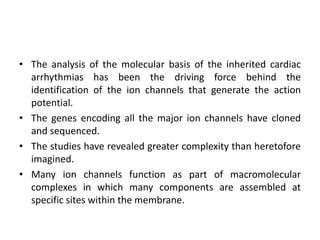

![• Phase 3 (repolarization) reflects the predominance of the delayed
outward rectifying currents after inactivation (closing) of the L-type
Ca2+ channels.
• Final repolarization during phase 3 is due to K+ efflux through the IK1
channels.
• In contrast to atrial and ventricular myocytes, SAN and AVN
myocytes demonstrate slow depolarization of the resting potential
during phase 4.
• This is mainly enabled by the absence of IK1, which allows inward
currents (e.g., pacemaker current [If]) to depolarize the membrane
potential.
• Slow depolarization during phase 4 inactivates most Na+ channels
and decreases their availability for phase 0.
• Consequently, in SAN and AVN myocytes, AP depolarization is
mainly achieved by ICa,L and the T-type Ca2+ current (ICa,T)](https://image.slidesharecdn.com/ionchannelopathy-160314170321/85/Ion-channelopathy-5-320.jpg)


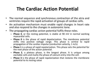
![• The generation of the action potential and the regional differences
that are observed throughout the heart are the result of the
selective permeability of ion channels distributed on the cell
membrane.
• The ion channels reduce the activation energy required for ion
movement across the lipophilic cell membrane.
• During the action potential, the permeability of ion channels
changes and each ion, eg, X, moves passively down its electro-
chemical gradients (ΔV=[Vm-Vx,] where Vm is the membrane
potential and Vx the reversal potential of ion X) to change the
membrane potential of the cell.
• The electrochemical gradient determines whether an ion moves
into the cell (depolarizing current for cations) or out of the cell
(repolarizing current for cations).
• Homeostasis of the intracellular ion concentrations is maintained by
active and coupled transport processes that are linked directly or
indirectly to ATP hydrolysis.](https://image.slidesharecdn.com/ionchannelopathy-160314170321/85/Ion-channelopathy-9-320.jpg)


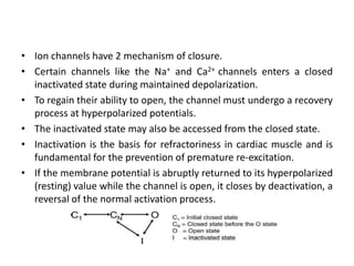
![• The open probability of this channel is proportional to the
[ADP]/[ATP] ratio.
• This channel couples the shape of the action potential to the
metabolic state of the cell.
• Energy depletion during ischemia increases the [ADP]/[ATP]
ratio, activates IK ATP, and abbreviates the action potential.
• The abbreviated action potential results in less force
generation and may be cardioprotective.
• This channel also plays a central role in ischemic
preconditioning.](https://image.slidesharecdn.com/ionchannelopathy-160314170321/85/Ion-channelopathy-16-320.jpg)


![Cardiac Ca2+ current (ICa) and intracellular
Ca2+ transients
• The L-type (long-lasting) inward Ca2+ current (ICa,L) is largely
responsible for the AP plateau.
• Ca2+ influx by ICa,L activates Ca2+ release channels (ryanodine
receptor [RyR2]), located in the sarcoplasmic reticulum
membrane.
• Sarcoplasmic reticulum Ca2+ release (Ca2+ transients) via RyR2
channels couples excitation to contraction in myocytes.
• CACNA1C encodes the α-subunit (Cav1.2) of L-type channels
• Beside ICa,L, T-type (tiny) Ca2+ current (ICa,T) is identified in SAN
and AVN myocytes.
• ICa,T is believed to contribute to AP formation in pacemaker
cells.](https://image.slidesharecdn.com/ionchannelopathy-160314170321/85/Ion-channelopathy-34-320.jpg)


The document discusses ion channelopathies and provides details on the cardiac action potential and the ion channels involved in each phase. It describes the major ion channels that generate the cardiac action potential, including sodium channels, transient outward potassium current, ultra-rapidly activating delayed outward rectifying current, and rapidly activating delayed outward rectifying current. It also discusses channelopathies associated with these ion channels like Brugada syndrome and long QT syndrome.