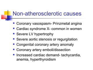
Coronary heart disease
Impaired cardiac function
due to inadequate
coronary circulation
Commonest cause- CAD
End result of accumulation of
atheromatous plaques in
coronary arteries
Non-atherosclerotic causes
 Coronary vasospasm- Prinzmetal angina
 Cardiac syndrome X- common in women
 Severe LV hypertrophy
 Severe aortic stenosis or regurgitation
 Congenital coronary artery anomaly
 Coronary artery emboli/dissection
 Increased cardiac demand- tachycardia,
anemia, hyperthyroidism
Risk factors for
atherosclerosis
 Non-modifiable- age, sex, family history
 Modifiable-
 Smoking
 Hypercholesterolemia- LDL, lipoprotein a
 Hypertension- systolic > diastolic
 Hyperglycemia- diabetes mellitus
 Type A behaviour- stress
 High fibrinogen, factor VII
 Hyperhomocysteinemia
 Obesity, sedentary lifestyle
 CRI
Pathophysiology
 Atherosclerosis is nearly universal & starts before
adulthood, leading to plaque formation
 Plaques cause narrowing of coronary arteries
 Stable plaque causes predictable angina
 Unstable plaque ruptures, activating clotting system &
thrombus formation, that impairs coronary blood flow
causing unstable angina or MI
 MI heals with scarring, causing impairing contractility
& increasing stiffness, leading to HF- acute/chronic
 Ischemic areas & scars are prone to cause
ventricular arrythmias, leading to sudden death
Manifestations
 Asymptomatic
 Angina-
 Acute- unstable- unpredictable
 Chronic- stable- predictable
 Myocardial infarction-
 Non ST elevated- NSTEMI
 ST elevated- STEMI
 Acute LVF
 Ischemic cardiomyopathy- CHF
 Sudden cardiac death
Clinical presentation
 Angina pectoris-
 Precordial/retrosternal/epigastric pain
 Described as tightness, squeezing, choking, indigestion
 Duration- <20 mins
 Radiation to left arm, shoulder, jaw
 Precipitated by exertion, stress, meal, cold, sex
 Relieved by rest or sublingual nitroglycerin
 Associated SOB, sweating, nausea, dizziness/syncope
 Unstable angina-
 Angina at rest, new-onset, more severe, increased frequency
 Myocardial infarction-
 Duration- >20 mins, not relieved by NTG
Evaluation
 Examination- HR, BP,
±S3/S4, murmur of MR
 Ix-
 Disease- ECG-ST elevation/depression, CxR,
Stress test-
TMT/radionuclide/ECHO,
ECHO- regional wall-motion abnormality,
Coronary angiography- CT or conventional, ±IVUS
 Risk factors- FBS, lipid profile, creatinine
 Precipitating factors- Hb, TSH
TMT- Treadmill test
 Bruce protocol-
 Increases treadmill speed & elevation every 3 minutes
 Indication-
 To confirm diagnosis of angina & determine severity
 To assess prognosis in patients with known CAD
 Screen those at high risk of CAD
 Interpretation-
 >1 mm flat or downsloping ST depression
 Severe disease- >2 mm depression, <6 mins. of exercise,
HR <70% predicted for age & hyper/hypotension
Coronary angiography
 For definitive diagnosis of CAD
 Indication- if PTCA/CABG an option-
 Limiting stable angina on adequate medical Rx
 High-risk disease- ACS or high-risk TMT
 Concomitant aortic valve disease
 Older patients undergoing valve surgery
 Recurrence of angina after PTCA/CABG
 Cardiac failure with surgically correctable lesion
 Survivors of SCD or VT
 Chest-pain or cardiomyopathy of unknown etiology
Treatment
 Medical-
 Aspirin- anti-thrombotic-1° & 2° prevention
 β-blockers- decrease cardiac workload- 2° prevention
 Statins- plaque stabilization & reduction- 2° prevention
 ACEI- cardiac remodelling- MI/HF
 Percutaneous- PTCA ± stent placement
 Bypass- CABG
 Experimental-
 Angiogenic growth factors- FGF-1, VEGF
 Stem-cell therapy
 Risk factor modification
Risk factor modification
 Quit smoking
 Control HT
 Control DM
 Control LDL
 Reduce stress
 Reduce weight
 Active lifestyle
Complication
 Recurrent ischemia- more after NSTEMI than STEMI
 Arrythmia- bradycardia, AV block, VT
 Shock- urgent PCI, ± IABP support
 Acute MR/VSD- supportsurgical correction
 Myocardial rupture- kills
 Heart failure- diuretics, nitrates, dobutamine
 Aneurysm- surgery, if required
 Mural thrombus ± embolization-
UFH/LMWHwarfarin
Chronic stable angina
 Angina occuring predictably on exertion &
relieved by rest or sublingual NTG
 Normal troponin & CK-MB
 ECG-
 Resting ECG- normal
 During anginal episode-
>1 mm ST depression ± T wave
flattening/inversion (ST
elevation seen in Prinzmetal angina)
 ECHO- for RWMA & LVEF
 Exercise testing- TMT
 Coronary angiography, if indicated
Treatment
 Sublingual NTG- for acute pain
 Prevention of attacks-
 Treat/avoid aggravating factors
 Aspirin (alternative- clopidogrel)
 Statins
 β-blockers
 ± long-acting nitrates
 ± CCB
 Risk factor modification
Revascularization
 Indication-
 Symptomatic despite adequate medical Rx
 Left main coronary artery stenosis
 Triple vessel disease with LVEF <50%
 Unstable angina
 Post-MI angina or +ve TMT
 Modalities-
 PCI- with stent- bare metal/drug eluting- placement
 CABG- preferred for L main/TVD with low
LVEF/T2DM
Acute coronary syndrome- ACS
 Unstable angina & myocardial infarction
 Unstable angina- cardiac markers- normal
 Angina at rest, new-onset, more severe, increased frequency
 With ST depression on ECG & normal Trop-T/I or CK-MB
 Myocardial infarction- cardiac markers- high
 Angina- lasts longer & not responsive to S/L NTG
 Rise of cardiac biomarkers- Trop-T/I & CK-MB
 With ECG changes- new Q waves/LBBB,
non-ST elevated-NSTEMI or ST elevated-
STEMI
 ECHO- new loss of viable myocardium or new RWMA
Recoverable myocardium
Hibernating- chronic ischemia
Stunned- post-MI
Evaluation- ECHO
Treatment of NSTE ACS
 Admit- rest, monitoring, ?oxygen
 Aspirin- 325 mg
 Clopidogrel- 300 mg stat75 mg OD
 Anticoagulation- UFH/LMWH
 Nitrates- for symptomatic relief
 β-blockers- as tolerated
 CCB- as add-on to nitrates & β-blockers
 Statins
 GP IIb/IIIa inhibitors- for intended early cath/PCI or
for high-risk patients- eptifibatide, tirofiban, abciximab
Indication for early angiography
All patients with ACS, except
those with normal stress test-
TMT/ECHO/radionuclide
STEMI
 Common in early morning
 ~1/2 have preceding angina- ignored
 1/3rd
without chest-pain,
specially diabetics
 e/o HF- poor prognosis
 Trop T/I- early MI, CK-MB- reinfarction
Treatment
 Admit- rest, morphine, ?oxygen, monitoring
 Aspirin + Clopidogrel
 β- blockers- early, if no contraindications
 ACEI- early, if no hypotension
 Statins
 Reperfusion-
within 12 hours of onset, sooner the better
 Options- for reperfusion
 1° angioplasty- with stenting & GP IIb/IIIa inhibitors
 Thrombolytic therapy- streptokinase, alteplase, tenecteplase-
followed by anticoagulation x 7 days
Post-infarction- no angiography
No complications
Preserved LVEF >50%
No exercise induced ischemia
Major differences
 Unstable angina-
 Trop T/I & CK-MB- normal
 Rx- Asp + Clopidogrel + UFH/LMWH ± GP IIb/IIIa inhibitors
 Early coronary angiography- Dx & Rx
 NSTEMI-
 Trop T/I & CK-MB- raised
 Rx- as for unstable angina
 Early coronary angiography
 STEMI-
 Trop T/I & CK-MB- raised
 Rx- Asp ± Clopidogrel + 1° PCI/Thrombolysis
 No angiography- post-MI normal LVEF & normal stress test

Ischemic heart disease

  • 1.
    Coronary heart disease Impairedcardiac function due to inadequate coronary circulation
  • 2.
    Commonest cause- CAD Endresult of accumulation of atheromatous plaques in coronary arteries
  • 3.
    Non-atherosclerotic causes  Coronaryvasospasm- Prinzmetal angina  Cardiac syndrome X- common in women  Severe LV hypertrophy  Severe aortic stenosis or regurgitation  Congenital coronary artery anomaly  Coronary artery emboli/dissection  Increased cardiac demand- tachycardia, anemia, hyperthyroidism
  • 4.
    Risk factors for atherosclerosis Non-modifiable- age, sex, family history  Modifiable-  Smoking  Hypercholesterolemia- LDL, lipoprotein a  Hypertension- systolic > diastolic  Hyperglycemia- diabetes mellitus  Type A behaviour- stress  High fibrinogen, factor VII  Hyperhomocysteinemia  Obesity, sedentary lifestyle  CRI
  • 5.
    Pathophysiology  Atherosclerosis isnearly universal & starts before adulthood, leading to plaque formation  Plaques cause narrowing of coronary arteries  Stable plaque causes predictable angina  Unstable plaque ruptures, activating clotting system & thrombus formation, that impairs coronary blood flow causing unstable angina or MI  MI heals with scarring, causing impairing contractility & increasing stiffness, leading to HF- acute/chronic  Ischemic areas & scars are prone to cause ventricular arrythmias, leading to sudden death
  • 6.
    Manifestations  Asymptomatic  Angina- Acute- unstable- unpredictable  Chronic- stable- predictable  Myocardial infarction-  Non ST elevated- NSTEMI  ST elevated- STEMI  Acute LVF  Ischemic cardiomyopathy- CHF  Sudden cardiac death
  • 7.
    Clinical presentation  Anginapectoris-  Precordial/retrosternal/epigastric pain  Described as tightness, squeezing, choking, indigestion  Duration- <20 mins  Radiation to left arm, shoulder, jaw  Precipitated by exertion, stress, meal, cold, sex  Relieved by rest or sublingual nitroglycerin  Associated SOB, sweating, nausea, dizziness/syncope  Unstable angina-  Angina at rest, new-onset, more severe, increased frequency  Myocardial infarction-  Duration- >20 mins, not relieved by NTG
  • 8.
    Evaluation  Examination- HR,BP, ±S3/S4, murmur of MR  Ix-  Disease- ECG-ST elevation/depression, CxR, Stress test- TMT/radionuclide/ECHO, ECHO- regional wall-motion abnormality, Coronary angiography- CT or conventional, ±IVUS  Risk factors- FBS, lipid profile, creatinine  Precipitating factors- Hb, TSH
  • 9.
    TMT- Treadmill test Bruce protocol-  Increases treadmill speed & elevation every 3 minutes  Indication-  To confirm diagnosis of angina & determine severity  To assess prognosis in patients with known CAD  Screen those at high risk of CAD  Interpretation-  >1 mm flat or downsloping ST depression  Severe disease- >2 mm depression, <6 mins. of exercise, HR <70% predicted for age & hyper/hypotension
  • 10.
    Coronary angiography  Fordefinitive diagnosis of CAD  Indication- if PTCA/CABG an option-  Limiting stable angina on adequate medical Rx  High-risk disease- ACS or high-risk TMT  Concomitant aortic valve disease  Older patients undergoing valve surgery  Recurrence of angina after PTCA/CABG  Cardiac failure with surgically correctable lesion  Survivors of SCD or VT  Chest-pain or cardiomyopathy of unknown etiology
  • 11.
    Treatment  Medical-  Aspirin-anti-thrombotic-1° & 2° prevention  β-blockers- decrease cardiac workload- 2° prevention  Statins- plaque stabilization & reduction- 2° prevention  ACEI- cardiac remodelling- MI/HF  Percutaneous- PTCA ± stent placement  Bypass- CABG  Experimental-  Angiogenic growth factors- FGF-1, VEGF  Stem-cell therapy  Risk factor modification
  • 12.
    Risk factor modification Quit smoking  Control HT  Control DM  Control LDL  Reduce stress  Reduce weight  Active lifestyle
  • 13.
    Complication  Recurrent ischemia-more after NSTEMI than STEMI  Arrythmia- bradycardia, AV block, VT  Shock- urgent PCI, ± IABP support  Acute MR/VSD- supportsurgical correction  Myocardial rupture- kills  Heart failure- diuretics, nitrates, dobutamine  Aneurysm- surgery, if required  Mural thrombus ± embolization- UFH/LMWHwarfarin
  • 14.
    Chronic stable angina Angina occuring predictably on exertion & relieved by rest or sublingual NTG  Normal troponin & CK-MB  ECG-  Resting ECG- normal  During anginal episode- >1 mm ST depression ± T wave flattening/inversion (ST elevation seen in Prinzmetal angina)  ECHO- for RWMA & LVEF  Exercise testing- TMT  Coronary angiography, if indicated
  • 15.
    Treatment  Sublingual NTG-for acute pain  Prevention of attacks-  Treat/avoid aggravating factors  Aspirin (alternative- clopidogrel)  Statins  β-blockers  ± long-acting nitrates  ± CCB  Risk factor modification
  • 16.
    Revascularization  Indication-  Symptomaticdespite adequate medical Rx  Left main coronary artery stenosis  Triple vessel disease with LVEF <50%  Unstable angina  Post-MI angina or +ve TMT  Modalities-  PCI- with stent- bare metal/drug eluting- placement  CABG- preferred for L main/TVD with low LVEF/T2DM
  • 17.
    Acute coronary syndrome-ACS  Unstable angina & myocardial infarction  Unstable angina- cardiac markers- normal  Angina at rest, new-onset, more severe, increased frequency  With ST depression on ECG & normal Trop-T/I or CK-MB  Myocardial infarction- cardiac markers- high  Angina- lasts longer & not responsive to S/L NTG  Rise of cardiac biomarkers- Trop-T/I & CK-MB  With ECG changes- new Q waves/LBBB, non-ST elevated-NSTEMI or ST elevated- STEMI  ECHO- new loss of viable myocardium or new RWMA
  • 18.
    Recoverable myocardium Hibernating- chronicischemia Stunned- post-MI Evaluation- ECHO
  • 19.
    Treatment of NSTEACS  Admit- rest, monitoring, ?oxygen  Aspirin- 325 mg  Clopidogrel- 300 mg stat75 mg OD  Anticoagulation- UFH/LMWH  Nitrates- for symptomatic relief  β-blockers- as tolerated  CCB- as add-on to nitrates & β-blockers  Statins  GP IIb/IIIa inhibitors- for intended early cath/PCI or for high-risk patients- eptifibatide, tirofiban, abciximab
  • 20.
    Indication for earlyangiography All patients with ACS, except those with normal stress test- TMT/ECHO/radionuclide
  • 21.
    STEMI  Common inearly morning  ~1/2 have preceding angina- ignored  1/3rd without chest-pain, specially diabetics  e/o HF- poor prognosis  Trop T/I- early MI, CK-MB- reinfarction
  • 22.
    Treatment  Admit- rest,morphine, ?oxygen, monitoring  Aspirin + Clopidogrel  β- blockers- early, if no contraindications  ACEI- early, if no hypotension  Statins  Reperfusion- within 12 hours of onset, sooner the better  Options- for reperfusion  1° angioplasty- with stenting & GP IIb/IIIa inhibitors  Thrombolytic therapy- streptokinase, alteplase, tenecteplase- followed by anticoagulation x 7 days
  • 23.
    Post-infarction- no angiography Nocomplications Preserved LVEF >50% No exercise induced ischemia
  • 24.
    Major differences  Unstableangina-  Trop T/I & CK-MB- normal  Rx- Asp + Clopidogrel + UFH/LMWH ± GP IIb/IIIa inhibitors  Early coronary angiography- Dx & Rx  NSTEMI-  Trop T/I & CK-MB- raised  Rx- as for unstable angina  Early coronary angiography  STEMI-  Trop T/I & CK-MB- raised  Rx- Asp ± Clopidogrel + 1° PCI/Thrombolysis  No angiography- post-MI normal LVEF & normal stress test