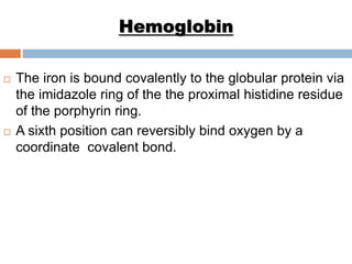
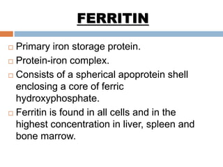
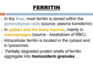
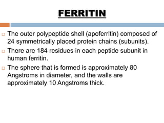
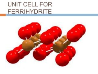
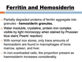
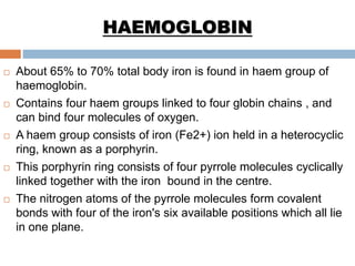
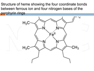
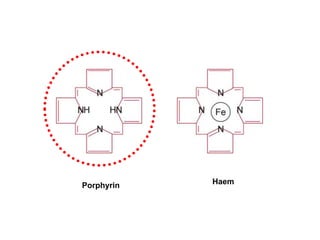
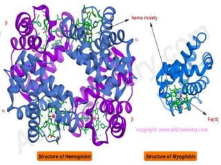
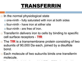
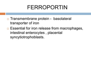
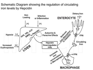
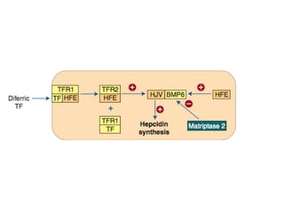
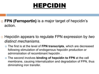
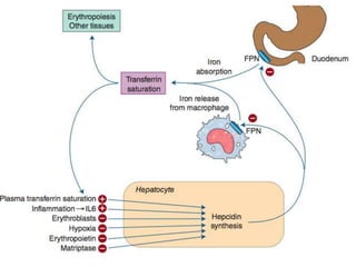
IRON METABOLISM I
DR AKSHAYA TOMAR
DEPT OF IMMUNOHEMATOLOGY AND
BLOOD TRANSFUSION
AFMC,PUNE
INTRODUCTION
 Iron is one of the most abundant element on earth, yet
only trace amounts are present in living cells.
 The total body iron content
 Adult male - 50mg/kg
 Adult female - 40 mg/kg
 Functional iron -> About 80% - Hemoglobin,
myoglobin , iron containing enzymes.
 Storage pool -> About 15% to 20%. Ferritin and
Hemosiderin.
DISTRIBUTION OF BODY
IRON
AMOUNT OF IRON
IN AVERAGE
ADULT
MALE(g) FEMALE(g) PERCENTAGE OF
TOTAL
HAEMOGLOBIN 2.4 1.7 65
FERRITIN AND
HAEMOSIDERIN
1.0(0.3-1.5) 0.3(0- 1.0) 30
MYOGLOBIN 0.15 0.12 3.5
HAEM
ENZYMES(eg
cytochrome,
catalase,
peroxidases
0.02 0.015 0.5
TRANSFERRIN
BOUND IRON
0.004 0.003 0.1
PROTEINS IMPORTANT IN
IRON METABOLISM
FERRITIN
 Primary iron storage protein.
 Protein-iron complex.
 Consists of a spherical apoprotein shell
enclosing a core of ferric
hydroxyphosphate.
 Ferritin is found in all cells and in the
highest concentration in liver, spleen and
bone marrow.
FERRITIN
 In the liver, most ferritin is stored within the
parenchymal cells (source- plasma transferrin)
 In spleen and the bone marrow, mainly in
macrophages (source - breakdown of RBC)
 Intracellular ferritin is located in the cytosol and
in lysosomes.
 Partially degraded protein shells of ferritin
aggregate into hemosiderin granules .
 The outer polypeptide shell (apoferritin) composed of
24 symmetrically placed protein chains (subunits).
 There are 184 residues in each peptide subunit in
human ferritin.
 The sphere that is formed is approximately 80
Angstroms in diameter, and the walls are
approximately 10 Angstroms thick.
FERRITIN
 The inner core contains an electron-dense
and chemically inert inorganic ferric iron-
core in the form of ferric hydroxy
phosphate.
 The ferritins are extremely large proteins
(450kDa) which can store up to 4500 iron
atoms as hydrous ferric oxide.
FERRITIN
Structure of ferritin showing the
outer polypeptide shell with inner
iron-core
FERRITIN
 The internal cavity of the ferritin molecule
communicates with the exterior via 6
channels through which the ferrous ions
may enter or leave.
 These channels maybe be three fold
protein channels or four fold protein
channels.
UNIT CELL FOR
FERRIHYDRITE
Ferritin and Hemosiderin
 Partially degraded proteins of ferritin aggregate into
granules - hemosiderin granules.
 Water insoluble, crystaline, protein iron complex
visible by light microscopy when stained by Prussian
blue stain( Pearls’ reaction)
 With normal iron stores, only trace amounts of
hemosiderin are found in macrophages of bone
marrow, spleen, and liver.
 In iron-overloaded cells, the proportion present as
haemosiderin increases considerably.
FERRITIN
 Plasma ferritin is derived largely from the
storage pool of body iron, its levels correlate
well with body iron stores.
 Normal Ferritin levels – 15to 300 μg/L. higher in
men ( median about 90μg/L) than in
premenopausal women ( 30μg/L)
 Storage iron depletion – < 15μg/L
 >300μg/L may indicate iron overload
HAEMOGLOBIN
 About 65% to 70% total body iron is found in haem group of
haemoglobin.
 Contains four haem groups linked to four globin chains , and
can bind four molecules of oxygen.
 A haem group consists of iron (Fe2+) ion held in a heterocyclic
ring, known as a porphyrin.
 This porphyrin ring consists of four pyrrole molecules cyclically
linked together with the iron bound in the centre.
 The nitrogen atoms of the pyrrole molecules form covalent
bonds with four of the iron's six available positions which all lie
in one plane.
Structure of heme showing the four coordinate bonds
between ferrous ion and four nitrogen bases of the
porphyrin rings.
Porphyrin Haem
Hemoglobin
 The iron is bound covalently to the globular protein via
the imidazole ring of the the proximal histidine residue
of the porphyrin ring.
 A sixth position can reversibly bind oxygen by a
coordinate covalent bond.
Structure of heme showing the square planar tetrapyrrole along
with the proximal and the distal histidine.
MYOGLOBIN
 Myoglobin is an iron- and oxygen-binding protein
found in the muscle tissue.
 It is a single-chain globular protein of 153 or 154
amino acids.
 Containing a haem prosthetic group in the center
around which the remaining apoprotein folds.
 It has eight alpha helices and a hydrophobic core.
 It is the primary oxygen-carrying pigment of muscle
tissues
TRANSFERRIN
 Iron is transported in plasma by an iron binding
glycoprotein called transferrin, which is synthesized
in liver, synthesis being inversely related to iron stores.
 Plasma half life – 8-11 days.
 The serum transferrin is 2.0–3.0 g/l and 1 mg of
transferrin binds 1.4 μg of iron.
 Two atoms of ferric iron bind to each molecule.
 Major function of transferrin is to deliver iron to the
tissues.
 Single chain polypeptide with molecular weight of
approximately 80,000 Da.
 Consisting of a single polypeptide chain of 680 to 700
amino acids.
 It has a bilobar structure with two Iron binding sites
 Each lobe contains an Iron binding site buried below
the surface of the protein in a hydrophilic environment.
TRANSFERRIN
Bi lobar structure of Human
transferrin
TRANSFERRIN
 In the normal physiological state
 one-ninth - fully saturated with iron at both sides
 four-ninth - have iron at either site
 four-ninth - are free of iron.
 Transferrin delivers iron to cells by binding to specific
cell surface receptors - TfR
 The TfR is a transmembrane protein consisting of two
subunits of 90,000 Da each, joined by a disulfide
bond.
 Each molecule of two subunits binds one transferrin
molecule.
OTHER HAEM PROTEINS
 Certain enzymes also contain haem as part of their
prosthetic group.
 Catalase
 Peroxidases
 Tryptophan pyrrolase
 Nitric oxide synthase
 Microsomal and mitochondrial cytochromes.
 Cyclooxygenase
DIVALENT METAL
TRANSPORTER 1
 Electrogenic pump that requires proton
cotransport in order to transfer Fe2+ across cell
membranes.
 Present at the apical membrane and subapical
endosomes of the duodenal enterocytes which
have a low pH.
 Transports iron from the gut lumen into the
labile iron pool.
FERROPORTIN
 Transmembrane protein - basolateral
transporter of iron
 Essential for iron release from macrophages,
intestinal enterocytes , placental
syncytiotrophoblasts.
HEPCIDIN
 Hepcidin was originally identified as an antimicrobial peptide
isolated from human urine.
 The liver is the predominant source of hepcidin
 84-amino-acid prepropeptide is synthesized
 Cleaved to yield 20-and 25-amino-acid peptides
 Released into the circulation
 Bound in plasma to α2 macroglobulin
 Filtered by the kidney.
 Hepcidin acts as a systemic iron-regulatory hormone
 Preferentially accumulates in the proximal duodenum and
spleen
 High expression of FPN in these areas.
 Hepatocytes release or down regulate hepcidin according to the
iron status of the body.
 The hepcidin response is remarkably rapid.
 In humans, iron ingestion results in a sharp increase in urinary
hepcidin excretion within 12 – 24 hours of starting treatment.
HEPCIDIN
Schematic Diagram showing the regulation of circulating
iron levels by Hepcidin
 FPN (Ferroportin) is a major target of hepcidin’s
action.
 Hepcidin appears to regulate FPN expression by two
distinct mechanisms.
 The first is at the level of FPN transcripts, which are decreased
following stimulation of endogenous hepcidin production or
administration of recombinant hepcidin .
 The second involves binding of hepcidin to FPN at the cell
membrane, causing internalization and degradation of FPN, thus
diminishing iron transfer.
HEPCIDIN
 Hepcidin might also directly inhibit erythroid-progenitor
proliferation and survival.
 Hepcidin synthesis is increased in response to raised serum
iron, iron overload and inflammation.
 Anemia, hypoxia and increased erythropoeitic activity are
associated with a dramatic decrease in liver hepcidin gene
expression.
 This may account for the increase in iron release from
reticuloendothelial cells and increase in iron absorption
HEPCIDIN
SCHEME OF NEXT PRESENTATION
IRON TURNOVER IN THE BODY
INTRACELLULAR IRON HOMEOSTASIS
DIAGNOSTIC METHODS FOR INVESTIGATING IRON METABOLISM
Iron metabolism PART I

Iron metabolism PART I

  • 1.
    IRON METABOLISM I DRAKSHAYA TOMAR DEPT OF IMMUNOHEMATOLOGY AND BLOOD TRANSFUSION AFMC,PUNE
  • 2.
    INTRODUCTION  Iron isone of the most abundant element on earth, yet only trace amounts are present in living cells.  The total body iron content  Adult male - 50mg/kg  Adult female - 40 mg/kg  Functional iron -> About 80% - Hemoglobin, myoglobin , iron containing enzymes.  Storage pool -> About 15% to 20%. Ferritin and Hemosiderin.
  • 3.
    DISTRIBUTION OF BODY IRON AMOUNTOF IRON IN AVERAGE ADULT MALE(g) FEMALE(g) PERCENTAGE OF TOTAL HAEMOGLOBIN 2.4 1.7 65 FERRITIN AND HAEMOSIDERIN 1.0(0.3-1.5) 0.3(0- 1.0) 30 MYOGLOBIN 0.15 0.12 3.5 HAEM ENZYMES(eg cytochrome, catalase, peroxidases 0.02 0.015 0.5 TRANSFERRIN BOUND IRON 0.004 0.003 0.1
  • 4.
  • 5.
    FERRITIN  Primary ironstorage protein.  Protein-iron complex.  Consists of a spherical apoprotein shell enclosing a core of ferric hydroxyphosphate.  Ferritin is found in all cells and in the highest concentration in liver, spleen and bone marrow.
  • 6.
    FERRITIN  In theliver, most ferritin is stored within the parenchymal cells (source- plasma transferrin)  In spleen and the bone marrow, mainly in macrophages (source - breakdown of RBC)  Intracellular ferritin is located in the cytosol and in lysosomes.  Partially degraded protein shells of ferritin aggregate into hemosiderin granules .
  • 7.
     The outerpolypeptide shell (apoferritin) composed of 24 symmetrically placed protein chains (subunits).  There are 184 residues in each peptide subunit in human ferritin.  The sphere that is formed is approximately 80 Angstroms in diameter, and the walls are approximately 10 Angstroms thick. FERRITIN
  • 8.
     The innercore contains an electron-dense and chemically inert inorganic ferric iron- core in the form of ferric hydroxy phosphate.  The ferritins are extremely large proteins (450kDa) which can store up to 4500 iron atoms as hydrous ferric oxide. FERRITIN
  • 9.
    Structure of ferritinshowing the outer polypeptide shell with inner iron-core
  • 10.
    FERRITIN  The internalcavity of the ferritin molecule communicates with the exterior via 6 channels through which the ferrous ions may enter or leave.  These channels maybe be three fold protein channels or four fold protein channels.
  • 12.
  • 15.
    Ferritin and Hemosiderin Partially degraded proteins of ferritin aggregate into granules - hemosiderin granules.  Water insoluble, crystaline, protein iron complex visible by light microscopy when stained by Prussian blue stain( Pearls’ reaction)  With normal iron stores, only trace amounts of hemosiderin are found in macrophages of bone marrow, spleen, and liver.  In iron-overloaded cells, the proportion present as haemosiderin increases considerably.
  • 16.
    FERRITIN  Plasma ferritinis derived largely from the storage pool of body iron, its levels correlate well with body iron stores.  Normal Ferritin levels – 15to 300 μg/L. higher in men ( median about 90μg/L) than in premenopausal women ( 30μg/L)  Storage iron depletion – < 15μg/L  >300μg/L may indicate iron overload
  • 17.
    HAEMOGLOBIN  About 65%to 70% total body iron is found in haem group of haemoglobin.  Contains four haem groups linked to four globin chains , and can bind four molecules of oxygen.  A haem group consists of iron (Fe2+) ion held in a heterocyclic ring, known as a porphyrin.  This porphyrin ring consists of four pyrrole molecules cyclically linked together with the iron bound in the centre.  The nitrogen atoms of the pyrrole molecules form covalent bonds with four of the iron's six available positions which all lie in one plane.
  • 18.
    Structure of hemeshowing the four coordinate bonds between ferrous ion and four nitrogen bases of the porphyrin rings.
  • 19.
  • 20.
    Hemoglobin  The ironis bound covalently to the globular protein via the imidazole ring of the the proximal histidine residue of the porphyrin ring.  A sixth position can reversibly bind oxygen by a coordinate covalent bond.
  • 21.
    Structure of hemeshowing the square planar tetrapyrrole along with the proximal and the distal histidine.
  • 23.
    MYOGLOBIN  Myoglobin isan iron- and oxygen-binding protein found in the muscle tissue.  It is a single-chain globular protein of 153 or 154 amino acids.  Containing a haem prosthetic group in the center around which the remaining apoprotein folds.  It has eight alpha helices and a hydrophobic core.  It is the primary oxygen-carrying pigment of muscle tissues
  • 25.
    TRANSFERRIN  Iron istransported in plasma by an iron binding glycoprotein called transferrin, which is synthesized in liver, synthesis being inversely related to iron stores.  Plasma half life – 8-11 days.  The serum transferrin is 2.0–3.0 g/l and 1 mg of transferrin binds 1.4 μg of iron.  Two atoms of ferric iron bind to each molecule.  Major function of transferrin is to deliver iron to the tissues.
  • 26.
     Single chainpolypeptide with molecular weight of approximately 80,000 Da.  Consisting of a single polypeptide chain of 680 to 700 amino acids.  It has a bilobar structure with two Iron binding sites  Each lobe contains an Iron binding site buried below the surface of the protein in a hydrophilic environment. TRANSFERRIN
  • 27.
    Bi lobar structureof Human transferrin
  • 28.
    TRANSFERRIN  In thenormal physiological state  one-ninth - fully saturated with iron at both sides  four-ninth - have iron at either site  four-ninth - are free of iron.  Transferrin delivers iron to cells by binding to specific cell surface receptors - TfR  The TfR is a transmembrane protein consisting of two subunits of 90,000 Da each, joined by a disulfide bond.  Each molecule of two subunits binds one transferrin molecule.
  • 29.
    OTHER HAEM PROTEINS Certain enzymes also contain haem as part of their prosthetic group.  Catalase  Peroxidases  Tryptophan pyrrolase  Nitric oxide synthase  Microsomal and mitochondrial cytochromes.  Cyclooxygenase
  • 30.
    DIVALENT METAL TRANSPORTER 1 Electrogenic pump that requires proton cotransport in order to transfer Fe2+ across cell membranes.  Present at the apical membrane and subapical endosomes of the duodenal enterocytes which have a low pH.  Transports iron from the gut lumen into the labile iron pool.
  • 31.
    FERROPORTIN  Transmembrane protein- basolateral transporter of iron  Essential for iron release from macrophages, intestinal enterocytes , placental syncytiotrophoblasts.
  • 33.
    HEPCIDIN  Hepcidin wasoriginally identified as an antimicrobial peptide isolated from human urine.  The liver is the predominant source of hepcidin  84-amino-acid prepropeptide is synthesized  Cleaved to yield 20-and 25-amino-acid peptides  Released into the circulation  Bound in plasma to α2 macroglobulin  Filtered by the kidney.  Hepcidin acts as a systemic iron-regulatory hormone
  • 34.
     Preferentially accumulatesin the proximal duodenum and spleen  High expression of FPN in these areas.  Hepatocytes release or down regulate hepcidin according to the iron status of the body.  The hepcidin response is remarkably rapid.  In humans, iron ingestion results in a sharp increase in urinary hepcidin excretion within 12 – 24 hours of starting treatment. HEPCIDIN
  • 35.
    Schematic Diagram showingthe regulation of circulating iron levels by Hepcidin
  • 37.
     FPN (Ferroportin)is a major target of hepcidin’s action.  Hepcidin appears to regulate FPN expression by two distinct mechanisms.  The first is at the level of FPN transcripts, which are decreased following stimulation of endogenous hepcidin production or administration of recombinant hepcidin .  The second involves binding of hepcidin to FPN at the cell membrane, causing internalization and degradation of FPN, thus diminishing iron transfer. HEPCIDIN
  • 39.
     Hepcidin mightalso directly inhibit erythroid-progenitor proliferation and survival.  Hepcidin synthesis is increased in response to raised serum iron, iron overload and inflammation.  Anemia, hypoxia and increased erythropoeitic activity are associated with a dramatic decrease in liver hepcidin gene expression.  This may account for the increase in iron release from reticuloendothelial cells and increase in iron absorption HEPCIDIN
  • 41.
    SCHEME OF NEXTPRESENTATION IRON TURNOVER IN THE BODY INTRACELLULAR IRON HOMEOSTASIS DIAGNOSTIC METHODS FOR INVESTIGATING IRON METABOLISM