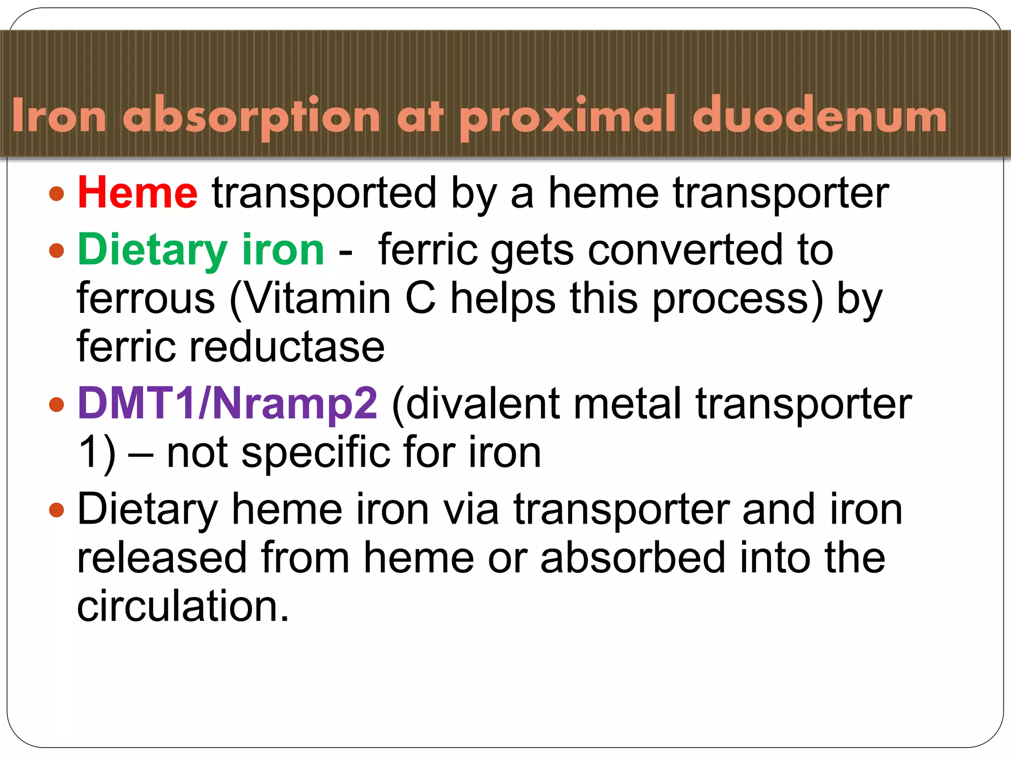
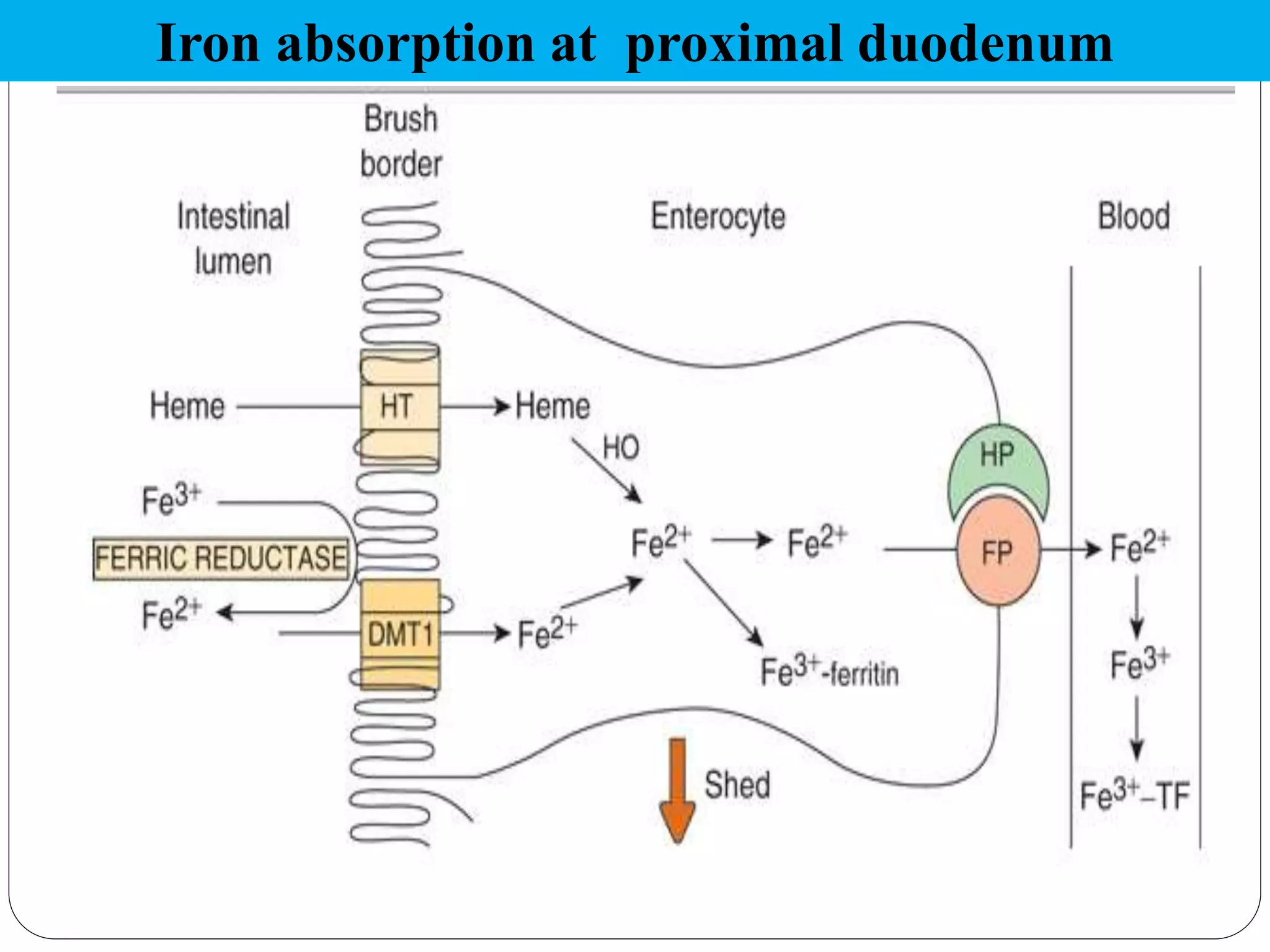
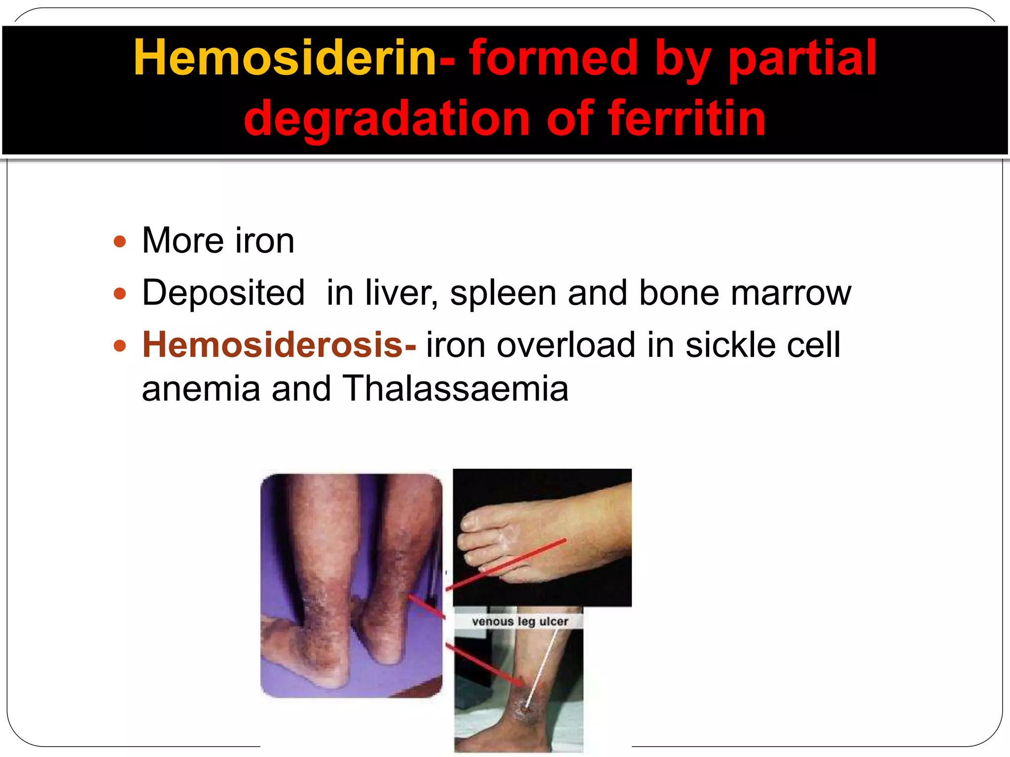
Iron Metabolism
Prof. Dr. V.P. Acharya
Why Iron??
 Micronutrient
 Iron deficiency and iron excess both are
deleterious
 Iron deficiency anemia is the most prevalent
cause of anemia in India- 65-75%
 Affects growth and mentation of fetus
Iron = 3-5 gms
65% as Hb
4-5% as
Myoglobin
1% as other
heme
compounds
15-30% as ferritin
in RES & liver
0.1 % as
Transferrin
75% in blood!
Adult female- amount of stores is 100-400 mg and the losses 1.5-2 mg/d
Strictly regulated
Minimal loss 1 – 2mg/d
Total body iron of 3 – 5g
Erythropoietic iron requirement only 20mg/d
Absorption is appropriately attenuated in dietary
iron excess
Important homeostatic mechanisms prevent
excessive iron absorption in duodenum and regulate
rate of iron release from RES
Iron is toxic to human cells and essential for
pathogens
Where do we find iron?
A. Heme containing proteins- Hb, Myoglobin,
Cytochromes, catalase, lactoperoxidase, Trp
pyrrolase, NO synthase
B. Non- heme iron containing proteins-
Aconitase, Phe-hydroxylase, Transferrin,
Ferritin, Hemosiderin
C. Fe-S complexes- Fe-S Complex III, Succinate
dehydrogenase, Xanthine oxidase
In Indian diet majority iron received from cereals
In premature infants early start of cereals recommended- 4th month
onwards
Hb iron turn over
Control of Iron Metabolism
Cellular Iron Homeostasis
Concerned with each cells
requirements for iron
Systemic Iron Homeostasis
Concerned with the body’s need for
iron- Mucosal block theory
Iron absorption at proximal duodenum
 Heme transported by a heme transporter
 Dietary iron - ferric gets converted to
ferrous (Vitamin C helps this process) by
ferric reductase
 DMT1/Nramp2 (divalent metal transporter
1) – not specific for iron
 Dietary heme iron via transporter and iron
released from heme or absorbed into the
circulation.
Iron absorption at proximal duodenum
 Iron then enters cytosol, binds to cytosolic
low molecular weight iron carriers and
proteins e.g. Mobilferrin (shuttles iron with
help of ATP) to basolateral membrane
 Export from basolateral membrane via
duodenal iron exporter – ferroportin
 Hephaestin (Cu containing protein) helps
ferroportin
 Hephaestin has a ferroxidase activity
Ferroportin
Present on:
basolateral membrane of
enterocytes
 macrophages and other RES cells
 hepatocytes
 β 1 globulin, a glycoprotein
 76 kDa
 Synthesized in liver
 Transport 2 Fe3+ in circulation – to BM or other organs
 Receptors – TfR1 & TfR2
 Internalized by receptor-mediated endocytosis
 Acid pH of Lysosome liberates iron that enters the
cytosol
 Affinity of Tf to Tfr1 falls ~ 500 times- complex
dissociates
 Defective glycosylation- congenital disorder of
glycosylation and Chronic alcohol abuse
 Total iron binding capacity
 Unsaturated iron binding capacity
 Actually we are measuring Transferrin saturation
 More useful when measured with total iron
 Iron deficiency or hemochromatosis can be
known
Ferritin- storage form
 Stores iron in cells
 Contains about 23% iron
 Made of 24 subunits – surround 3000-
4500 ferric atoms in a micellar form
 Normally very less; in Hemochromatosis
its level elevated
 Measure of iron store in body
 Transferrin - TIBC
Ferritin
 L and H chains (chromosome 19, 11).
 Synthesis controlled at 2 levels –
DNA transcription via its promoter.
mRNA translation via interactions with
iron regulatory proteins.
 Acute phase reactant.
Hemosiderin- formed by partial
degradation of ferritin
 More iron
 Deposited in liver, spleen and bone marrow
 Hemosiderosis- iron overload in sickle cell
anemia and Thalassaemia
Synthesis of TfR and Ferritin are reciprocally
linked to cellular iron content
 Ferritin in erythroid precursors may be of special
importance in haem synthesis especially at beginning
of Hb accumulation, when Tf-TfR pathway still
insufficient.
 When ferritin accumulates, it aggregates, proteolyzed
by lysosomal enzymes, , then converted to iron-rich,
poorly characterised haemosiderin, which releases
iron slowly.
 M-ferritin – present in mitochondria. Expression
correlated with tissues that have high mitochondrial
number, rather than those involved in iron storage.
Iron absorption regulated by many
stimuli –
Iron stores.
Degree of erythropoiesis (increased with
increased erythropoiesis,
reticulocytosis).
Ineffective erythropoiesis.
Mobilferrin – mechanism of loss in iron
replete state.
Role of specific proteins
 Transferrin and TfR.
 Ferritin.
 Iron responsive element-binding protein (IRE-BP)
aka iron regulatory protein/factor (IRP/IRF).
 HFE.
 Divalent metal transporter (DMT1, Nramp2,
DCT1,Slc11a) – duodenal iron transporter.
 Ferroportin and hephaestin, iron export proteins.
 Hepcidin.
Iron is conserved
 Hb released from lysed RBCs- conserved
by Haptoglobin (Hp) in urine
 Heme released from Hb- conserved by
Hemopexin in urine
Iron loss is through only mucosal shedding
 Regulation at absorption level
 Mucosal block theory
 Women lose through menstrual bleeding 1mg/d
 Loss through GIT mucosa
 Skin cells contain iron too
 Iron deficiency anemia
 Hemosiderosis
 Hemochromatosis- Bronze
diabetes
Fe deficiency anaemia FAQs
 51% of population in developing countries
 India- 65-75%
 India contributes to 80% maternal deaths due to
this
 Prevalence in preschool children- 70%
 Prevalence in pregnant women & adolescent
girls- 70%
Microcytic hypochromic anaemia
Causes:
 Nutritional deficiency
 Lack of absorption- Hypochlorhydria, subtotal
gastrectomy
 Hookworm infestation- 0.3 ml/day blood loss
 Repeated pregnancies- 1g loss /pregnancy
 Chronic blood loss- hemorrhoids, melaena, APD,
menorrhagia
 Nephrosis- important proteins lost
 Lead poisoning- Fe absorption and Hb synthesis ↓
Treatment
 100 mg Fe + 500 mg Folic acid-
Pregnancy
 20mg Fe + 100 mg FA- Children
 Vitamin C- enhances absorption
 Vitamin E- quenches ROS
Hemosiderosis
 Iron overload
 Hereditary hemosiderosis
 Bantu siderosis
 Iron vessels
Multiple organ affected
Hemochromatosis/ Bronze diabetes
 Hemosiderin deposition in liver- cirrhosis
 In pancreas- Diabetes
 Yellow-brown discolouration of skin
Hepcidin – the chief regulator
25 aa peptide. Identified 2000
Antimicrobial activity. Hepatic bacteriocidal protein
MHC I protein
Factors regulating intestinal iron absorption also
regulate the expression of hepcidin
Decreased iron stores
Increased erythropoietic activity
Anaemia
Hypoxia
Hepcidin regulation by erythropoietic
activity
 Hepcidin decreased in iron-deficiency anaemia,
hereditary anaemias with ineffective erythropoiesis,
and mouse models of anaemia from bleeding and
haemolysis. Response not seen when erythropoiesis
suppressed.
 Allows greater availability of iron for erythropoiesis.
 Degree of anaemia by itself doesn’t seem as
important.
 Nature of erythropoietic regulator of hepcidin is
unknown – proteins secreted by developing
erythrocytes?
 Intestinal iron absorption varies
inversely with liver hepcidin expression
 Hepcidin decrease the functional
activity of ferroportin by directly
binding to it and causing it to be
internalised from the cell surface and
deregulated
Decreases basolateral iron transfer and
thus dietary iron absorption
Decrease in iron export by hepatocyte
and macrophage and a resultant
increase in stored iron
 Human hemochromatosis protein (High iron Fe)
 Gene located at Chr 6
 Expression in GIT limited to cells in deep crypts in
proximity to site of iron absorption.
 HFE protein associated with TfR, acts to modulate
uptake of Tf-bound iron into crypt cells.
 Along with hepcidin, acts as iron sensor.
 Hereditary haemochromatosis with HFE gene
mutation - inability to bind beta 2-microglobulin,
impaired cellular trafficking, reduced incorporation
into the cell membrane, reduced association with
TfR1.
 For more ppt on medical
Biochemistry please visit my
website
 www.vpacharya.com

Iron metabolism

  • 1.
  • 2.
    Why Iron??  Micronutrient Iron deficiency and iron excess both are deleterious  Iron deficiency anemia is the most prevalent cause of anemia in India- 65-75%  Affects growth and mentation of fetus
  • 3.
    Iron = 3-5gms 65% as Hb 4-5% as Myoglobin 1% as other heme compounds 15-30% as ferritin in RES & liver 0.1 % as Transferrin
  • 4.
    75% in blood! Adultfemale- amount of stores is 100-400 mg and the losses 1.5-2 mg/d
  • 5.
    Strictly regulated Minimal loss1 – 2mg/d Total body iron of 3 – 5g Erythropoietic iron requirement only 20mg/d Absorption is appropriately attenuated in dietary iron excess Important homeostatic mechanisms prevent excessive iron absorption in duodenum and regulate rate of iron release from RES Iron is toxic to human cells and essential for pathogens
  • 6.
    Where do wefind iron? A. Heme containing proteins- Hb, Myoglobin, Cytochromes, catalase, lactoperoxidase, Trp pyrrolase, NO synthase B. Non- heme iron containing proteins- Aconitase, Phe-hydroxylase, Transferrin, Ferritin, Hemosiderin C. Fe-S complexes- Fe-S Complex III, Succinate dehydrogenase, Xanthine oxidase
  • 7.
    In Indian dietmajority iron received from cereals In premature infants early start of cereals recommended- 4th month onwards
  • 8.
  • 9.
    Control of IronMetabolism Cellular Iron Homeostasis Concerned with each cells requirements for iron Systemic Iron Homeostasis Concerned with the body’s need for iron- Mucosal block theory
  • 10.
    Iron absorption atproximal duodenum  Heme transported by a heme transporter  Dietary iron - ferric gets converted to ferrous (Vitamin C helps this process) by ferric reductase  DMT1/Nramp2 (divalent metal transporter 1) – not specific for iron  Dietary heme iron via transporter and iron released from heme or absorbed into the circulation.
  • 11.
    Iron absorption atproximal duodenum
  • 13.
     Iron thenenters cytosol, binds to cytosolic low molecular weight iron carriers and proteins e.g. Mobilferrin (shuttles iron with help of ATP) to basolateral membrane  Export from basolateral membrane via duodenal iron exporter – ferroportin  Hephaestin (Cu containing protein) helps ferroportin  Hephaestin has a ferroxidase activity
  • 15.
    Ferroportin Present on: basolateral membraneof enterocytes  macrophages and other RES cells  hepatocytes
  • 16.
     β 1globulin, a glycoprotein  76 kDa  Synthesized in liver  Transport 2 Fe3+ in circulation – to BM or other organs  Receptors – TfR1 & TfR2  Internalized by receptor-mediated endocytosis  Acid pH of Lysosome liberates iron that enters the cytosol  Affinity of Tf to Tfr1 falls ~ 500 times- complex dissociates  Defective glycosylation- congenital disorder of glycosylation and Chronic alcohol abuse
  • 17.
     Total ironbinding capacity  Unsaturated iron binding capacity  Actually we are measuring Transferrin saturation  More useful when measured with total iron  Iron deficiency or hemochromatosis can be known
  • 19.
    Ferritin- storage form Stores iron in cells  Contains about 23% iron  Made of 24 subunits – surround 3000- 4500 ferric atoms in a micellar form  Normally very less; in Hemochromatosis its level elevated  Measure of iron store in body  Transferrin - TIBC
  • 20.
    Ferritin  L andH chains (chromosome 19, 11).  Synthesis controlled at 2 levels – DNA transcription via its promoter. mRNA translation via interactions with iron regulatory proteins.  Acute phase reactant.
  • 21.
    Hemosiderin- formed bypartial degradation of ferritin  More iron  Deposited in liver, spleen and bone marrow  Hemosiderosis- iron overload in sickle cell anemia and Thalassaemia
  • 22.
    Synthesis of TfRand Ferritin are reciprocally linked to cellular iron content
  • 23.
     Ferritin inerythroid precursors may be of special importance in haem synthesis especially at beginning of Hb accumulation, when Tf-TfR pathway still insufficient.  When ferritin accumulates, it aggregates, proteolyzed by lysosomal enzymes, , then converted to iron-rich, poorly characterised haemosiderin, which releases iron slowly.  M-ferritin – present in mitochondria. Expression correlated with tissues that have high mitochondrial number, rather than those involved in iron storage.
  • 24.
    Iron absorption regulatedby many stimuli – Iron stores. Degree of erythropoiesis (increased with increased erythropoiesis, reticulocytosis). Ineffective erythropoiesis. Mobilferrin – mechanism of loss in iron replete state.
  • 25.
    Role of specificproteins  Transferrin and TfR.  Ferritin.  Iron responsive element-binding protein (IRE-BP) aka iron regulatory protein/factor (IRP/IRF).  HFE.  Divalent metal transporter (DMT1, Nramp2, DCT1,Slc11a) – duodenal iron transporter.  Ferroportin and hephaestin, iron export proteins.  Hepcidin.
  • 26.
    Iron is conserved Hb released from lysed RBCs- conserved by Haptoglobin (Hp) in urine  Heme released from Hb- conserved by Hemopexin in urine
  • 27.
    Iron loss isthrough only mucosal shedding  Regulation at absorption level  Mucosal block theory  Women lose through menstrual bleeding 1mg/d  Loss through GIT mucosa  Skin cells contain iron too
  • 28.
     Iron deficiencyanemia  Hemosiderosis  Hemochromatosis- Bronze diabetes
  • 29.
    Fe deficiency anaemiaFAQs  51% of population in developing countries  India- 65-75%  India contributes to 80% maternal deaths due to this  Prevalence in preschool children- 70%  Prevalence in pregnant women & adolescent girls- 70%
  • 30.
    Microcytic hypochromic anaemia Causes: Nutritional deficiency  Lack of absorption- Hypochlorhydria, subtotal gastrectomy  Hookworm infestation- 0.3 ml/day blood loss  Repeated pregnancies- 1g loss /pregnancy  Chronic blood loss- hemorrhoids, melaena, APD, menorrhagia  Nephrosis- important proteins lost  Lead poisoning- Fe absorption and Hb synthesis ↓
  • 32.
    Treatment  100 mgFe + 500 mg Folic acid- Pregnancy  20mg Fe + 100 mg FA- Children  Vitamin C- enhances absorption  Vitamin E- quenches ROS
  • 33.
    Hemosiderosis  Iron overload Hereditary hemosiderosis  Bantu siderosis  Iron vessels Multiple organ affected
  • 34.
    Hemochromatosis/ Bronze diabetes Hemosiderin deposition in liver- cirrhosis  In pancreas- Diabetes  Yellow-brown discolouration of skin
  • 36.
    Hepcidin – thechief regulator 25 aa peptide. Identified 2000 Antimicrobial activity. Hepatic bacteriocidal protein MHC I protein Factors regulating intestinal iron absorption also regulate the expression of hepcidin Decreased iron stores Increased erythropoietic activity Anaemia Hypoxia
  • 37.
    Hepcidin regulation byerythropoietic activity  Hepcidin decreased in iron-deficiency anaemia, hereditary anaemias with ineffective erythropoiesis, and mouse models of anaemia from bleeding and haemolysis. Response not seen when erythropoiesis suppressed.  Allows greater availability of iron for erythropoiesis.  Degree of anaemia by itself doesn’t seem as important.  Nature of erythropoietic regulator of hepcidin is unknown – proteins secreted by developing erythrocytes?
  • 38.
     Intestinal ironabsorption varies inversely with liver hepcidin expression  Hepcidin decrease the functional activity of ferroportin by directly binding to it and causing it to be internalised from the cell surface and deregulated Decreases basolateral iron transfer and thus dietary iron absorption Decrease in iron export by hepatocyte and macrophage and a resultant increase in stored iron
  • 39.
     Human hemochromatosisprotein (High iron Fe)  Gene located at Chr 6  Expression in GIT limited to cells in deep crypts in proximity to site of iron absorption.  HFE protein associated with TfR, acts to modulate uptake of Tf-bound iron into crypt cells.  Along with hepcidin, acts as iron sensor.  Hereditary haemochromatosis with HFE gene mutation - inability to bind beta 2-microglobulin, impaired cellular trafficking, reduced incorporation into the cell membrane, reduced association with TfR1.
  • 40.
     For moreppt on medical Biochemistry please visit my website  www.vpacharya.com