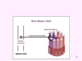
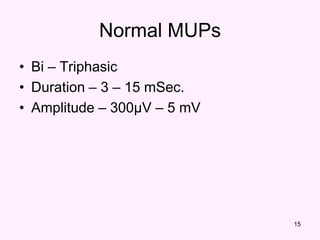
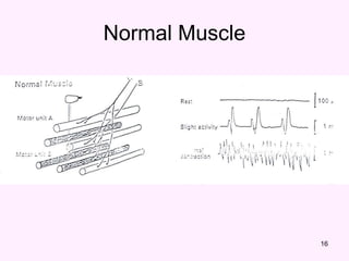
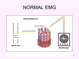
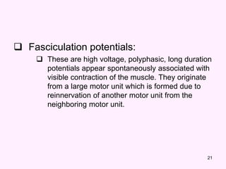
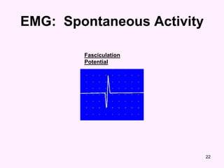
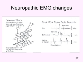
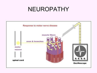
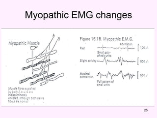
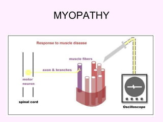
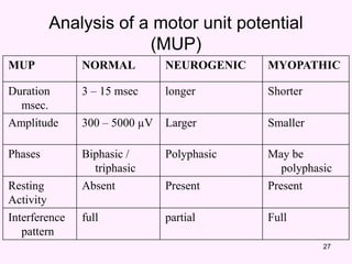
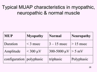
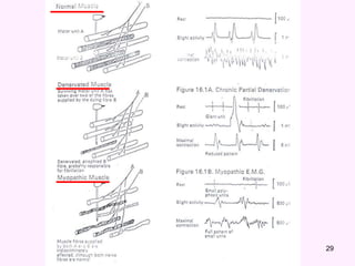
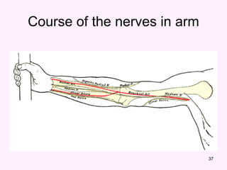
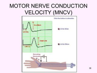
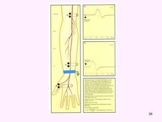
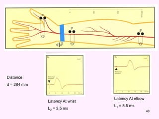
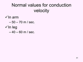
ELECTROMYOGRAPHY AND
MOTOR NERVE CONDUCTION
VELOCITY
2
ELECTROMYOGRAPHY (EMG)
• It’s a recording of electrical activity of the
muscle by inserting needle electrode in the
belly of the muscles or by applying the
surface electrodes.
• The potentials recorded on volitional effort
are derived from motor units of the
muscle, hence known as motor unit
potentials (MUPs).
3
• Electromyography (EMG) is a technique
for evaluating and recording physiologic
properties of muscles at rest and while
contracting.
4
• A motor unit is defined as one motor
neuron and all of the muscle fibers it
innervates.
5
6
Motor nerve conduction velocity
• Motor nerve conduction velocity of
peripheral nerves may be closely
correlated to their functional integrity or to
their structural abnormalities.
• Based on the nature of conduction
abnormalities two principal types of
peripheral nerve lesions can be identified:
Axonal degeneration and segmental
demyelination.
7
• In the patients of muscular weakness,
muscle atrophy, traumatic or metabolic
neuropathy, these tests are considered as
an extension of the physical examination
rather than a simple laboratory procedure.
8
OBJECTIVES
At the end of the session the students
should be able to:
• Acquire a skill to perform the test by
themselves.
• Analyze the motor unit potentials and
states their uses in health and diseases.
• Determine and calculate motor conduction
velocities of the peripheral nerves.
9
Requirements
• Machine.
• Electrodes.
• Electrode jelly
• Adhesive tape
• Saline & antiseptic (70 % alcohol)
10
Instrument set up
EMG
• Sweep time 10msec / cm
• Amplitude 1µV / cm
• Audioamplifier on
11
Instrument set up
Nerve conduction velocity
• Sweep time 2msec / cm
• Amplitude 1µV / cm
• Stimulator set up
• Frequency 1 / sec.
• Duration 0.2 msec.
• Intensity gradually increasing (MAM)
12
Procedure EMG
• Select a volunteer and explain him the
procedure.
• Put the ground electrode over the forearm
after soaking with saline.
• Clean the skin over the selected muscle.
• Apply the surface electrodes with the
electrode jelly and reference electrode
over bony point at least 3 cm apart.
13
Cont…
• Put the sweep run (continuous).
• Ask the subject to relax to evaluate any
resting activity.
• Ask the subject to exert mild voluntary
effort then moderate effort while continue
recording.
• Change the sweep speed to 100msec/cm
and then ask the subject to exert
maximum effort to determine interference
pattern.
14
Analysis
EMG
• Spontaneous activity
– The skeletal muscle is silent at rest, hence
spontaneous activity is absent.
15
Normal MUPs
• Bi – Triphasic
• Duration – 3 – 15 mSec.
• Amplitude – 300μV – 5 mV
16
Normal Muscle
17
NORMAL EMG
18
In neurogenic lesion or in active myositis,
the following spontaneous activity is
noted
 Positive sharp wave:
 A small potential of 50 to 100 µV, 5 to 10 msec
duration with abrupt onset and slow outset.
Abnormal MUPs
19
Fibrillation Potentials Positive Sharp Waves
20
 Fibrillation potential:
 these are randomly occurring small amplitude
potentials or may appear in runs. The
audioamplifier gives sounds, as if somebody
listen sounds of rains in a tin shade house.
These potentials are generated from the single
muscle fiber of a denervated muscle, possibly
due to denervation hypersensitivity to acetyl
choline.
21
 Fasciculation potentials:
 These are high voltage, polyphasic, long duration
potentials appear spontaneously associated with
visible contraction of the muscle. They originate
from a large motor unit which is formed due to
reinnervation of another motor unit from the
neighboring motor unit.
EMG: Spontaneous Activity
22
Fasciculation
Potential
23
Neuropathic EMG changes
24
NEUROPATHY
25
Myopathic EMG changes
26
MYOPATHY
27
MUP NORMAL NEUROGENIC MYOPATHIC
Duration
msec.
3 – 15 msec longer Shorter
Amplitude 300 – 5000 µV Larger Smaller
Phases Biphasic /
triphasic
Polyphasic May be
polyphasic
Resting
Activity
Absent Present Present
Interference
pattern
full partial Full
Analysis of a motor unit potential
(MUP)
28
MUP Myopathy Normal Neuropathy
Duration < 3 msec 3 – 15 msec > 15 msec
Amplitude < 300 µV 300-5000 µV > 5 mV
configuration polyphasic triphasic Polyphasic
Typical MUAP characteristics in myopathic,
neuropathic & normal muscle
29
30
Nerve Conduction studies
• A nerve conduction study (NCS) is a test
commonly used to evaluate the function,
especially the ability of electrical
conduction, of the motor and sensory
nerves of the human body. Nerve
conduction velocity (NCV) is a common
measurement made during this test.
31
Procedure for MNCV
• Give assurance to the subject about the
short harmless electric stimulation.
• Adjust the sweep speed to 2msec / cm.
• Adjust stimulus duration to 0.2 msec and
stimulus frequency to 1 / sec.
• Apply electrode jelly on plate electrode.
32
Cont…
• Put recording electrode over the thenar
eminence for median nerve conduction
velocity.
• Fix the reference electrode 3 cm away &
over a boney point.
33
Cont..
• Soak the stimulating electrode with saline
and put it over median nerve at elbow.
• Increase the stimulus intensity in steps. In
each step give stimulation manually by
pressing the stimulation switch once or
twice until a visible muscle contraction is
seen and a reproducible compound action
potential (CMAP) is recorded.
• Store the CMAP in the first channel.
34
Cont…
• Change the stimulating site i.e. from elbow
to wrist.
• Stimulate the nerve & record the CMAP for
median nerve at wrist.
• Measure the distance from elbow to wrist
with a measuring tape.
• Measure the latency in first CMAP & in the
next CAMP.
• Enter the distance between the elbow and
wrist.
35
MNCV
• MNCV will appear.
• It can also be
calculated by formula
• MNCV (m/sec)=
• l1 = latency at elbow.
• l2 = latency at wrist
sec)
(
2
1
)
(
tan
m
L
L
mm
ce
Dis

36
Duration
Amplitude
Analysis of MNCV
37
Course of the nerves in arm
38
MOTOR NERVE CONDUCTION
VELOCITY (MNCV)
39
40
Latency At wrist
L2 = 3.5 ms
Latency At elbow
L1 = 8.5 ms
Distance
d = 284 mm
41
Normal values for conduction
velocity
In arm
– 50 – 70 m / sec.
In leg
– 40 – 60 m / sec.
THANK YOU

L1-_EMGMNCV.power point presentation for ncv

  • 1.
  • 2.
    2 ELECTROMYOGRAPHY (EMG) • It’sa recording of electrical activity of the muscle by inserting needle electrode in the belly of the muscles or by applying the surface electrodes. • The potentials recorded on volitional effort are derived from motor units of the muscle, hence known as motor unit potentials (MUPs).
  • 3.
    3 • Electromyography (EMG)is a technique for evaluating and recording physiologic properties of muscles at rest and while contracting.
  • 4.
    4 • A motorunit is defined as one motor neuron and all of the muscle fibers it innervates.
  • 5.
  • 6.
    6 Motor nerve conductionvelocity • Motor nerve conduction velocity of peripheral nerves may be closely correlated to their functional integrity or to their structural abnormalities. • Based on the nature of conduction abnormalities two principal types of peripheral nerve lesions can be identified: Axonal degeneration and segmental demyelination.
  • 7.
    7 • In thepatients of muscular weakness, muscle atrophy, traumatic or metabolic neuropathy, these tests are considered as an extension of the physical examination rather than a simple laboratory procedure.
  • 8.
    8 OBJECTIVES At the endof the session the students should be able to: • Acquire a skill to perform the test by themselves. • Analyze the motor unit potentials and states their uses in health and diseases. • Determine and calculate motor conduction velocities of the peripheral nerves.
  • 9.
    9 Requirements • Machine. • Electrodes. •Electrode jelly • Adhesive tape • Saline & antiseptic (70 % alcohol)
  • 10.
    10 Instrument set up EMG •Sweep time 10msec / cm • Amplitude 1µV / cm • Audioamplifier on
  • 11.
    11 Instrument set up Nerveconduction velocity • Sweep time 2msec / cm • Amplitude 1µV / cm • Stimulator set up • Frequency 1 / sec. • Duration 0.2 msec. • Intensity gradually increasing (MAM)
  • 12.
    12 Procedure EMG • Selecta volunteer and explain him the procedure. • Put the ground electrode over the forearm after soaking with saline. • Clean the skin over the selected muscle. • Apply the surface electrodes with the electrode jelly and reference electrode over bony point at least 3 cm apart.
  • 13.
    13 Cont… • Put thesweep run (continuous). • Ask the subject to relax to evaluate any resting activity. • Ask the subject to exert mild voluntary effort then moderate effort while continue recording. • Change the sweep speed to 100msec/cm and then ask the subject to exert maximum effort to determine interference pattern.
  • 14.
    14 Analysis EMG • Spontaneous activity –The skeletal muscle is silent at rest, hence spontaneous activity is absent.
  • 15.
    15 Normal MUPs • Bi– Triphasic • Duration – 3 – 15 mSec. • Amplitude – 300μV – 5 mV
  • 16.
  • 17.
  • 18.
    18 In neurogenic lesionor in active myositis, the following spontaneous activity is noted  Positive sharp wave:  A small potential of 50 to 100 µV, 5 to 10 msec duration with abrupt onset and slow outset. Abnormal MUPs
  • 19.
  • 20.
    20  Fibrillation potential: these are randomly occurring small amplitude potentials or may appear in runs. The audioamplifier gives sounds, as if somebody listen sounds of rains in a tin shade house. These potentials are generated from the single muscle fiber of a denervated muscle, possibly due to denervation hypersensitivity to acetyl choline.
  • 21.
    21  Fasciculation potentials: These are high voltage, polyphasic, long duration potentials appear spontaneously associated with visible contraction of the muscle. They originate from a large motor unit which is formed due to reinnervation of another motor unit from the neighboring motor unit.
  • 22.
  • 23.
  • 24.
  • 25.
  • 26.
  • 27.
    27 MUP NORMAL NEUROGENICMYOPATHIC Duration msec. 3 – 15 msec longer Shorter Amplitude 300 – 5000 µV Larger Smaller Phases Biphasic / triphasic Polyphasic May be polyphasic Resting Activity Absent Present Present Interference pattern full partial Full Analysis of a motor unit potential (MUP)
  • 28.
    28 MUP Myopathy NormalNeuropathy Duration < 3 msec 3 – 15 msec > 15 msec Amplitude < 300 µV 300-5000 µV > 5 mV configuration polyphasic triphasic Polyphasic Typical MUAP characteristics in myopathic, neuropathic & normal muscle
  • 29.
  • 30.
    30 Nerve Conduction studies •A nerve conduction study (NCS) is a test commonly used to evaluate the function, especially the ability of electrical conduction, of the motor and sensory nerves of the human body. Nerve conduction velocity (NCV) is a common measurement made during this test.
  • 31.
    31 Procedure for MNCV •Give assurance to the subject about the short harmless electric stimulation. • Adjust the sweep speed to 2msec / cm. • Adjust stimulus duration to 0.2 msec and stimulus frequency to 1 / sec. • Apply electrode jelly on plate electrode.
  • 32.
    32 Cont… • Put recordingelectrode over the thenar eminence for median nerve conduction velocity. • Fix the reference electrode 3 cm away & over a boney point.
  • 33.
    33 Cont.. • Soak thestimulating electrode with saline and put it over median nerve at elbow. • Increase the stimulus intensity in steps. In each step give stimulation manually by pressing the stimulation switch once or twice until a visible muscle contraction is seen and a reproducible compound action potential (CMAP) is recorded. • Store the CMAP in the first channel.
  • 34.
    34 Cont… • Change thestimulating site i.e. from elbow to wrist. • Stimulate the nerve & record the CMAP for median nerve at wrist. • Measure the distance from elbow to wrist with a measuring tape. • Measure the latency in first CMAP & in the next CAMP. • Enter the distance between the elbow and wrist.
  • 35.
    35 MNCV • MNCV willappear. • It can also be calculated by formula • MNCV (m/sec)= • l1 = latency at elbow. • l2 = latency at wrist sec) ( 2 1 ) ( tan m L L mm ce Dis 
  • 36.
  • 37.
    37 Course of thenerves in arm
  • 38.
  • 39.
  • 40.
    40 Latency At wrist L2= 3.5 ms Latency At elbow L1 = 8.5 ms Distance d = 284 mm
  • 41.
    41 Normal values forconduction velocity In arm – 50 – 70 m / sec. In leg – 40 – 60 m / sec.
  • 42.