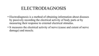
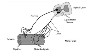
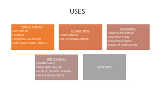
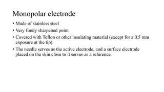
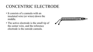
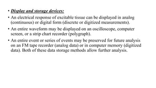
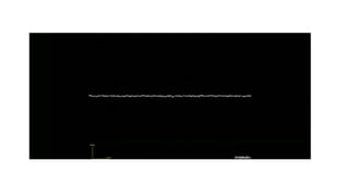
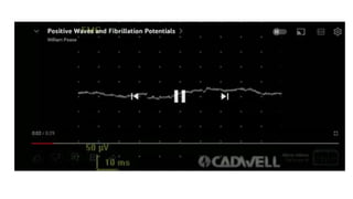
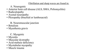
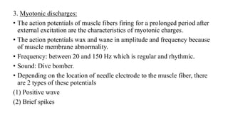
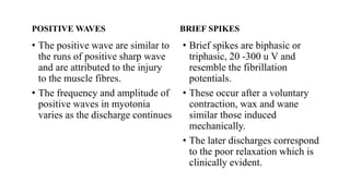
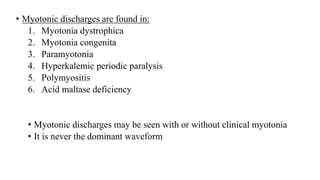
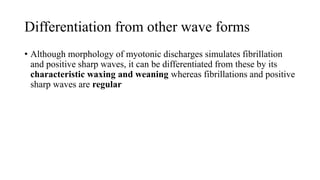
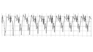
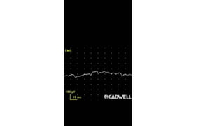
SlideShare a Scribd company logo
1 of 96
ELECTROMYOGRAPHY
-Batul Dawoodi (MPTh musculoskeletal disorders)
-Guided by: Dr. Kairavi Trivedi (PhD Scholar)
ELECTRODIAGNOSIS
• Electrodiagnosis is a method of obtaining information about diseases
by passively recording the electrical activity of body parts or by
measuring their response to external electrical stimulus.
• It measures the electrical activity of nerve (cause and extent of nerve
damage) and muscle.
Benefits of testing
• Correlating with physical examination
• Definitive diagnosis in nerve/muscle disorders
• Localisation of lesion
• Determination of physiological status of lesion
• Data for clinical or lab research
• Regeneration status of nerve
• Temporal parameters: acute, subacute, chronic
Goals
localization
muscle
Fibre type
Pathology
Temporal course
Neuromuscular
junction (NMJ)
Nerve Anterior Horn cell
severity
ANATOMY OF MUSCLE
• SKELETAL MUSCLE
• MUSCLE FIBRE
ELECTROMYOGRAPHY
• Electromyography refers to
recording of action potentials
of muscle fibers firing singly or
in groups near the needle
electrode in a muscle
• The muscle action potential
when recorded by a needle
appears triphasic as the action
potential approaches, crosses
and leaves the recording
electrode.
• Recording from an area incapable of
propagating the impulse, therefore,
results in large positivity with a low
and long negativity.
USES
MEDICAL RESEARCH:
1:ORTHOPEDIC
2:SURGERY
3:FUNTIONAL NEUROLOGY
4:GAIT AND POSTURAL ANALYSIS
REHABILITATION:
1:POST SURGICAL
2:NEURO-REHABILITATION
ERGONOMICS:
1:ANALYSIS OF DEMAND
2:RISK PREVENTION
3:ERGONOMIC DESIGN
4:PRODUCT CERTIFICATION
SPORTS SCIENCES:
1:BIOMECHANICS
2:MOVEMENT ANALYSIS
3:ATHLETIC STRENGTH TRAINING
4:SPORTS REHABILITATION
BIOFEEDBACK
RELATIVE CONTRAINDICATIONS
1. Coagulopathy
2. Risk of infection
3. Chest or abdominal area- risk of pneumothorax or peritoneal
insertion
Instrumentation
Stimulator/
electrodes
Patient Electrodes Preamplifier Amplifier
Display
(visual and
sound)
Input Phase Processing phase(amplified *1000 times)
Processing phase Output phase
TYPES OF EMG
NEEDLE
ELECTRODES
NEEDLE
ELECTRODE
CONCENTRIC MONOPOLAR SINGLE FIBRE MACROELECTRODE
BIPOLAR
ELECTRODE
SURFACE
ELECTRODE
Monopolar electrode
• Made of stainless steel
• Very finely sharpened point
• Covered with Teflon or other insulating material (except for a 0.5 mm
exposure at the tip).
• The needle serves as the active electrode, and a surface electrode
placed on the skin close to it serves as a reference.
• ADVANTAGE:
• patients accept them better because they are of small diameter and Teflon
covering allows them to slide in and out of the muscle easily.
• DISADVANTAGES:
• With repeated use, the size of the bare tip changes, thereby limiting the
number of examinations for which that needle can be used.
• The Teflon peels back, exposing a larger area that then changes the recorded
characteristics of the motor unit potentials.
• Because the active electrode tip and the surface electrode are separated by
some distance, the background noise becomes much greater as remote
muscle contractions may be picked up easier.
CONCENTRIC ELECTRODE
• It consists of a cannula with an
insulated wire (or wires) down the
middle.
• The active electrode is the small tip of
the center wire, and the reference
electrode is the outside cannula.
• ADVANTAGES:
• Because the active and reference electrodes are closer together, using
the concentric electrode minimizes background noise.
• The electrode picks up motor units from only a very small distance.
• No (reference) surface electrode is needed.
• DISADVANTAGES:
• On comparison with other needles, its larger diameter can cause more
pain, and moving the electrode around is uncomfortable.
BIPOLAR NEEDLE ELECTRODE
Concentric needles may have two
central wires (bipolar), in which case
the active and reference electrodes
are at the tip and the outside cannula
acts as the ground.
SINGLE FIBER NEEDLE ELECTRODE
• Used for special studies, this needle consists of a 0.5-0.6 mm stainless
steel cannula with a 25 µm fine platinum wire inside its hollow shaft.
In a side port 3 mm behind its tip, the cut end of the platinum wire is
exposed.
• Used in diagnosis of myasthenia gravis and NMJ disorders
SURFACE ELECTRODES
• Because they can pick up gross motor unit activities, the evaluation of
single motor unit potentials with surface electrodes is difficult. They
are best used as reference electrodes when monopolar needles are
used.
• They can also be used however, in kinesiology to obtain gross
indications about muscle activity and in gait analysis.
SIGNAL PROCESSING COMPONENTS
• When electrical potential differences are recorded in response to activating
excitable tissue, the voltage changes are generally too small to be
immediately visualized on common display devices
• The amplitudes of biological electrical signals recorded from nerve and
muscle are in the range of several microvolts to a few millivolts.
• Adequate display of the responses requires some signal processing by other
electronic devices such as
1. Amplifiers
2. Filters
3. Signal averages
4. Integrators
5. analog-to-digital converters
1. Amplifiers:
• It increases the amplitude of electrical voltages monitored in electrophysiological
examination.
• Such devices are used to make small electrical signals larger on signal display
instruments
• In some cases when low-voltage signals are being monitored, the amplifier built
into the display device increases the signal size enough for accurate measurements.
• A feature characteristic of all amplifiers is the device's gain.
• If the peak voltage of a monophasic input signal is doubled from I mV to 2 mV, the
gain of the amplifier is two.
• Amplifiers used in electrophysiological examination should uniformly increase
signal size for all signals within a specified frequency range (or bandwidth) from 2
to 10 Hz to 10,000 Hz.
Amplifiers
Simple Differential
• 2. Filters:
• Physiological responses of nerves and muscles have characteristic frequency ranges.
• The "noise" of the external environment can be electronically removed from the excitable
tissue signal (as long as the frequency ranges do not overlap) by an electronic device
called a filter.
• Several types of bandpass filters are commonly found on modern electrophysiological
testing instruments.
1. Notch filter: 60-Hz AC line noise can be eliminated from higher frequency signals by
using a notch filter. This type of filter can be set to reject signals at 60 Hz and pass (or
keep) a band of frequencies both less than and greater than 60 Hz.
2. Low frequency filter: It filters out the low-frequency signals (like those associated
with movement of the electrode) and passes (keeps) the higher frequency signals (this
type of filter is also known as a high pass filter).
3. High-frequency filter: (also known as a low pass filter), rejects higher frequencies
and passes the lower frequency signals.
• The low- and high frequency filters of ENMG devices are generally adjusted to 2 to 10
Hz to 10,000 Hz for motor nerve conduction studies, 10 Hz to 10,000 Hz for needle
electromyography, and 2 to 30 Hz to 2000 Hz for sensory nerve conduction studies.
• 3. Signal averagers:
• If a single response to an electrical stimulus is very small, and the electrical noise
recorded from the environment is large, visualization of the electrical signal of interest
may not be immediately possible.
• To overcome this problem of an important electrical signal being buried in noise, signal
averaging was developed.
• In signal averaging, an electrical stimulus is applied to evoke electrical activity in an
excitable tissue. For a short period of time following the stimulus, all electrical activity
(voltage changes) from the tissue is recorded and stored.
• When a second stimulus is applied, the electrical response is again stored and
electronically added to the record from the first stimulus.
• This process is repeated, with each successive record being added to the sum of all
previous records.
• Because the elicited potentials are time locked to the stimulus and reoccur at a fixed time
after the stimulus, signal averaging will enhance these signals.
• Electrical interference (noise) is not time locked to the stimulus, and the averaged noise
voltage transients will be eliminated by the average
• The signal averaging technique is used to record sensory nerve action potentials and
somatosensory evoked potentials.
• 4. Integrators:
• Integration of an electrical signal is the calculation of the area under a
signal waveform or curve.
• The units of the output of an electronic integrator are volts-seconds.
5. Analog to digital converters:
• For many clinical applications, the physiologic electrical responses from
nerve or muscle are recorded, processed (e.g., amplified and filtered),
and displayed as an analog output.
• This is the form of the output signal displayed on strip charts and many
oscilloscopes and stored on FM tape recorders.
• Display and storage devices:
• An electrical response of excitable tissue can be displayed in analog
(continuous) or digital form (discrete or digitized measurements).
• An entire waveform may be displayed on an oscilloscope, computer
screen, or a strip chart recorder (polygraph).
• An entire event or series of events may be preserved for future analysis
on an FM tape recorder (analog data) or in computer memory (digitized
data). Both of these data storage methods allow further analysis.
CROSS TALK
• An undesired EMG signal from a muscle group that is not commonly
monitored is called “crosstalk”
• Crosstalk contaminates the signal and can cause an incorrect interpretation of
the signal information.
• Crosstalk depends on the many physiological parameters and can be
minimized by choosing electrode size and inter-electrode distances (typically
1–2 cm or the radius of the electrode) carefully.
• Crosstalk increases with increasing subcutaneous fat thickness.
• Minimal crosstalk area (MCA) is defined as a surface where crosstalk versus
co-contraction of muscles is minimal. The precise location and measurements
of the distance between two bony landmarks are the keys to finding the
“minimal crosstalk area” of the targeted muscle. MCA helps to limit or avoid
crosstalk from neighboring muscles
Steps for performing EMG
1. Select the muscle as per the suspected clinical diagnosis- proximal muscles in
myopathy, both distal and proximal muscles supplied by different roots and nerves in
motor neuron disease and the specific muscles innervated by the suspected nerve or
radicular involvement
2. Instruct the patient how to contract and relax the muscles
3. Identify the muscle while the patient is contracting and relaxing the muscle
4. Locate the needle insertion point slightly away from the motor point to prevent end
plate noise.
5. Insert the needle quickly while the muscle is relaxed to minimize the pain.
6. Sharp MUP’s on minimal contraction confirm that the needle is in proper position. If
MUPs are not sharp, needle should be repositioned.
Recording
technique
Insertional activity
Spontaneous
activity
Normal
spontaneous
activity originating
from NMJ or
terminal axon
Abnormal
spontaneous
activity originating
from muscle fibre
Fibrillations
Positive sharp waves
Myotonic discharges
Complex repetitive
discharges
Abnormal
spontaneous
activities from
motor neuron or
axon
1.Fasciculations
2.Doublets, triplets and
multiplets
3.Myokymic discharges
4.Neuromyotonia
5.Cramp potentials
Voluntary activity
Minimal voluntary
contraction
Maximal voluntary
contraction
INSERTIONALACTIVITY
• Introduction of the needle into the muscle normally produces a brief
burst of electrical activity due to mechanical damage by needle
movement and it lasts slightly exceeding the needle movement
• Duration: 50-200 ms
• It appears as positive or negative high frequency spike in a cluster
Insertional activity
Normal Decreased
In chronic cases where
very few viable muscle
fibers are left
Eg: Fibrosis
Increased
Prolonged
In acute cases when
membrane stability is
compromised
Absent
Complete chronic
denervation
• THE MUSCLE AT REST:
• Following cessation of insertional activity, a normal relaxed muscle
will exhibit electrical silence which is the absence of electrical
potentials
• It is often difficult for the patient to completely relax sufficiently to
observe complete electrical silence
SPONTANEOUS ACTIVITY
• Normal spontaneous activity originating from NMJ or terminal axon:
• Once the insertion activity decays after a second in normal individuals,
there is spontaneous electrical activity.
• In the end plate zone however miniature end plate potentials are
spontaneously recorded.
• The end plate potentials are usually seen with an irregular baseline and
called as end plate noise. In the end plate region, action potentials which
are brief, rapid, and irregular with an initial negative deflection are known
as end plate spike.
• Sound: sputtering fat in a frying pan.
• Endplate spikes are due to mechanical activation of nerve terminals by the
needle. To avoid the normally occurring spontaneous and plate activities,
the needle should be introduced slightly away the motor point.
• Miniature end plate potentials:
• Miniature end plate potentials are the small (~0.4mV) depolarizations of the
postsynaptic terminal caused by the release of a single vesicle into the
synaptic cleft.
• Neurotransmitter vesicles containing acetylcholine collide spontaneously
with the nerve terminal and release acetylcholine into the neuromuscular
junction even without a signal from the axon.
• These small depolarizations are not enough to reach threshold and so an
action potential in the postsynaptic membrane does not occur.
• During experimentation with MEPPs, it was noticed that often spontaneous
action potentials would occur, called end plate spikes in normal striated
muscle without any stimulus.
• Sound: sea shell heard near to ear
• Sellin LC, Molgo J, Thornquist K, Hansson B, Thesleff S (1996). "On the possible origin of giant or slow-rising miniature end plate potentials at the
neuromuscular junction". Pflügers Archiv: European Journal of Physiology. 431 (3): 325–334. doi:10.1007/BF02207269. PMID 8584425. S2CID 8748384.
• Abnormal spontaneous activities from muscle fiber:
1. Fibrillation:
• Fibrillation are spontaneously occurring action potentials from a single
muscle fiber.
• Frequency: 0.5 – 15 Hz
• Amplitude of 20-200 u V
• Duration of 1-5 ms when recorded by a concentric needle (Danner 1982).
• Sound: rain drop on roof
• Fibrillation are biphasic or triphasic waves with initial positivity which is
an important differentiating feature from end plate spike.
2. Positive sharp waves
• long duration biphasic potentials with initial sharp positivity followed by a long duration
negative phase resulting in a saw tooth appearance.
• amplitude of 20 – 200 µ V
• Duration of 10- 30 ms.
• Sound: dull pop
• The denervated muscle fibres develop not only hypersensitivity to acetylcholine but the
number of ach receptors also increase which leads to depolarization and is responsible
for fibrillation and positive sharp waves.
Fibrillation and sharp waves are found in:
A. Neurogenic
• Anterior horn cell disease (ALS, SMA, Poliomyelitis)
• Radiculopathy
• Axonal neuropathy
• Plexopathy (brachial or lumbosacral)
B. Neuromuscular junction
• Botulism
• Myasthenia gravis
C. Myogenic
• Myositis
• Muscular dystrophy
• Acid maltase deficiency
• Myotubular myopathy
• Muscle trauma
• Fibrillations and positive sharp waves are graded as follows:
0= none
+1= persistent single train of potential (2-3 sec) in at least two areas
+2= moderate number of potentials in 3 or more areas
+3= many fibrillations or sharp waves in all areas
+4= full interference pattern of fibrillations or sharp waves
• Sharp waves: Seen early stage of denervation before the appearance of
fibrillation, myotonic disorders, and polymyositis and rarely in normal
individuals.
• Sharp waves are more commonly recorded with monopolar needles
compared to concentric ones.
• The mechanisms responsible for sharp waves are:
• Distant recording from fibrillating fibres
• Different degrees of membrane instability. (Sharp waves probably appear at a
lesser degree of membrane instability.)
3. Myotonic discharges:
• The action potentials of muscle fibers firing for a prolonged period after
external excitation are the characteristics of myotonic charges.
• The action potentials wax and wane in amplitude and frequency because
of muscle membrane abnormality.
• Frequency: between 20 and 150 Hz which is regular and rhythmic.
• Sound: Dive bomber.
• Depending on the location of needle electrode to the muscle fiber, there
are 2 types of these potentials
(1) Positive wave
(2) Brief spikes
POSITIVE WAVES
• The positive wave are similar to
the runs of positive sharp wave
and are attributed to the injury
to the muscle fibres.
• The frequency and amplitude of
positive waves in myotonia
varies as the discharge continues
BRIEF SPIKES
• Brief spikes are biphasic or
triphasic, 20 -300 u V and
resemble the fibrillation
potentials.
• These occur after a voluntary
contraction, wax and wane
similar those induced
mechanically.
• The later discharges correspond
to the poor relaxation which is
clinically evident.
• Myotonic discharges are found in:
1. Myotonia dystrophica
2. Myotonia congenita
3. Paramyotonia
4. Hyperkalemic periodic paralysis
5. Polymyositis
6. Acid maltase deficiency
• Myotonic discharges may be seen with or without clinical myotonia
• It is never the dominant waveform
Differentiation from other wave forms
• Although morphology of myotonic discharges simulates fibrillation
and positive sharp waves, it can be differentiated from these by its
characteristic waxing and weaning whereas fibrillations and positive
sharp waves are regular
4. Complex repetitive discharges (CRDs):
• Complex repetitive discharges refer to repetitive and synchronous firing of a
group of muscle fibers spontaneously or following needle movement.
• Amplitude: 50 µV to 1mv
• Morphology of all CRDs are identical from one discharge to another
• Duration:50 to100 ms.
• Frequency: 5-100 Hz.
• Sound: machine like sound
• Their unique repetitive pattern bears a superficial resemblance to myotonic
discharges and these are also called pseudomyotonic discharges.
• Complex repetitive discharges do not have a waxing and weaning
character. On the contrary these appear and disappear suddenly and this
feature helps in differentiating it from myotonic discharges
Ephaptic transmission
• CRDs are found in:
• Myogenic:
• Polymyositis
• Muscular dystrophies
• Myxoedema
• Schwartz-Jampel syndrome
• Neurogenic:
• Poliomyelitis
• Amyotrophic lateral sclerosis
• Chronic radiculopathies
• Chronic neuropathies
• Abnormal spontaneous activities from motor neuron or axon:
• Fasciculations:
• Fasciculation potentials are spontaneous contractions of a number of
muscle fibers belonging to whole or a part of motor unit.
• Fasciculations occur randomly and irregularly at variable rates ranging
between 1 and 500/min.
• The size and shape of fasciculations depend on the distance of
recording electrode from the motor unit.
• Fasciculations may have the appearance of normal or abnormal MUP
and can be recognized by their slower firing rare compared to MUP.
• Fasciculations are of two types:
1. Benign:
• Not accompanied by muscle weakness, wasting and reflex changes.
• Normal MUP morphology
• Fire at faster rate and occur at same site repetitively
2. Pathological
• Fasciculations are found in:
• Neurogenic disorders:
• Amyotrophic lateral sclerosis
• Spinal muscle atrophy
• Radiculopathy
• Syringomyelia
• Peripheral neuropathies
• Metabolic disorders:
• Tetany
• Thyrotoxicosis
• Anticholinesterase drugs
• Uremia
• Normal:
• Benign fasciculation
• Muscle cramps
• Doublets, Triplets And Multiplets:
• Spontaneous MUPs that fire in group of two, three or multiple potentials
• They originate because of spontaneous depolarisation of motor unit or
its axon similar to fasciculations
• Found in:
• Hyperventilation
• Tetany
• Motor neuron disease
• Metabolic diseases
• Ischemia can also induce doublets or multiplets
• Myokymic discharges:
• Spontaneous muscle potentials associated with fine, worm like quivering
of facial muscles are known as myokymic discharges.
• On EMG, these discharges reveal normal MUPs which fire in fixed
pattern and rhythm, occurring in bursts of 2-10 potentials firing at 40-60
Hz.
• The bursts recur at regular intervals of 0.1 to 10 sec creating a marching
sound.
• It appears as a result of spontaneous depolarization or ephaptic
transmission along demyelinated segment of nerve
• Myokymia can be differentiated from fasciculations by their distinct
pattern and different clinical significance.
• Facial Myokymia is found in
• Multiple sclerosis
• Brainstem neoplasm
• Polyradiculopathy
• Facial nerve palsy.
• Limb Myokymia is found in
• radiation plexopathy
• chronic nerve compression.
• Neuromyotonia:
• Neuromyotonia refers to continuous motor unit potentials firing at a
rate of 100-300 Hz which continue for long runs or recur in bursts.
• These are not influenced by voluntary activity.
• Neuromyotonia is found in syndrome of continuous muscle fiber,
activity, anticholinesterase poisoning. and spinal muscular atrophy.
• Ischemia augments or precipitates the Neuromyotonia in Tetany.
• Neuro myotonic discharges also occur in peripheral nerve lrritation
during surgery and serve as valuable guide of possible damage.
• Cramp potentials:
• In cramps, spontaneous discharges of potentials occur at 40-60 Hz
usually with an abrupt onset and cessation. Increasing number of
potentials are recruited as the cramp develops and drops out it subsides.
• The high frequency discharges of MUPs in cramp suggests its origin in
nerve rather than muscle
• The cramps occur in
• Salt depletion,
• Chronic neurogenic atrophy,
• Myxoedema,
• Pregnancy,
• Uremia
• Normal individuals.
• Motor unit potential (MUP)
• After evaluation of insertional and spontaneous activities, the next step
of EMG is evaluation of MUP on voluntary contraction.
• The basic unit of peripheral nervous system is the motor unit, which
comprises individual lower motor neuron, its axon, NMJ and muscle
fibers
• Depolarization of motor neuron to the threshold results in impulse
propagation through axon, which in turn results in excitation of all
muscle fibers more or less simultaneously supplied by that motor
neuron in normal situation
• Recruitment of MUPs refers to additional firing of MUPs during
muscle contraction which depends on size principle.
• Size principle:
• The size of the motor unit is dependent on size of motor neuron,
diameter of axon, thickness of myelin, conduction velocity,
depolarization threshold and metabolic type of muscle fibre
• The larger motor neuron has larger axon and thicker myelin hence
faster conduction velocity, higher threshold for depolarisation and
usually supplies type II muscle fibres which are required for fast twitch
• According to size principle, the motor neurons are recruited in order of
size from small to large (Henneman1974)
• In routine EMG, most MUPs analyzed are the smaller low threshold
representing type I muscle fibre
• The MUP represents the sum of the muscle action potentials supplied
by an anterior horn cell.
• The MUP therefore has a higher amplitude and longer duration than
action potential produced by a single muscle fiber.
• MUPs are assessed for their morphology, stability and firing pattern
• Amplitude, duration, number of phases, rise
time, and firing rates characterize the
morphology of motor unit potential.
• Traditionally one measures the amplitude
from peak to peak
• the duration from the first deflection of the
baseline to the last return to it
• the number of phases by counting the
number of times the components of the
motor unit potential cross the baseline plus
one
• rise time as that elapsed between the peak of
the initial positive (down) deflection to the
peak of the highest negative (up) deflection.
• The number of fibers contained in a motor unit and their degree of synchrony
affect those characteristics.
• The number of phases a motor unit contains depends largely on the synchrony of
depolarization of its muscle fibers and can be affected either by nerve disease
causing differential slowing in impulse conduction, or muscle disease where the
conduction characteristics of the muscle fibers themselves have changed.
• The rise time, strictly a function of the proximity of the needle tip to the muscle
fibers of the contracting unit, is usually between 200 and 300 µsec.
• The firing rates of motor units depend on their type and size. Smaller units are
recruited early, with weak effort, and fire faster than large units which are
recruited later as effort is increased.
• All the above characteristics vary with age, with the muscle under study, and with
muscle temperature. Minute changes in needle position can greatly affect the
shape of the motor unit potential.
• Temperature Effect: At lower temperatures the motor unit duration and its
amplitude are increased
• DURATION:
• The duration of MUP is a measure
of conduction velocity, length of
muscle fiber, membrane excitability
and synchrony of different muscle
fibers of a motor unit.
• The duration of MUP, is much less
influenced by the distance of
recoding electrode compared to the
amplitude
• SOUND:
• Low frequency long duration: dull
and thud like
• High frequency short duration: crisp
and sharp
• RISE TIME:
• The rise time of MUP is the duration from initial positive to subsequent
negative peak.
• It is an indicator of the distance of needle electrode from the muscle
fiber.
• A slower rise time is attributed to resistance and capacitance of the
intervening tissue which acts as high frequency filter and results in a
dull sound on the loudspeaker of EMG equipment.
• This indicates the need to reposition the needle closer to the muscle
fibers.
• AMPLITUDE:
• The amplitude of MUP is measured peak to peak
• It depends upon size and density of muscle fibers, synchrony of firing,
proximity of needle to the muscle fiber, age of the subject, muscle
examined and muscle temperature.
• Decreasing muscle temperature results in higher amplitude and longer
duration of MUPs
• PHASE OF MUP:
• Motor unit potential recorded by a concentric or monopolar needle
reveals as inverted triphasic potential (Positive- negative – positive).
• The phase is defined as the portion of MUP between departure and
return to the baseline i.e. number of Baseline crossings + 1.
• It is a measure of how synchronously muscle fibers in a motor unit fire
• A motor unit potential with more than 4 phases is called as polyphasic
potentials.
• Some potentials show directional changes without crossing the
baseline and these are known as turns.
• Polyphasia and turns suggest de-synchronisation or drop out of muscle
fibres.
Normal motor unit parameters
• Amplitude: 200 µV-5 Mv
• Duration:5-15 ms
• Rise time:100-200 μs (<500 μs)
• Frequency: 5-15/s (<60/s)
*Measured from initial positive peak to negative peak for triphasic
potentials
• Satellite potential:
• Satellite potentials are the
late potentials which are
time – locked to the main
motor unit potential.
• Seen in early innervation
• STABILITY OF MUP:
• Normally, if the needle electrode is fixed, there is no variation in the MUP
morphology of an individual motor unit.
• Variation in MUP is is a feature of abnormal NMJ transmission
VOLUNTARY CONTRACTION OF MUSCLE
• FIRING PATTERN OF MUP:
• On initiation of voluntary contraction of muscle, the motor units are
recruited in an orderly manner; the smallest appearing first, larger ones
later and the largest still later.
• It is based on size principle
• Key points to study firing pattern is activation and recruitment.
• Activation denotes the firing rate and is dependent on central mechanism
• Recruitment refers to the ability to recruit new MUPs as the firing rate
increases
EVALUATION OF MUP
• The classic method of MUP measurement is the isolation and
recording of at least 20 individual potentials and then measuring their
duration, phases, turns and amplitude which compared with the control
values for the muscle for the corresponding age.
ABNORMALITIES OF MUP
1. Short duration MUPs
2. Long duration MUPs
3. Polyphasic MUPs
4. Mixed pattern
1. SHORT DURATION MOTOR UNIT POTENTIALS (MUPS):
• The short duration MUPs are those with a duration shorter than for the
muscle of corresponding age.
• Short duration MUPs are usually of low amplitude and have rapid
recruitment at minimal effort
• The short duration MUPs are found in the disorders associated with loss
OR segmentation of muscle fibres.
• Short duration MUPs that found in
• myopathies and neuromuscular junction disorders
• early stage of reinnervation after nerve damage.
• In certain myopathies such as metabolic and endocrine, only a few or no short
duration MUPs are recorded.
2. LONG DURATION MUPs:
• The duration of MUPs if exceeds the normal value for the corresponding
muscle and age, constitutes the long duration MUPs.
• The long duration MUPs are generally associated with high amplitude and
poor recruitment; but sometimes the amplitude may be normal or low.
• Long duration MUPs are seen in
• motor neuron disease
• axonal neuropathies with collateral sprouting
• Chronic radiculopathies
• chronic Myositis such as polymyositis and inclusion body Myositis.
• The long duration MUPs are attributed to increase in fiber density. loss of
synchrony of firing of muscle fibres and increase in number of muscle fibres
in a motor unit.
3. POLYPHASIC MUP:
• Polyphasic MUPs are diagnosed when there are 4 or more phases in a
MUP.
• The individual components of a polyphasic potential represent the action
potentials recorded from a single muscle fiber.
• These are most frequently seen in myopathies where there is regeneration
of fibers and increased fiber density.
• Regeneration of axons in neurogenic diseases can also result in polyphasic
MUPs.
• If MUP is large, an increase in fiber density results in more turns without
extra phases, such a MUP is called complex MUP and it may be found in
any myopathy or neurogenic atrophy.
• Some polyphasic potentials may have very late satellite potential which is
known as linked potential or parasite potential. This makes the MUP
duration very long.
4. MIXED PATTERN:
• Occasionally instead of a polar from of short or long duration MUPs, a
mixed variety, comprising of short, long and polyphasic MUPs is
found.
• This pattern may be found in both myopathies and neurogenic
abnormalities.
Maximal voluntary contraction
• INTERFERENCE:
• During strong voluntary contraction, normally there is a dense pattern
of multiple superimposed potentials which is called as interference
pattern.
• Less dense pattern may occur with a loss of motor units, poor effort
and upper motor neuron lesions or a strong muscle.
ELECTROMYOGRAPHY.pptx

More Related Content

What's hot

Somatosensory evoked potentials
Somatosensory evoked potentialsSomatosensory evoked potentials
Somatosensory evoked potentialsManideep Malaka
 
H reflex (Hoffmann's Reflex)
H reflex (Hoffmann's Reflex)H reflex (Hoffmann's Reflex)
H reflex (Hoffmann's Reflex)Murtaza Syed
 
Cortical stimulation and mapping
Cortical stimulation and mappingCortical stimulation and mapping
Cortical stimulation and mappingPramod Krishnan
 
Muscle plasticity in response to electrical stimulation
Muscle plasticity in response to electrical stimulationMuscle plasticity in response to electrical stimulation
Muscle plasticity in response to electrical stimulationDrPriyanka PT
 
Emg and ncs slides chapter 1 and 2
Emg and ncs slides chapter 1 and 2Emg and ncs slides chapter 1 and 2
Emg and ncs slides chapter 1 and 2romegonzal
 
Electrotherapy intro.. 5 th semester
Electrotherapy intro.. 5 th semesterElectrotherapy intro.. 5 th semester
Electrotherapy intro.. 5 th semesterRiaz Ahmed
 
Somatosensory evoked potential
Somatosensory evoked potentialSomatosensory evoked potential
Somatosensory evoked potentialdahmed hamed
 
Neuromuscular electrical stimulation
Neuromuscular electrical stimulation Neuromuscular electrical stimulation
Neuromuscular electrical stimulation HemangiParmar4
 
Evoked potentials
Evoked potentialsEvoked potentials
Evoked potentialsKeerthi Priya
 
Medium Frequency Currents in Physiotherapy SRS
Medium Frequency Currents in  Physiotherapy SRSMedium Frequency Currents in  Physiotherapy SRS
Medium Frequency Currents in Physiotherapy SRSSreeraj S R
 
Nerves conduction study
Nerves conduction study Nerves conduction study
Nerves conduction study Sachin Adukia
 
Electro diagnostic tests ppt
Electro diagnostic tests pptElectro diagnostic tests ppt
Electro diagnostic tests pptKanchan Sharma
 

What's hot (20)

Emg biofeedback
Emg biofeedbackEmg biofeedback
Emg biofeedback
 
Emg
EmgEmg
Emg
 
Somatosensory evoked potentials
Somatosensory evoked potentialsSomatosensory evoked potentials
Somatosensory evoked potentials
 
H reflex (Hoffmann's Reflex)
H reflex (Hoffmann's Reflex)H reflex (Hoffmann's Reflex)
H reflex (Hoffmann's Reflex)
 
Emg presentation
Emg presentationEmg presentation
Emg presentation
 
PNF
PNFPNF
PNF
 
Cortical stimulation and mapping
Cortical stimulation and mappingCortical stimulation and mapping
Cortical stimulation and mapping
 
Muscle plasticity in response to electrical stimulation
Muscle plasticity in response to electrical stimulationMuscle plasticity in response to electrical stimulation
Muscle plasticity in response to electrical stimulation
 
Emg and ncs slides chapter 1 and 2
Emg and ncs slides chapter 1 and 2Emg and ncs slides chapter 1 and 2
Emg and ncs slides chapter 1 and 2
 
Electrotherapy intro.. 5 th semester
Electrotherapy intro.. 5 th semesterElectrotherapy intro.. 5 th semester
Electrotherapy intro.. 5 th semester
 
EEG & Evoked potentials
EEG & Evoked potentialsEEG & Evoked potentials
EEG & Evoked potentials
 
Somatosensory evoked potential
Somatosensory evoked potentialSomatosensory evoked potential
Somatosensory evoked potential
 
Neuromuscular electrical stimulation
Neuromuscular electrical stimulation Neuromuscular electrical stimulation
Neuromuscular electrical stimulation
 
Evoked potentials
Evoked potentialsEvoked potentials
Evoked potentials
 
Electrodiagnosis 2
Electrodiagnosis 2Electrodiagnosis 2
Electrodiagnosis 2
 
Ift russian
Ift russianIft russian
Ift russian
 
Medium Frequency Currents in Physiotherapy SRS
Medium Frequency Currents in  Physiotherapy SRSMedium Frequency Currents in  Physiotherapy SRS
Medium Frequency Currents in Physiotherapy SRS
 
Nerves conduction study
Nerves conduction study Nerves conduction study
Nerves conduction study
 
Electrical stimulation
Electrical stimulationElectrical stimulation
Electrical stimulation
 
Electro diagnostic tests ppt
Electro diagnostic tests pptElectro diagnostic tests ppt
Electro diagnostic tests ppt
 

Similar to ELECTROMYOGRAPHY.pptx

Electromyography (EMG)
Electromyography (EMG)Electromyography (EMG)
Electromyography (EMG)Dr Usha (Physio)
 
PERIPHERAL NERVES.pptx
PERIPHERAL NERVES.pptxPERIPHERAL NERVES.pptx
PERIPHERAL NERVES.pptxNainaJoshi9
 
Electromyography
ElectromyographyElectromyography
ElectromyographyGanesh Kushwah
 
Emg changes during fatigue and contraction
Emg changes during fatigue and contractionEmg changes during fatigue and contraction
Emg changes during fatigue and contractionDr Akshay RAj Chandra PT
 
Medical Instrumentation- EMG,ERG,EOG,PCG
Medical Instrumentation- EMG,ERG,EOG,PCGMedical Instrumentation- EMG,ERG,EOG,PCG
Medical Instrumentation- EMG,ERG,EOG,PCGPoornima D
 
emgpresentation-161114163\359.pptx abcdefghijh
emgpresentation-161114163\359.pptx abcdefghijhemgpresentation-161114163\359.pptx abcdefghijh
emgpresentation-161114163\359.pptx abcdefghijhpiyushbhardwaj93
 
EMG Final year bpT.pptx
EMG Final year bpT.pptxEMG Final year bpT.pptx
EMG Final year bpT.pptxDrYeshaVashi
 
Basics of peripheral nerve stimulation
Basics of peripheral nerve stimulationBasics of peripheral nerve stimulation
Basics of peripheral nerve stimulationDrYaminiVS
 
Full field electroretinogram
Full field electroretinogramFull field electroretinogram
Full field electroretinogramSmriti Ranabhat
 
MEDICAL ELECTRONICS_EC8073_Unit 1session 8
MEDICAL ELECTRONICS_EC8073_Unit 1session 8MEDICAL ELECTRONICS_EC8073_Unit 1session 8
MEDICAL ELECTRONICS_EC8073_Unit 1session 8SIVALAKSHMIPANNEERSE
 
Myoelectric Leg for Transfemoral Amputee
Myoelectric Leg for Transfemoral AmputeeMyoelectric Leg for Transfemoral Amputee
Myoelectric Leg for Transfemoral Amputeeijsrd.com
 
STSN1132 Radiation Detection #3.pptx
STSN1132 Radiation Detection #3.pptxSTSN1132 Radiation Detection #3.pptx
STSN1132 Radiation Detection #3.pptxNurmaizatulAtikah
 
ANESTHESIA for MRI procedures
ANESTHESIA for MRI proceduresANESTHESIA for MRI procedures
ANESTHESIA for MRI proceduresprateek gupta
 
SWD (Short wave diathermy)- Physiotherapy.ppt
SWD (Short wave diathermy)- Physiotherapy.pptSWD (Short wave diathermy)- Physiotherapy.ppt
SWD (Short wave diathermy)- Physiotherapy.pptMumux Mirani
 

Similar to ELECTROMYOGRAPHY.pptx (20)

Electromyography (EMG)
Electromyography (EMG)Electromyography (EMG)
Electromyography (EMG)
 
PERIPHERAL NERVES.pptx
PERIPHERAL NERVES.pptxPERIPHERAL NERVES.pptx
PERIPHERAL NERVES.pptx
 
Electromyography
ElectromyographyElectromyography
Electromyography
 
Electromyogram
ElectromyogramElectromyogram
Electromyogram
 
Emg changes during fatigue and contraction
Emg changes during fatigue and contractionEmg changes during fatigue and contraction
Emg changes during fatigue and contraction
 
Medical Instrumentation- EMG,ERG,EOG,PCG
Medical Instrumentation- EMG,ERG,EOG,PCGMedical Instrumentation- EMG,ERG,EOG,PCG
Medical Instrumentation- EMG,ERG,EOG,PCG
 
emgpresentation-161114163\359.pptx abcdefghijh
emgpresentation-161114163\359.pptx abcdefghijhemgpresentation-161114163\359.pptx abcdefghijh
emgpresentation-161114163\359.pptx abcdefghijh
 
L1- EMG+MNCV.ppt
L1- EMG+MNCV.pptL1- EMG+MNCV.ppt
L1- EMG+MNCV.ppt
 
EMG Final year bpT.pptx
EMG Final year bpT.pptxEMG Final year bpT.pptx
EMG Final year bpT.pptx
 
Power sources
Power sourcesPower sources
Power sources
 
Nanoknife
NanoknifeNanoknife
Nanoknife
 
Basics of peripheral nerve stimulation
Basics of peripheral nerve stimulationBasics of peripheral nerve stimulation
Basics of peripheral nerve stimulation
 
Full field electroretinogram
Full field electroretinogramFull field electroretinogram
Full field electroretinogram
 
Electrocardiogram pptx
Electrocardiogram pptxElectrocardiogram pptx
Electrocardiogram pptx
 
MEDICAL ELECTRONICS_EC8073_Unit 1session 8
MEDICAL ELECTRONICS_EC8073_Unit 1session 8MEDICAL ELECTRONICS_EC8073_Unit 1session 8
MEDICAL ELECTRONICS_EC8073_Unit 1session 8
 
Myoelectric Leg for Transfemoral Amputee
Myoelectric Leg for Transfemoral AmputeeMyoelectric Leg for Transfemoral Amputee
Myoelectric Leg for Transfemoral Amputee
 
STSN1132 Radiation Detection #3.pptx
STSN1132 Radiation Detection #3.pptxSTSN1132 Radiation Detection #3.pptx
STSN1132 Radiation Detection #3.pptx
 
ANESTHESIA for MRI procedures
ANESTHESIA for MRI proceduresANESTHESIA for MRI procedures
ANESTHESIA for MRI procedures
 
SWD (Short wave diathermy)- Physiotherapy.ppt
SWD (Short wave diathermy)- Physiotherapy.pptSWD (Short wave diathermy)- Physiotherapy.ppt
SWD (Short wave diathermy)- Physiotherapy.ppt
 
Electrosurgery
ElectrosurgeryElectrosurgery
Electrosurgery
 

Recently uploaded

Call Girls Jp Nagar Just Call 7001305949 Top Class Call Girl Service Available
Call Girls Jp Nagar Just Call 7001305949 Top Class Call Girl Service AvailableCall Girls Jp Nagar Just Call 7001305949 Top Class Call Girl Service Available
Call Girls Jp Nagar Just Call 7001305949 Top Class Call Girl Service Availablenarwatsonia7
 
Russian Call Girl Brookfield - 7001305949 Escorts Service 50% Off with Cash O...
Russian Call Girl Brookfield - 7001305949 Escorts Service 50% Off with Cash O...Russian Call Girl Brookfield - 7001305949 Escorts Service 50% Off with Cash O...
Russian Call Girl Brookfield - 7001305949 Escorts Service 50% Off with Cash O...narwatsonia7
 
VIP Call Girls Mumbai Arpita 9910780858 Independent Escort Service Mumbai
VIP Call Girls Mumbai Arpita 9910780858 Independent Escort Service MumbaiVIP Call Girls Mumbai Arpita 9910780858 Independent Escort Service Mumbai
VIP Call Girls Mumbai Arpita 9910780858 Independent Escort Service Mumbaisonalikaur4
 
Call Girl Surat Madhuri 7001305949 Independent Escort Service Surat
Call Girl Surat Madhuri 7001305949 Independent Escort Service SuratCall Girl Surat Madhuri 7001305949 Independent Escort Service Surat
Call Girl Surat Madhuri 7001305949 Independent Escort Service Suratnarwatsonia7
 
Call Girls Service Nandiambakkam | 7001305949 At Low Cost Cash Payment Booking
Call Girls Service Nandiambakkam | 7001305949 At Low Cost Cash Payment BookingCall Girls Service Nandiambakkam | 7001305949 At Low Cost Cash Payment Booking
Call Girls Service Nandiambakkam | 7001305949 At Low Cost Cash Payment BookingNehru place Escorts
 
Bangalore Call Girls Majestic đź“ž 9907093804 High Profile Service 100% Safe
Bangalore Call Girls Majestic đź“ž 9907093804 High Profile Service 100% SafeBangalore Call Girls Majestic đź“ž 9907093804 High Profile Service 100% Safe
Bangalore Call Girls Majestic đź“ž 9907093804 High Profile Service 100% Safenarwatsonia7
 
Russian Call Girls Chickpet - 7001305949 Booking and charges genuine rate for...
Russian Call Girls Chickpet - 7001305949 Booking and charges genuine rate for...Russian Call Girls Chickpet - 7001305949 Booking and charges genuine rate for...
Russian Call Girls Chickpet - 7001305949 Booking and charges genuine rate for...narwatsonia7
 
Call Girls Service Chennai Jiya 7001305949 Independent Escort Service Chennai
Call Girls Service Chennai Jiya 7001305949 Independent Escort Service ChennaiCall Girls Service Chennai Jiya 7001305949 Independent Escort Service Chennai
Call Girls Service Chennai Jiya 7001305949 Independent Escort Service ChennaiNehru place Escorts
 
Call Girls Hosur Just Call 7001305949 Top Class Call Girl Service Available
Call Girls Hosur Just Call 7001305949 Top Class Call Girl Service AvailableCall Girls Hosur Just Call 7001305949 Top Class Call Girl Service Available
Call Girls Hosur Just Call 7001305949 Top Class Call Girl Service Availablenarwatsonia7
 
Housewife Call Girls Hoskote | 7001305949 At Low Cost Cash Payment Booking
Housewife Call Girls Hoskote | 7001305949 At Low Cost Cash Payment BookingHousewife Call Girls Hoskote | 7001305949 At Low Cost Cash Payment Booking
Housewife Call Girls Hoskote | 7001305949 At Low Cost Cash Payment Bookingnarwatsonia7
 
Call Girls Service In Shyam Nagar Whatsapp 8445551418 Independent Escort Service
Call Girls Service In Shyam Nagar Whatsapp 8445551418 Independent Escort ServiceCall Girls Service In Shyam Nagar Whatsapp 8445551418 Independent Escort Service
Call Girls Service In Shyam Nagar Whatsapp 8445551418 Independent Escort Serviceparulsinha
 
Kolkata Call Girls Services 9907093804 @24x7 High Class Babes Here Call Now
Kolkata Call Girls Services 9907093804 @24x7 High Class Babes Here Call NowKolkata Call Girls Services 9907093804 @24x7 High Class Babes Here Call Now
Kolkata Call Girls Services 9907093804 @24x7 High Class Babes Here Call NowNehru place Escorts
 
Russian Call Girls in Pune Riya 9907093804 Short 1500 Night 6000 Best call gi...
Russian Call Girls in Pune Riya 9907093804 Short 1500 Night 6000 Best call gi...Russian Call Girls in Pune Riya 9907093804 Short 1500 Night 6000 Best call gi...
Russian Call Girls in Pune Riya 9907093804 Short 1500 Night 6000 Best call gi...Miss joya
 
Artifacts in Nuclear Medicine with Identifying and resolving artifacts.
Artifacts in Nuclear Medicine with Identifying and resolving artifacts.Artifacts in Nuclear Medicine with Identifying and resolving artifacts.
Artifacts in Nuclear Medicine with Identifying and resolving artifacts.MiadAlsulami
 
College Call Girls Vyasarpadi Whatsapp 7001305949 Independent Escort Service
College Call Girls Vyasarpadi Whatsapp 7001305949 Independent Escort ServiceCollege Call Girls Vyasarpadi Whatsapp 7001305949 Independent Escort Service
College Call Girls Vyasarpadi Whatsapp 7001305949 Independent Escort ServiceNehru place Escorts
 
Bangalore Call Girls Marathahalli đź“ž 9907093804 High Profile Service 100% Safe
Bangalore Call Girls Marathahalli đź“ž 9907093804 High Profile Service 100% SafeBangalore Call Girls Marathahalli đź“ž 9907093804 High Profile Service 100% Safe
Bangalore Call Girls Marathahalli đź“ž 9907093804 High Profile Service 100% Safenarwatsonia7
 
Call Girl Lucknow Mallika 7001305949 Independent Escort Service Lucknow
Call Girl Lucknow Mallika 7001305949 Independent Escort Service LucknowCall Girl Lucknow Mallika 7001305949 Independent Escort Service Lucknow
Call Girl Lucknow Mallika 7001305949 Independent Escort Service Lucknownarwatsonia7
 
Call Girls Service in Bommanahalli - 7001305949 with real photos and phone nu...
Call Girls Service in Bommanahalli - 7001305949 with real photos and phone nu...Call Girls Service in Bommanahalli - 7001305949 with real photos and phone nu...
Call Girls Service in Bommanahalli - 7001305949 with real photos and phone nu...narwatsonia7
 
High Profile Call Girls Jaipur Vani 8445551418 Independent Escort Service Jaipur
High Profile Call Girls Jaipur Vani 8445551418 Independent Escort Service JaipurHigh Profile Call Girls Jaipur Vani 8445551418 Independent Escort Service Jaipur
High Profile Call Girls Jaipur Vani 8445551418 Independent Escort Service Jaipurparulsinha
 

Recently uploaded (20)

Call Girls Jp Nagar Just Call 7001305949 Top Class Call Girl Service Available
Call Girls Jp Nagar Just Call 7001305949 Top Class Call Girl Service AvailableCall Girls Jp Nagar Just Call 7001305949 Top Class Call Girl Service Available
Call Girls Jp Nagar Just Call 7001305949 Top Class Call Girl Service Available
 
Russian Call Girl Brookfield - 7001305949 Escorts Service 50% Off with Cash O...
Russian Call Girl Brookfield - 7001305949 Escorts Service 50% Off with Cash O...Russian Call Girl Brookfield - 7001305949 Escorts Service 50% Off with Cash O...
Russian Call Girl Brookfield - 7001305949 Escorts Service 50% Off with Cash O...
 
VIP Call Girls Mumbai Arpita 9910780858 Independent Escort Service Mumbai
VIP Call Girls Mumbai Arpita 9910780858 Independent Escort Service MumbaiVIP Call Girls Mumbai Arpita 9910780858 Independent Escort Service Mumbai
VIP Call Girls Mumbai Arpita 9910780858 Independent Escort Service Mumbai
 
Call Girl Surat Madhuri 7001305949 Independent Escort Service Surat
Call Girl Surat Madhuri 7001305949 Independent Escort Service SuratCall Girl Surat Madhuri 7001305949 Independent Escort Service Surat
Call Girl Surat Madhuri 7001305949 Independent Escort Service Surat
 
Call Girls Service Nandiambakkam | 7001305949 At Low Cost Cash Payment Booking
Call Girls Service Nandiambakkam | 7001305949 At Low Cost Cash Payment BookingCall Girls Service Nandiambakkam | 7001305949 At Low Cost Cash Payment Booking
Call Girls Service Nandiambakkam | 7001305949 At Low Cost Cash Payment Booking
 
Bangalore Call Girls Majestic đź“ž 9907093804 High Profile Service 100% Safe
Bangalore Call Girls Majestic đź“ž 9907093804 High Profile Service 100% SafeBangalore Call Girls Majestic đź“ž 9907093804 High Profile Service 100% Safe
Bangalore Call Girls Majestic đź“ž 9907093804 High Profile Service 100% Safe
 
Russian Call Girls Chickpet - 7001305949 Booking and charges genuine rate for...
Russian Call Girls Chickpet - 7001305949 Booking and charges genuine rate for...Russian Call Girls Chickpet - 7001305949 Booking and charges genuine rate for...
Russian Call Girls Chickpet - 7001305949 Booking and charges genuine rate for...
 
Call Girls Service Chennai Jiya 7001305949 Independent Escort Service Chennai
Call Girls Service Chennai Jiya 7001305949 Independent Escort Service ChennaiCall Girls Service Chennai Jiya 7001305949 Independent Escort Service Chennai
Call Girls Service Chennai Jiya 7001305949 Independent Escort Service Chennai
 
Call Girls Hosur Just Call 7001305949 Top Class Call Girl Service Available
Call Girls Hosur Just Call 7001305949 Top Class Call Girl Service AvailableCall Girls Hosur Just Call 7001305949 Top Class Call Girl Service Available
Call Girls Hosur Just Call 7001305949 Top Class Call Girl Service Available
 
Housewife Call Girls Hoskote | 7001305949 At Low Cost Cash Payment Booking
Housewife Call Girls Hoskote | 7001305949 At Low Cost Cash Payment BookingHousewife Call Girls Hoskote | 7001305949 At Low Cost Cash Payment Booking
Housewife Call Girls Hoskote | 7001305949 At Low Cost Cash Payment Booking
 
Call Girls Service In Shyam Nagar Whatsapp 8445551418 Independent Escort Service
Call Girls Service In Shyam Nagar Whatsapp 8445551418 Independent Escort ServiceCall Girls Service In Shyam Nagar Whatsapp 8445551418 Independent Escort Service
Call Girls Service In Shyam Nagar Whatsapp 8445551418 Independent Escort Service
 
Kolkata Call Girls Services 9907093804 @24x7 High Class Babes Here Call Now
Kolkata Call Girls Services 9907093804 @24x7 High Class Babes Here Call NowKolkata Call Girls Services 9907093804 @24x7 High Class Babes Here Call Now
Kolkata Call Girls Services 9907093804 @24x7 High Class Babes Here Call Now
 
Russian Call Girls in Pune Riya 9907093804 Short 1500 Night 6000 Best call gi...
Russian Call Girls in Pune Riya 9907093804 Short 1500 Night 6000 Best call gi...Russian Call Girls in Pune Riya 9907093804 Short 1500 Night 6000 Best call gi...
Russian Call Girls in Pune Riya 9907093804 Short 1500 Night 6000 Best call gi...
 
sauth delhi call girls in Bhajanpura 🔝 9953056974 🔝 escort Service
sauth delhi call girls in Bhajanpura 🔝 9953056974 🔝 escort Servicesauth delhi call girls in Bhajanpura 🔝 9953056974 🔝 escort Service
sauth delhi call girls in Bhajanpura 🔝 9953056974 🔝 escort Service
 
Artifacts in Nuclear Medicine with Identifying and resolving artifacts.
Artifacts in Nuclear Medicine with Identifying and resolving artifacts.Artifacts in Nuclear Medicine with Identifying and resolving artifacts.
Artifacts in Nuclear Medicine with Identifying and resolving artifacts.
 
College Call Girls Vyasarpadi Whatsapp 7001305949 Independent Escort Service
College Call Girls Vyasarpadi Whatsapp 7001305949 Independent Escort ServiceCollege Call Girls Vyasarpadi Whatsapp 7001305949 Independent Escort Service
College Call Girls Vyasarpadi Whatsapp 7001305949 Independent Escort Service
 
Bangalore Call Girls Marathahalli đź“ž 9907093804 High Profile Service 100% Safe
Bangalore Call Girls Marathahalli đź“ž 9907093804 High Profile Service 100% SafeBangalore Call Girls Marathahalli đź“ž 9907093804 High Profile Service 100% Safe
Bangalore Call Girls Marathahalli đź“ž 9907093804 High Profile Service 100% Safe
 
Call Girl Lucknow Mallika 7001305949 Independent Escort Service Lucknow
Call Girl Lucknow Mallika 7001305949 Independent Escort Service LucknowCall Girl Lucknow Mallika 7001305949 Independent Escort Service Lucknow
Call Girl Lucknow Mallika 7001305949 Independent Escort Service Lucknow
 
Call Girls Service in Bommanahalli - 7001305949 with real photos and phone nu...
Call Girls Service in Bommanahalli - 7001305949 with real photos and phone nu...Call Girls Service in Bommanahalli - 7001305949 with real photos and phone nu...
Call Girls Service in Bommanahalli - 7001305949 with real photos and phone nu...
 
High Profile Call Girls Jaipur Vani 8445551418 Independent Escort Service Jaipur
High Profile Call Girls Jaipur Vani 8445551418 Independent Escort Service JaipurHigh Profile Call Girls Jaipur Vani 8445551418 Independent Escort Service Jaipur
High Profile Call Girls Jaipur Vani 8445551418 Independent Escort Service Jaipur
 

ELECTROMYOGRAPHY.pptx

  • 1. ELECTROMYOGRAPHY -Batul Dawoodi (MPTh musculoskeletal disorders) -Guided by: Dr. Kairavi Trivedi (PhD Scholar)
  • 2. ELECTRODIAGNOSIS • Electrodiagnosis is a method of obtaining information about diseases by passively recording the electrical activity of body parts or by measuring their response to external electrical stimulus. • It measures the electrical activity of nerve (cause and extent of nerve damage) and muscle.
  • 3. Benefits of testing • Correlating with physical examination • Definitive diagnosis in nerve/muscle disorders • Localisation of lesion • Determination of physiological status of lesion • Data for clinical or lab research • Regeneration status of nerve • Temporal parameters: acute, subacute, chronic
  • 5. ANATOMY OF MUSCLE • SKELETAL MUSCLE
  • 7.
  • 8. ELECTROMYOGRAPHY • Electromyography refers to recording of action potentials of muscle fibers firing singly or in groups near the needle electrode in a muscle • The muscle action potential when recorded by a needle appears triphasic as the action potential approaches, crosses and leaves the recording electrode.
  • 9. • Recording from an area incapable of propagating the impulse, therefore, results in large positivity with a low and long negativity.
  • 10. USES MEDICAL RESEARCH: 1:ORTHOPEDIC 2:SURGERY 3:FUNTIONAL NEUROLOGY 4:GAIT AND POSTURAL ANALYSIS REHABILITATION: 1:POST SURGICAL 2:NEURO-REHABILITATION ERGONOMICS: 1:ANALYSIS OF DEMAND 2:RISK PREVENTION 3:ERGONOMIC DESIGN 4:PRODUCT CERTIFICATION SPORTS SCIENCES: 1:BIOMECHANICS 2:MOVEMENT ANALYSIS 3:ATHLETIC STRENGTH TRAINING 4:SPORTS REHABILITATION BIOFEEDBACK
  • 11. RELATIVE CONTRAINDICATIONS 1. Coagulopathy 2. Risk of infection 3. Chest or abdominal area- risk of pneumothorax or peritoneal insertion
  • 12. Instrumentation Stimulator/ electrodes Patient Electrodes Preamplifier Amplifier Display (visual and sound) Input Phase Processing phase(amplified *1000 times) Processing phase Output phase
  • 13. TYPES OF EMG NEEDLE ELECTRODES NEEDLE ELECTRODE CONCENTRIC MONOPOLAR SINGLE FIBRE MACROELECTRODE BIPOLAR ELECTRODE SURFACE ELECTRODE
  • 14. Monopolar electrode • Made of stainless steel • Very finely sharpened point • Covered with Teflon or other insulating material (except for a 0.5 mm exposure at the tip). • The needle serves as the active electrode, and a surface electrode placed on the skin close to it serves as a reference.
  • 15. • ADVANTAGE: • patients accept them better because they are of small diameter and Teflon covering allows them to slide in and out of the muscle easily. • DISADVANTAGES: • With repeated use, the size of the bare tip changes, thereby limiting the number of examinations for which that needle can be used. • The Teflon peels back, exposing a larger area that then changes the recorded characteristics of the motor unit potentials. • Because the active electrode tip and the surface electrode are separated by some distance, the background noise becomes much greater as remote muscle contractions may be picked up easier.
  • 16. CONCENTRIC ELECTRODE • It consists of a cannula with an insulated wire (or wires) down the middle. • The active electrode is the small tip of the center wire, and the reference electrode is the outside cannula.
  • 17. • ADVANTAGES: • Because the active and reference electrodes are closer together, using the concentric electrode minimizes background noise. • The electrode picks up motor units from only a very small distance. • No (reference) surface electrode is needed. • DISADVANTAGES: • On comparison with other needles, its larger diameter can cause more pain, and moving the electrode around is uncomfortable.
  • 18. BIPOLAR NEEDLE ELECTRODE Concentric needles may have two central wires (bipolar), in which case the active and reference electrodes are at the tip and the outside cannula acts as the ground.
  • 19.
  • 20. SINGLE FIBER NEEDLE ELECTRODE • Used for special studies, this needle consists of a 0.5-0.6 mm stainless steel cannula with a 25 µm fine platinum wire inside its hollow shaft. In a side port 3 mm behind its tip, the cut end of the platinum wire is exposed. • Used in diagnosis of myasthenia gravis and NMJ disorders
  • 21. SURFACE ELECTRODES • Because they can pick up gross motor unit activities, the evaluation of single motor unit potentials with surface electrodes is difficult. They are best used as reference electrodes when monopolar needles are used. • They can also be used however, in kinesiology to obtain gross indications about muscle activity and in gait analysis.
  • 22. SIGNAL PROCESSING COMPONENTS • When electrical potential differences are recorded in response to activating excitable tissue, the voltage changes are generally too small to be immediately visualized on common display devices • The amplitudes of biological electrical signals recorded from nerve and muscle are in the range of several microvolts to a few millivolts. • Adequate display of the responses requires some signal processing by other electronic devices such as 1. Amplifiers 2. Filters 3. Signal averages 4. Integrators 5. analog-to-digital converters
  • 23. 1. Amplifiers: • It increases the amplitude of electrical voltages monitored in electrophysiological examination. • Such devices are used to make small electrical signals larger on signal display instruments • In some cases when low-voltage signals are being monitored, the amplifier built into the display device increases the signal size enough for accurate measurements. • A feature characteristic of all amplifiers is the device's gain. • If the peak voltage of a monophasic input signal is doubled from I mV to 2 mV, the gain of the amplifier is two. • Amplifiers used in electrophysiological examination should uniformly increase signal size for all signals within a specified frequency range (or bandwidth) from 2 to 10 Hz to 10,000 Hz.
  • 24.
  • 26. • 2. Filters: • Physiological responses of nerves and muscles have characteristic frequency ranges. • The "noise" of the external environment can be electronically removed from the excitable tissue signal (as long as the frequency ranges do not overlap) by an electronic device called a filter. • Several types of bandpass filters are commonly found on modern electrophysiological testing instruments. 1. Notch filter: 60-Hz AC line noise can be eliminated from higher frequency signals by using a notch filter. This type of filter can be set to reject signals at 60 Hz and pass (or keep) a band of frequencies both less than and greater than 60 Hz. 2. Low frequency filter: It filters out the low-frequency signals (like those associated with movement of the electrode) and passes (keeps) the higher frequency signals (this type of filter is also known as a high pass filter). 3. High-frequency filter: (also known as a low pass filter), rejects higher frequencies and passes the lower frequency signals. • The low- and high frequency filters of ENMG devices are generally adjusted to 2 to 10 Hz to 10,000 Hz for motor nerve conduction studies, 10 Hz to 10,000 Hz for needle electromyography, and 2 to 30 Hz to 2000 Hz for sensory nerve conduction studies.
  • 27. • 3. Signal averagers: • If a single response to an electrical stimulus is very small, and the electrical noise recorded from the environment is large, visualization of the electrical signal of interest may not be immediately possible. • To overcome this problem of an important electrical signal being buried in noise, signal averaging was developed. • In signal averaging, an electrical stimulus is applied to evoke electrical activity in an excitable tissue. For a short period of time following the stimulus, all electrical activity (voltage changes) from the tissue is recorded and stored. • When a second stimulus is applied, the electrical response is again stored and electronically added to the record from the first stimulus. • This process is repeated, with each successive record being added to the sum of all previous records. • Because the elicited potentials are time locked to the stimulus and reoccur at a fixed time after the stimulus, signal averaging will enhance these signals. • Electrical interference (noise) is not time locked to the stimulus, and the averaged noise voltage transients will be eliminated by the average • The signal averaging technique is used to record sensory nerve action potentials and somatosensory evoked potentials.
  • 28. • 4. Integrators: • Integration of an electrical signal is the calculation of the area under a signal waveform or curve. • The units of the output of an electronic integrator are volts-seconds.
  • 29. 5. Analog to digital converters: • For many clinical applications, the physiologic electrical responses from nerve or muscle are recorded, processed (e.g., amplified and filtered), and displayed as an analog output. • This is the form of the output signal displayed on strip charts and many oscilloscopes and stored on FM tape recorders.
  • 30. • Display and storage devices: • An electrical response of excitable tissue can be displayed in analog (continuous) or digital form (discrete or digitized measurements). • An entire waveform may be displayed on an oscilloscope, computer screen, or a strip chart recorder (polygraph). • An entire event or series of events may be preserved for future analysis on an FM tape recorder (analog data) or in computer memory (digitized data). Both of these data storage methods allow further analysis.
  • 31. CROSS TALK • An undesired EMG signal from a muscle group that is not commonly monitored is called “crosstalk” • Crosstalk contaminates the signal and can cause an incorrect interpretation of the signal information. • Crosstalk depends on the many physiological parameters and can be minimized by choosing electrode size and inter-electrode distances (typically 1–2 cm or the radius of the electrode) carefully. • Crosstalk increases with increasing subcutaneous fat thickness. • Minimal crosstalk area (MCA) is defined as a surface where crosstalk versus co-contraction of muscles is minimal. The precise location and measurements of the distance between two bony landmarks are the keys to finding the “minimal crosstalk area” of the targeted muscle. MCA helps to limit or avoid crosstalk from neighboring muscles
  • 32. Steps for performing EMG 1. Select the muscle as per the suspected clinical diagnosis- proximal muscles in myopathy, both distal and proximal muscles supplied by different roots and nerves in motor neuron disease and the specific muscles innervated by the suspected nerve or radicular involvement 2. Instruct the patient how to contract and relax the muscles 3. Identify the muscle while the patient is contracting and relaxing the muscle 4. Locate the needle insertion point slightly away from the motor point to prevent end plate noise. 5. Insert the needle quickly while the muscle is relaxed to minimize the pain. 6. Sharp MUP’s on minimal contraction confirm that the needle is in proper position. If MUPs are not sharp, needle should be repositioned.
  • 33. Recording technique Insertional activity Spontaneous activity Normal spontaneous activity originating from NMJ or terminal axon Abnormal spontaneous activity originating from muscle fibre Fibrillations Positive sharp waves Myotonic discharges Complex repetitive discharges Abnormal spontaneous activities from motor neuron or axon 1.Fasciculations 2.Doublets, triplets and multiplets 3.Myokymic discharges 4.Neuromyotonia 5.Cramp potentials Voluntary activity Minimal voluntary contraction Maximal voluntary contraction
  • 34. INSERTIONALACTIVITY • Introduction of the needle into the muscle normally produces a brief burst of electrical activity due to mechanical damage by needle movement and it lasts slightly exceeding the needle movement • Duration: 50-200 ms • It appears as positive or negative high frequency spike in a cluster
  • 35. Insertional activity Normal Decreased In chronic cases where very few viable muscle fibers are left Eg: Fibrosis Increased Prolonged In acute cases when membrane stability is compromised Absent Complete chronic denervation
  • 36.
  • 37. • THE MUSCLE AT REST: • Following cessation of insertional activity, a normal relaxed muscle will exhibit electrical silence which is the absence of electrical potentials • It is often difficult for the patient to completely relax sufficiently to observe complete electrical silence
  • 38. SPONTANEOUS ACTIVITY • Normal spontaneous activity originating from NMJ or terminal axon: • Once the insertion activity decays after a second in normal individuals, there is spontaneous electrical activity. • In the end plate zone however miniature end plate potentials are spontaneously recorded. • The end plate potentials are usually seen with an irregular baseline and called as end plate noise. In the end plate region, action potentials which are brief, rapid, and irregular with an initial negative deflection are known as end plate spike. • Sound: sputtering fat in a frying pan. • Endplate spikes are due to mechanical activation of nerve terminals by the needle. To avoid the normally occurring spontaneous and plate activities, the needle should be introduced slightly away the motor point.
  • 39.
  • 40.
  • 41. • Miniature end plate potentials: • Miniature end plate potentials are the small (~0.4mV) depolarizations of the postsynaptic terminal caused by the release of a single vesicle into the synaptic cleft. • Neurotransmitter vesicles containing acetylcholine collide spontaneously with the nerve terminal and release acetylcholine into the neuromuscular junction even without a signal from the axon. • These small depolarizations are not enough to reach threshold and so an action potential in the postsynaptic membrane does not occur. • During experimentation with MEPPs, it was noticed that often spontaneous action potentials would occur, called end plate spikes in normal striated muscle without any stimulus. • Sound: sea shell heard near to ear • Sellin LC, Molgo J, Thornquist K, Hansson B, Thesleff S (1996). "On the possible origin of giant or slow-rising miniature end plate potentials at the neuromuscular junction". PflĂĽgers Archiv: European Journal of Physiology. 431 (3): 325–334. doi:10.1007/BF02207269. PMID 8584425. S2CID 8748384.
  • 42. • Abnormal spontaneous activities from muscle fiber: 1. Fibrillation: • Fibrillation are spontaneously occurring action potentials from a single muscle fiber. • Frequency: 0.5 – 15 Hz • Amplitude of 20-200 u V • Duration of 1-5 ms when recorded by a concentric needle (Danner 1982). • Sound: rain drop on roof • Fibrillation are biphasic or triphasic waves with initial positivity which is an important differentiating feature from end plate spike.
  • 43.
  • 44. 2. Positive sharp waves • long duration biphasic potentials with initial sharp positivity followed by a long duration negative phase resulting in a saw tooth appearance. • amplitude of 20 – 200 µ V • Duration of 10- 30 ms. • Sound: dull pop • The denervated muscle fibres develop not only hypersensitivity to acetylcholine but the number of ach receptors also increase which leads to depolarization and is responsible for fibrillation and positive sharp waves.
  • 45.
  • 46. Fibrillation and sharp waves are found in: A. Neurogenic • Anterior horn cell disease (ALS, SMA, Poliomyelitis) • Radiculopathy • Axonal neuropathy • Plexopathy (brachial or lumbosacral) B. Neuromuscular junction • Botulism • Myasthenia gravis C. Myogenic • Myositis • Muscular dystrophy • Acid maltase deficiency • Myotubular myopathy • Muscle trauma
  • 47. • Fibrillations and positive sharp waves are graded as follows: 0= none +1= persistent single train of potential (2-3 sec) in at least two areas +2= moderate number of potentials in 3 or more areas +3= many fibrillations or sharp waves in all areas +4= full interference pattern of fibrillations or sharp waves
  • 48. • Sharp waves: Seen early stage of denervation before the appearance of fibrillation, myotonic disorders, and polymyositis and rarely in normal individuals. • Sharp waves are more commonly recorded with monopolar needles compared to concentric ones. • The mechanisms responsible for sharp waves are: • Distant recording from fibrillating fibres • Different degrees of membrane instability. (Sharp waves probably appear at a lesser degree of membrane instability.)
  • 49.
  • 50. 3. Myotonic discharges: • The action potentials of muscle fibers firing for a prolonged period after external excitation are the characteristics of myotonic charges. • The action potentials wax and wane in amplitude and frequency because of muscle membrane abnormality. • Frequency: between 20 and 150 Hz which is regular and rhythmic. • Sound: Dive bomber. • Depending on the location of needle electrode to the muscle fiber, there are 2 types of these potentials (1) Positive wave (2) Brief spikes
  • 51. POSITIVE WAVES • The positive wave are similar to the runs of positive sharp wave and are attributed to the injury to the muscle fibres. • The frequency and amplitude of positive waves in myotonia varies as the discharge continues BRIEF SPIKES • Brief spikes are biphasic or triphasic, 20 -300 u V and resemble the fibrillation potentials. • These occur after a voluntary contraction, wax and wane similar those induced mechanically. • The later discharges correspond to the poor relaxation which is clinically evident.
  • 52. • Myotonic discharges are found in: 1. Myotonia dystrophica 2. Myotonia congenita 3. Paramyotonia 4. Hyperkalemic periodic paralysis 5. Polymyositis 6. Acid maltase deficiency • Myotonic discharges may be seen with or without clinical myotonia • It is never the dominant waveform
  • 53. Differentiation from other wave forms • Although morphology of myotonic discharges simulates fibrillation and positive sharp waves, it can be differentiated from these by its characteristic waxing and weaning whereas fibrillations and positive sharp waves are regular
  • 54.
  • 55.
  • 56. 4. Complex repetitive discharges (CRDs): • Complex repetitive discharges refer to repetitive and synchronous firing of a group of muscle fibers spontaneously or following needle movement. • Amplitude: 50 µV to 1mv • Morphology of all CRDs are identical from one discharge to another • Duration:50 to100 ms. • Frequency: 5-100 Hz. • Sound: machine like sound • Their unique repetitive pattern bears a superficial resemblance to myotonic discharges and these are also called pseudomyotonic discharges. • Complex repetitive discharges do not have a waxing and weaning character. On the contrary these appear and disappear suddenly and this feature helps in differentiating it from myotonic discharges
  • 58.
  • 59.
  • 60. • CRDs are found in: • Myogenic: • Polymyositis • Muscular dystrophies • Myxoedema • Schwartz-Jampel syndrome • Neurogenic: • Poliomyelitis • Amyotrophic lateral sclerosis • Chronic radiculopathies • Chronic neuropathies
  • 61. • Abnormal spontaneous activities from motor neuron or axon: • Fasciculations: • Fasciculation potentials are spontaneous contractions of a number of muscle fibers belonging to whole or a part of motor unit. • Fasciculations occur randomly and irregularly at variable rates ranging between 1 and 500/min. • The size and shape of fasciculations depend on the distance of recording electrode from the motor unit. • Fasciculations may have the appearance of normal or abnormal MUP and can be recognized by their slower firing rare compared to MUP.
  • 62. • Fasciculations are of two types: 1. Benign: • Not accompanied by muscle weakness, wasting and reflex changes. • Normal MUP morphology • Fire at faster rate and occur at same site repetitively 2. Pathological
  • 63. • Fasciculations are found in: • Neurogenic disorders: • Amyotrophic lateral sclerosis • Spinal muscle atrophy • Radiculopathy • Syringomyelia • Peripheral neuropathies • Metabolic disorders: • Tetany • Thyrotoxicosis • Anticholinesterase drugs • Uremia • Normal: • Benign fasciculation • Muscle cramps
  • 64.
  • 65. • Doublets, Triplets And Multiplets: • Spontaneous MUPs that fire in group of two, three or multiple potentials • They originate because of spontaneous depolarisation of motor unit or its axon similar to fasciculations • Found in: • Hyperventilation • Tetany • Motor neuron disease • Metabolic diseases • Ischemia can also induce doublets or multiplets
  • 66.
  • 67. • Myokymic discharges: • Spontaneous muscle potentials associated with fine, worm like quivering of facial muscles are known as myokymic discharges. • On EMG, these discharges reveal normal MUPs which fire in fixed pattern and rhythm, occurring in bursts of 2-10 potentials firing at 40-60 Hz. • The bursts recur at regular intervals of 0.1 to 10 sec creating a marching sound. • It appears as a result of spontaneous depolarization or ephaptic transmission along demyelinated segment of nerve
  • 68. • Myokymia can be differentiated from fasciculations by their distinct pattern and different clinical significance. • Facial Myokymia is found in • Multiple sclerosis • Brainstem neoplasm • Polyradiculopathy • Facial nerve palsy. • Limb Myokymia is found in • radiation plexopathy • chronic nerve compression.
  • 69. • Neuromyotonia: • Neuromyotonia refers to continuous motor unit potentials firing at a rate of 100-300 Hz which continue for long runs or recur in bursts. • These are not influenced by voluntary activity. • Neuromyotonia is found in syndrome of continuous muscle fiber, activity, anticholinesterase poisoning. and spinal muscular atrophy. • Ischemia augments or precipitates the Neuromyotonia in Tetany. • Neuro myotonic discharges also occur in peripheral nerve lrritation during surgery and serve as valuable guide of possible damage.
  • 70.
  • 71. • Cramp potentials: • In cramps, spontaneous discharges of potentials occur at 40-60 Hz usually with an abrupt onset and cessation. Increasing number of potentials are recruited as the cramp develops and drops out it subsides. • The high frequency discharges of MUPs in cramp suggests its origin in nerve rather than muscle • The cramps occur in • Salt depletion, • Chronic neurogenic atrophy, • Myxoedema, • Pregnancy, • Uremia • Normal individuals.
  • 72.
  • 73.
  • 74. • Motor unit potential (MUP) • After evaluation of insertional and spontaneous activities, the next step of EMG is evaluation of MUP on voluntary contraction. • The basic unit of peripheral nervous system is the motor unit, which comprises individual lower motor neuron, its axon, NMJ and muscle fibers • Depolarization of motor neuron to the threshold results in impulse propagation through axon, which in turn results in excitation of all muscle fibers more or less simultaneously supplied by that motor neuron in normal situation • Recruitment of MUPs refers to additional firing of MUPs during muscle contraction which depends on size principle.
  • 75. • Size principle: • The size of the motor unit is dependent on size of motor neuron, diameter of axon, thickness of myelin, conduction velocity, depolarization threshold and metabolic type of muscle fibre • The larger motor neuron has larger axon and thicker myelin hence faster conduction velocity, higher threshold for depolarisation and usually supplies type II muscle fibres which are required for fast twitch • According to size principle, the motor neurons are recruited in order of size from small to large (Henneman1974)
  • 76. • In routine EMG, most MUPs analyzed are the smaller low threshold representing type I muscle fibre • The MUP represents the sum of the muscle action potentials supplied by an anterior horn cell. • The MUP therefore has a higher amplitude and longer duration than action potential produced by a single muscle fiber. • MUPs are assessed for their morphology, stability and firing pattern
  • 77. • Amplitude, duration, number of phases, rise time, and firing rates characterize the morphology of motor unit potential. • Traditionally one measures the amplitude from peak to peak • the duration from the first deflection of the baseline to the last return to it • the number of phases by counting the number of times the components of the motor unit potential cross the baseline plus one • rise time as that elapsed between the peak of the initial positive (down) deflection to the peak of the highest negative (up) deflection.
  • 78. • The number of fibers contained in a motor unit and their degree of synchrony affect those characteristics. • The number of phases a motor unit contains depends largely on the synchrony of depolarization of its muscle fibers and can be affected either by nerve disease causing differential slowing in impulse conduction, or muscle disease where the conduction characteristics of the muscle fibers themselves have changed. • The rise time, strictly a function of the proximity of the needle tip to the muscle fibers of the contracting unit, is usually between 200 and 300 µsec. • The firing rates of motor units depend on their type and size. Smaller units are recruited early, with weak effort, and fire faster than large units which are recruited later as effort is increased. • All the above characteristics vary with age, with the muscle under study, and with muscle temperature. Minute changes in needle position can greatly affect the shape of the motor unit potential. • Temperature Effect: At lower temperatures the motor unit duration and its amplitude are increased
  • 79. • DURATION: • The duration of MUP is a measure of conduction velocity, length of muscle fiber, membrane excitability and synchrony of different muscle fibers of a motor unit. • The duration of MUP, is much less influenced by the distance of recoding electrode compared to the amplitude • SOUND: • Low frequency long duration: dull and thud like • High frequency short duration: crisp and sharp
  • 80. • RISE TIME: • The rise time of MUP is the duration from initial positive to subsequent negative peak. • It is an indicator of the distance of needle electrode from the muscle fiber. • A slower rise time is attributed to resistance and capacitance of the intervening tissue which acts as high frequency filter and results in a dull sound on the loudspeaker of EMG equipment. • This indicates the need to reposition the needle closer to the muscle fibers.
  • 81. • AMPLITUDE: • The amplitude of MUP is measured peak to peak • It depends upon size and density of muscle fibers, synchrony of firing, proximity of needle to the muscle fiber, age of the subject, muscle examined and muscle temperature. • Decreasing muscle temperature results in higher amplitude and longer duration of MUPs
  • 82. • PHASE OF MUP: • Motor unit potential recorded by a concentric or monopolar needle reveals as inverted triphasic potential (Positive- negative – positive). • The phase is defined as the portion of MUP between departure and return to the baseline i.e. number of Baseline crossings + 1. • It is a measure of how synchronously muscle fibers in a motor unit fire • A motor unit potential with more than 4 phases is called as polyphasic potentials. • Some potentials show directional changes without crossing the baseline and these are known as turns. • Polyphasia and turns suggest de-synchronisation or drop out of muscle fibres.
  • 83. Normal motor unit parameters • Amplitude: 200 µV-5 Mv • Duration:5-15 ms • Rise time:100-200 ÎĽs (<500 ÎĽs) • Frequency: 5-15/s (<60/s) *Measured from initial positive peak to negative peak for triphasic potentials
  • 84. • Satellite potential: • Satellite potentials are the late potentials which are time – locked to the main motor unit potential. • Seen in early innervation
  • 85. • STABILITY OF MUP: • Normally, if the needle electrode is fixed, there is no variation in the MUP morphology of an individual motor unit. • Variation in MUP is is a feature of abnormal NMJ transmission
  • 86. VOLUNTARY CONTRACTION OF MUSCLE • FIRING PATTERN OF MUP: • On initiation of voluntary contraction of muscle, the motor units are recruited in an orderly manner; the smallest appearing first, larger ones later and the largest still later. • It is based on size principle • Key points to study firing pattern is activation and recruitment. • Activation denotes the firing rate and is dependent on central mechanism • Recruitment refers to the ability to recruit new MUPs as the firing rate increases
  • 87. EVALUATION OF MUP • The classic method of MUP measurement is the isolation and recording of at least 20 individual potentials and then measuring their duration, phases, turns and amplitude which compared with the control values for the muscle for the corresponding age.
  • 88.
  • 89. ABNORMALITIES OF MUP 1. Short duration MUPs 2. Long duration MUPs 3. Polyphasic MUPs 4. Mixed pattern
  • 90. 1. SHORT DURATION MOTOR UNIT POTENTIALS (MUPS): • The short duration MUPs are those with a duration shorter than for the muscle of corresponding age. • Short duration MUPs are usually of low amplitude and have rapid recruitment at minimal effort • The short duration MUPs are found in the disorders associated with loss OR segmentation of muscle fibres. • Short duration MUPs that found in • myopathies and neuromuscular junction disorders • early stage of reinnervation after nerve damage. • In certain myopathies such as metabolic and endocrine, only a few or no short duration MUPs are recorded.
  • 91. 2. LONG DURATION MUPs: • The duration of MUPs if exceeds the normal value for the corresponding muscle and age, constitutes the long duration MUPs. • The long duration MUPs are generally associated with high amplitude and poor recruitment; but sometimes the amplitude may be normal or low. • Long duration MUPs are seen in • motor neuron disease • axonal neuropathies with collateral sprouting • Chronic radiculopathies • chronic Myositis such as polymyositis and inclusion body Myositis. • The long duration MUPs are attributed to increase in fiber density. loss of synchrony of firing of muscle fibres and increase in number of muscle fibres in a motor unit.
  • 92.
  • 93. 3. POLYPHASIC MUP: • Polyphasic MUPs are diagnosed when there are 4 or more phases in a MUP. • The individual components of a polyphasic potential represent the action potentials recorded from a single muscle fiber. • These are most frequently seen in myopathies where there is regeneration of fibers and increased fiber density. • Regeneration of axons in neurogenic diseases can also result in polyphasic MUPs. • If MUP is large, an increase in fiber density results in more turns without extra phases, such a MUP is called complex MUP and it may be found in any myopathy or neurogenic atrophy. • Some polyphasic potentials may have very late satellite potential which is known as linked potential or parasite potential. This makes the MUP duration very long.
  • 94. 4. MIXED PATTERN: • Occasionally instead of a polar from of short or long duration MUPs, a mixed variety, comprising of short, long and polyphasic MUPs is found. • This pattern may be found in both myopathies and neurogenic abnormalities.
  • 95. Maximal voluntary contraction • INTERFERENCE: • During strong voluntary contraction, normally there is a dense pattern of multiple superimposed potentials which is called as interference pattern. • Less dense pattern may occur with a loss of motor units, poor effort and upper motor neuron lesions or a strong muscle.