SlideShare a Scribd company logo
1 of 32
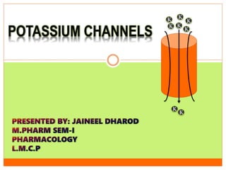
K K
K
KK K
K
K
POTASSIUM CHANNELS
INTRODUCTION
 Potassium channels are the most widely distributed type of ion channel and
are found in all living organisms.
 They form potassium-selective pores that span cell membranes.
 Furthermore potassium channels are found in most cell types and control a
wide variety of cell functions.
Functions
 Potassium channels function to conduct potassium ions down their electrochemical
gradient, doing so both rapidly and selectively.
 Biologically, these channels act to set or reset the resting potential in many cells. In
excitable cells, such as neurons, the delayed counter flow of potassium ions shapes the
action potential.
 By contributing to the regulation of the action potential duration in cardiac muscle,
malfunction of potassium channels may cause life-threatening arrhythmias. Potassium
channels may also be involved in maintaining vascular tone.
 They also regulate cellular processes such as the secretion of hormones (e.g., insulin release
from beta-cells in the pancreas) so their malfunction can lead to diseases (such as diabetes).
STRUCTURE
• Potassium channels have a tetrameric structure
in which four identical protein subunits
associate to form a fourfold symmetric (C4)
complex arranged around a central ion
conducting pore (i.e., a homotetramer).
• Alternatively four related but not identical
protein subunits may associate to form
heterotetrameric complexes with pseudo C4
symmetry.
• All potassium channel subunits have a
distinctive pore-loop structure that lines the top
of the pore and is responsible for potassium
selective permeability.
SELECTIVITY FILTER
 Using X-ray crystallography, profound insights
have been gained into how potassium ions pass
through these channels and why (smaller) sodium
ions do not. The 2003 Nobel Prize for Chemistry
was awarded to Rod MacKinnon for his pioneering
work in this area.
 Crystallographic structure of the bacterial: The
protein is displayed as a green cartoon diagram. In
addition backbone carbonyl groups and threonine
sidechain protein atoms (oxygen = red, carbon =
green) are displayed. Finally potassium ions
(occupying the S2 and S4 sites) and the oxygen
atoms of water molecules (S1 and S3) are depicted
as purple and red spheres respectively.
 Potassium ion channels remove the hydration shell from the ion when it enters the
selectivity filter.
 The selectivity filter is formed by a five residue sequence, TVGYG, termed the signature
sequence, within each of the four subunits.
 This signature sequence is within a loop between the pore helix and TM2/6, historically
termed the P-loop.
 This signature sequence is highly conserved, with the exception that a valine residue in
prokaryotic potassium channels is often substituted with an isoleucine residue in
eukaryotic channels.
 This sequence adopts a unique main chain structure, structurally analogous to a nest
protein structural motif.
 The four sets of electronegative carbonyl oxygen atoms are aligned toward the centre of the
filter pore and form a square anti-prism similar to a water-solvating shell around each
potassium binding site.
SELECTIVITY FILTER
 The distance between the carbonyl oxygens and potassium ions in the binding sites of the
selectivity filter is the same as between water oxygens in the first hydration shell and a
potassium ion in water solution, providing an energetically favourable route for de-
solvation of the ions.
 This width appears to be maintained by hydrogen bonding and van der Waals forces
within a sheet of aromatic amino acid residues surrounding the selectivity filter.
 The selectivity filter opens towards the extracellular solution, exposing four carbonyl
oxygens in a glycine residue.
 The next residue toward the extracellular side of the protein is the negatively charged
Asp80 (KcsA).
 This residue together with the five filter residues form the pore that connects the water-
filled cavity in the centre of the protein with the extracellular solution.
SELECTIVITY FILTER
SELECTIVITY FILTER
REGULATION
 The flux of ions through the potassium channel pore is regulated by two related processes,
termed gating and inactivation. Gating is the opening or closing of the channel in response
to stimuli, while inactivation is the rapid cessation of current from an open potassium
channel and the suppression of the channel's ability to resume conducting.
 Generally, gating is thought to be mediated by additional structural domains which sense
stimuli and in turn open the channel pore. These domains include the RCK domains of BK
channels, and voltage sensor domains of voltage gated K+ channels. These domains are
thought to respond to the stimuli by physically opening the intracellular gate of the pore
domain, thereby allowing potassium ions to traverse the membrane.
REGULATION
 Some channels have multiple regulatory domains or accessory proteins, which can act to
modulate the response to stimulus.
 N-type inactivation is typically the faster inactivation mechanism, and is termed the "ball
and chain" model. N-type inactivation involves interaction of the N-terminus of the
channel, or an associated protein, which interacts with the pore domain and occludes the
ion conduction pathway like a "ball".
 C-type inactivation is thought to occur within the selectivity filter itself, where structural
changes within the filter render it non-conductive. There are a number of structural models
of C-type inactivated K+ channel filters.
TYPES
 Calcium-activated potassium channel - open in response to the presence of calcium
ions or other signalling molecules.
 Inwardly rectifying potassium channel - passes current (positive charge) more
easily in the inward direction (into the cell).
 Tandem pore domain potassium channel - are constitutively open or possess high
basal activation, such as the "resting potassium channels" or "leak channels" that
set the negative membrane potential of neurons.
 Voltage-gated potassium channel - are voltage-gated ion channels that open or
close in response to changes in the transmembrane voltage.
Ca+2 ACTIVATED K+ CHANNELS
 Calcium-activated potassium channels are potassium channels gated by calcium, or
that are structurally or phylogenetically related to calcium gated channels.
 They were first discovered in 1958 by Gardos who saw that Calcium levels inside of a
cell could affect the permeability of potassium through that cell membrane.
 Then in 1970, Meech was the first to observe that intracellular calcium could trigger
potassium currents.
 In humans they are divided into three subtypes large conductance or BK channels,
which have very high conductance which range from 100 to 300 pS, intermediate
conductance or IK channels, with intermediate conductance ranging from 25 to 100
pS, and small conductance or SK channels with small conductance from 2-25 pS.
 BK channels (Big Potassium), are characterized by large conductance of potassium ions
(K+) through cell membranes.
 Channels are activated by changes in membrane electrical potential and/or by increases in
intracellular [Ca2+].
 Essential for the regulation of several physiological processes including smooth muscle tone
and neuronal excitability.
 A) Structure of the α and β1-subunits of the BK channel. The β1-subunit consists of 2
transmembrane domains and the α-subunit of 11 (S0–S10) hydrophobic domains, with
S0–S6 located in the cytoplasmic membrane and the pore region between S5 and S6.
 B) Association of four α and four β1-subunits forms the native BK channel.
BK Potassium Channel
Figure: BK potassium channel structure.
BK Potassium Channel
 BK channels are pharmacological targets for the treatment of several medical disorders including
stroke and overactive bladder.
 However, BK channels are reduced in patients suffering from the Fragile X syndrome and the
agonist, BMS-204352, corrects some of the deficits observed in Fmr1 knockout mice, a model of
Fragile X syndrome.
 BK channels have also been found to be activated by exogenous pollutants and endogenous gas
transmitters carbon monoxide and hydrogen sulphide.
 BK channels can be readily inhibited by a range of compounds including tetraethyl ammonium
(TEA), paxilline, Limbatustoxin, and iberiotoxin.
 The BK-channel blocker GAL-021 has been investigated for potential use in inhibiting opioid
induced respiratory depression without affecting analgesia.
IK Potassium Channel
 The IK channel (KCa3.1) is expressed mainly in peripheral tissues such as those of the
haematopoietic system, colon, placenta, lung and pancreas.
 Has been implicated in a wide range of cell functions, including vasodilation of the
microvasculature, K+ flux across endothelial cells of brain capillaries and phagocytic
activity of neutrophils.
 In comparison with the large-conductance (BK) channels, KCa3.1 is much more sensitive
to Ca2+ than BK channels. This high affinity is due to the presence of 4 calmodulin
molecules bound to the cytoplasmic tails of the four pore-forming α-subunits.
 Before the channel can open, Ca2+ must bind to each of the calmodulins to induce the co-
operative conformational change that opens the gate.
IK Potassium Channel
Cross-section of the IK channel. Binding of calcium ion to CaM produces conformational change that opens the pore.
 Small single channel conductance . SK channels allow potassium ions to cross the cell
membrane and are activated (opened) by an increase in the intracellular [Ca2+]
through N-type calcium channels.
 SK channels are thought to be involved in synaptic plasticity and therefore play
important roles in learning and memory.
 SK channels are Ca-activated K channels, gated solely by intracellular Ca ions.
 SK channels are constitutively associated with calmodulin (CaM) that binds to the
CaMBD in the intracellular C-terminus of the channels.
 When Ca ions bind to the N-lobe E-F hands of CaM, a gating transition is initiated,
the gate of the channel opens and K flows through the channel pore, exerting a
repolarizing influence on the membrane potential.
SK Potassium Channel
 In dendritic spines, SK channels are directly coupled to NMDA receptors. In addition to being
activated by calcium flow through voltage-gated calcium channels, SK channels can be activated by
calcium flowing through NMDA receptors, which occurs after depolarization of the postsynaptic
membrane. Experiments using apamin have shown that specifically blocking SK channels can
increase learning and long-term potentiation.
 SK channels are widely expressed in midbrain dopaminergic neurons. Multiple pharmacological
techniques have been used to adjust SK affinity for calcium ions, thereby modulating the excitability
of substantia nigra dopaminergic neurons. Blockage of SK channels in vivo increases the firing rate
of substantia nigra cells, which increases the amount of dopamine released from the synaptic
terminals. When a large amount of dopamine accumulates in the cytosol, cell damage is induced due
to the build-up of free radicals and damage to mitochondria.
 SK channel blockers: bicuculline, apamin, scorpion venom, charybdotoxin, TEA, D-tubocurarine,
Cyproheptadine, pancuronium, Atracurium, etc.
SK Potassium Channel
Inward-rectifier potassium ion channel
 A channel that is "inwardly-rectifying" is one that passes current (positive charge) more easily in the
inward direction (into the cell) than in the outward direction (out of the cell). It is thought that this
current may play an important role in regulating neuronal activity, by helping to stabilize the
resting membrane potential of the cell.
 The phenomenon of inward rectification of Kir channels is the result of high-affinity block by
endogenous polyamines, namely spermine, as well as magnesium ions, that plug the channel pore at
positive potentials, resulting in a decrease in outward currents. This voltage-dependent block by
polyamines results in efficient conduction of current only in the inward direction.
 All Kir channels require phosphatidylinositol 4,5-bisphosphate (PIP2) for activation. PIP2 binds to
and directly activates Kir 2.2 with agonist-like properties. In this regard Kir channels are PIP2
ligand-gated ion channels.
Location Function
Cardiac
myocytes
Kir channels close upon depolarization, slowing membrane repolarization and
helping maintain a more prolonged cardiac action potential.
Endothelial
cells
Kir channels are involved in regulation of nitric oxide synthase.
Kidneys
Kir export surplus potassium into collecting tubules for removal in the urine,
or alternatively may be involved in the reuptake of potassium back into the
body.
Neurons
and in
heart cells
G-protein activated IRKs (Kir3) are important regulators, modulated by
neurotransmitters. A mutation in the GIRK2 channel leads to the weaver
mouse mutation. "Weaver" mutant mice are ataxic and display a
neuroinflammation-mediated degeneration of their dopaminergic neurons.
Relative to non-ataxic controls, Weaver mutants have deficits in motor
coordination and changes in regional brain metabolism. Weaver mice have
been examined in labs interested in neural development and disease for over
30 years.
Pancreatic
beta cells
KATP channels control insulin release.
Inward-rectifier potassium ion channel
Diseases related to Kir channels
 Persistent hyperinsulinemic hypoglycaemia of infancy is related to autosomal recessive mutations in
Kir6.2. It diminish the channel's ability to regulate insulin secretion, leading to hypoglycaemia.
 Bartter's syndrome can be caused by mutations in Kir channels. This condition is characterized by the
inability of kidneys to recycle potassium, causing low levels of potassium in the body.
 Andersen's syndrome is a rare condition caused by multiple mutations of Kir2.1. Depending on the
mutation, it can be dominant or recessive. It is characterized by periodic paralysis, cardiac arrhythmias
and dysmorphic features.
 Barium poisoning is likely due to its ability to block Kir channels.
 Atherosclerosis may be related to Kir channels. The loss of Kir currents in endothelial cells is one of the
first known indicators of atherogenesis.
 Thyrotoxic hypokalaemic periodic paralysis has been linked to altered Kir2.6 function
Tandem pore domain potassium Channel
 Tandem pore domain potassium channels are a family of 15 members form what is known as
"leak channels“.
 Leak channels allow potassium to exit the cell in order to reduce positive charge within the cell
and maintain stable resting membrane potential.
 Channels are regulated by several mechanisms including:
 oxygen tension
 pH
 mechanical stretch
 G-proteins
 For most, the α subunits consist of 4 transmembrane segments, each containing 2 pore loops.
 They structurally correspond to two inward-rectifier α subunits and thus form dimers in the
membrane. K2P dimer
Tandem pore domain potassium Channel
Voltage-gated potassium (Kv) channels
 Present in all animal cells. Open and close based on voltage changes in the cell's membrane
potential.
 During action potentials, these channels open and allow passive flow of K+ ions from the
cell to return the depolarized cell to a resting state.
 Kv channels are one of the key components in generation and propagation of electrical
impulses in nervous system.
Voltage-gated potassium (Kv) channels
 The tetrameric structure of Kv channels is made of two functionally and structurally independent
domains: an ion conduction pore, and voltage-sensor domains.
 The ion conduction pore is made of subunits which are arranged symmetrically around the
conduction pathway.
 Voltage-sensor domains are positioned at the periphery of the channel and consist of four
transmembrane segments (S1-S4).
 Structural rearrangement of the voltage-sensor domains in response to changes in the membrane
potential, and in particular S4, which includes positively charged amino acids at every third
position, results in conformational changes in the conduction pore, which could open or close the ion
conduction pathway.
Class Subclasses Function Blockers Activators
Calcium-activated
6T & 1P
•BK channel
•SK channel
•IK channel
•inhibition in
response to rising
intracellular calcium
•charybdotoxin,
iberiotoxin
•apamin
•1-EBIO
•NS309
•CyPPA
Inwardly rectifying
2T & 1P
•ROMK (Kir1.1)
•recycling and
secretion of
potassium in
nephrons
•Nonselective: Ba2+,
Cs+ •none
•GPCR regulated
(Kir3.x)
•mediate the
inhibitory effect of
many GPCRs
•GPCR antagonists
•ifenprodil
•GPCR agonists
•ATP-sensitive
(Kir6.x)
•close when ATP is
high to promote
insulin secretion
•glibenclamide
•tolbutamide
•diazoxide
•pinacidil
•minoxidil
•nicorandil
Tandem pore
domain
4T & 2P
•Contribute to
resting potential
•bupivacaine
•quinidine
•halothane
Voltage-gated
6T & 1P
•hERG (Kv11.1)
•KvLQT1 (Kv7.1)
•action potential
repolarization
•limits frequency of
action potentials
(disturbances cause
dysrhythmia)
•tetraethylammoniu
m
•4-aminopyridine
•dendrotoxins (some
types)
•Retigabine (Kv7)
REFERENCES
1. Doyle DA, Morais Cabral J, Pfuetzner RA, Kuo A, Gulbis JM, Cohen SL, Chait BT, MacKinnon R (Apr
1998). "The structure of the potassium channel: molecular basis of K+ conduction and selectivity".
Science. 280 (5360): 69–77. Bibcode:1998Sci...280...69D. PMID 9525859.
doi:10.1126/science.280.5360.69
2. MacKinnon R, Cohen SL, Kuo A, Lee A, Chait BT (Apr 1998). "Structural conservation in prokaryotic
and eukaryotic potassium channels". Science. 280 (5360): 106–9. Bibcode:1998Sci...280..106M.
PMID 9525854. doi:10.1126/science.280.5360.106
3. Armstrong C (Apr 1998). "The vision of the pore". Science. 280 (5360): 56–7. PMID 9556453.
doi:10.1126/science.280.5360.56
4. Lim, Carmay; Dudev, Todor (2016). "Chapter 10. Potassium Versus Sodium Selectivity in Monovalent
Ion Channel Selectivity Filters". In Astrid, Sigel; Helmut, Sigel; Roland K.O., Sigel. The Alkali Metal
Ions: Their Role in Life. Metal Ions in Life Sciences. 16. Springer. pp. 325–347. doi:10.1007/978-4-
319-21756-7_9
5. ‘The complete drug reference’, thirty sixth edition by Martindale.
Roderick MacKinnon commissioned Birth of an Idea, a 5-foot
(1.5 m) tall sculpture based on the KcsA potassium channel. The
artwork contains a wire object representing the channel's interior
with a blown glass object representing the main cavity of the
channel structure.

More Related Content

What's hot

Sodium Ion Channel
Sodium Ion ChannelSodium Ion Channel
Sodium Ion ChannelUshaYadav24
 
G protein coupled receptors and their Signaling Mechanism
G protein coupled receptors and their Signaling MechanismG protein coupled receptors and their Signaling Mechanism
G protein coupled receptors and their Signaling MechanismFarazaJaved
 
Ion channels, types and their importace in managment of diseases
Ion channels, types and their importace in managment of diseasesIon channels, types and their importace in managment of diseases
Ion channels, types and their importace in managment of diseasesFarazaJaved
 
Second messengers cAMP and cGMP
Second messengers cAMP and cGMPSecond messengers cAMP and cGMP
Second messengers cAMP and cGMPFarazaJaved
 
JAK STAT SIGNALLING PATHWAY
JAK STAT SIGNALLING PATHWAYJAK STAT SIGNALLING PATHWAY
JAK STAT SIGNALLING PATHWAYLolzz Fernandes
 
Jak stat signalling pathway
Jak stat signalling pathwayJak stat signalling pathway
Jak stat signalling pathwayAmit Sahoo
 
Second messenger system
Second messenger systemSecond messenger system
Second messenger systemdamarisb
 
Slideshare second messengers aj
Slideshare second messengers ajSlideshare second messengers aj
Slideshare second messengers ajAnu Priya
 
Tyrosine kinase receptors & Nuclear receptors
Tyrosine kinase receptors & Nuclear receptorsTyrosine kinase receptors & Nuclear receptors
Tyrosine kinase receptors & Nuclear receptorsAaqib Naseer
 
Receptor tyrosine kinase
Receptor tyrosine kinaseReceptor tyrosine kinase
Receptor tyrosine kinaseSobia
 
Receptor tyrosine kinases.ppt
Receptor tyrosine kinases.pptReceptor tyrosine kinases.ppt
Receptor tyrosine kinases.pptDr. Khuram Aziz
 
G protein coupled receptor
G protein coupled receptorG protein coupled receptor
G protein coupled receptorSumit Kumar
 

What's hot (20)

Sodium Ion Channel
Sodium Ion ChannelSodium Ion Channel
Sodium Ion Channel
 
MAPK pathway,
MAPK pathway, MAPK pathway,
MAPK pathway,
 
Cell signaling
Cell signaling Cell signaling
Cell signaling
 
G protein coupled receptors and their Signaling Mechanism
G protein coupled receptors and their Signaling MechanismG protein coupled receptors and their Signaling Mechanism
G protein coupled receptors and their Signaling Mechanism
 
Ion channels, types and their importace in managment of diseases
Ion channels, types and their importace in managment of diseasesIon channels, types and their importace in managment of diseases
Ion channels, types and their importace in managment of diseases
 
Second messengers cAMP and cGMP
Second messengers cAMP and cGMPSecond messengers cAMP and cGMP
Second messengers cAMP and cGMP
 
Jak stat
Jak statJak stat
Jak stat
 
Na channel
Na channelNa channel
Na channel
 
Mapk pathways
Mapk pathwaysMapk pathways
Mapk pathways
 
JAK STAT SIGNALLING PATHWAY
JAK STAT SIGNALLING PATHWAYJAK STAT SIGNALLING PATHWAY
JAK STAT SIGNALLING PATHWAY
 
Receptors
ReceptorsReceptors
Receptors
 
Jak stat signalling pathway
Jak stat signalling pathwayJak stat signalling pathway
Jak stat signalling pathway
 
Receptors
ReceptorsReceptors
Receptors
 
Second messenger system
Second messenger systemSecond messenger system
Second messenger system
 
Slideshare second messengers aj
Slideshare second messengers ajSlideshare second messengers aj
Slideshare second messengers aj
 
Tyrosine kinase receptors & Nuclear receptors
Tyrosine kinase receptors & Nuclear receptorsTyrosine kinase receptors & Nuclear receptors
Tyrosine kinase receptors & Nuclear receptors
 
Cyclic amp
Cyclic ampCyclic amp
Cyclic amp
 
Receptor tyrosine kinase
Receptor tyrosine kinaseReceptor tyrosine kinase
Receptor tyrosine kinase
 
Receptor tyrosine kinases.ppt
Receptor tyrosine kinases.pptReceptor tyrosine kinases.ppt
Receptor tyrosine kinases.ppt
 
G protein coupled receptor
G protein coupled receptorG protein coupled receptor
G protein coupled receptor
 

Similar to Potassium Channel Structure and Function Guide

Ion gating plants
Ion gating plantsIon gating plants
Ion gating plantssunilbanu1
 
cmb(1) calcium.pptx
cmb(1) calcium.pptxcmb(1) calcium.pptx
cmb(1) calcium.pptxPriyashreeK1
 
Unit 5 ion channel receptor
Unit  5 ion channel receptorUnit  5 ion channel receptor
Unit 5 ion channel receptorPriyansha Singh
 
ion channel and carrier protein By KK Sahu Sir
ion channel and carrier protein By KK Sahu Sirion channel and carrier protein By KK Sahu Sir
ion channel and carrier protein By KK Sahu SirKAUSHAL SAHU
 
Transport across cell membrane
Transport across cell membraneTransport across cell membrane
Transport across cell membranemuti ullah
 
Re absorption and secretion by nephron
Re absorption and secretion by nephronRe absorption and secretion by nephron
Re absorption and secretion by nephronAmir Bahadur
 
Transport across cell membrane
Transport across cell membraneTransport across cell membrane
Transport across cell membranemuti ullah
 
Second messengers in Signal transduction (Calcium ions)
Second messengers in Signal transduction (Calcium ions) Second messengers in Signal transduction (Calcium ions)
Second messengers in Signal transduction (Calcium ions) vibhakhanna1
 
Sodium Ion Channel
Sodium Ion ChannelSodium Ion Channel
Sodium Ion Channelnisha rani
 
Transmembrane transport of ions and small molecules by Kainat Ramzan
Transmembrane transport of ions and small molecules by Kainat RamzanTransmembrane transport of ions and small molecules by Kainat Ramzan
Transmembrane transport of ions and small molecules by Kainat RamzanKainatRamzan3
 
POTASSIUM PHYSIOLOGY.pptx
POTASSIUM PHYSIOLOGY.pptxPOTASSIUM PHYSIOLOGY.pptx
POTASSIUM PHYSIOLOGY.pptxMSrujanaDevi
 
Day 4-TRANSPORT ACROSS THE CELL MEMBRANE (5)b.pptx
Day 4-TRANSPORT ACROSS THE CELL MEMBRANE (5)b.pptxDay 4-TRANSPORT ACROSS THE CELL MEMBRANE (5)b.pptx
Day 4-TRANSPORT ACROSS THE CELL MEMBRANE (5)b.pptxMkindi Mkindi
 
Transport across cell membranes
Transport across cell membranesTransport across cell membranes
Transport across cell membranesBhavya Oberoi
 
Ion channelopathy
Ion channelopathyIon channelopathy
Ion channelopathyVivek Rana
 
Recent role of calcium in cell signaling
Recent role of calcium in cell signalingRecent role of calcium in cell signaling
Recent role of calcium in cell signalingDr Muhammad Mustansar
 

Similar to Potassium Channel Structure and Function Guide (20)

Channels
ChannelsChannels
Channels
 
Ion gating plants
Ion gating plantsIon gating plants
Ion gating plants
 
cmb(1) calcium.pptx
cmb(1) calcium.pptxcmb(1) calcium.pptx
cmb(1) calcium.pptx
 
Unit 5 ion channel receptor
Unit  5 ion channel receptorUnit  5 ion channel receptor
Unit 5 ion channel receptor
 
ion channel and carrier protein By KK Sahu Sir
ion channel and carrier protein By KK Sahu Sirion channel and carrier protein By KK Sahu Sir
ion channel and carrier protein By KK Sahu Sir
 
Transport across cell membrane
Transport across cell membraneTransport across cell membrane
Transport across cell membrane
 
Calcium as a Regulatory and Signalling ion
Calcium as a Regulatory and Signalling ionCalcium as a Regulatory and Signalling ion
Calcium as a Regulatory and Signalling ion
 
Re absorption and secretion by nephron
Re absorption and secretion by nephronRe absorption and secretion by nephron
Re absorption and secretion by nephron
 
Transport across cell membrane
Transport across cell membraneTransport across cell membrane
Transport across cell membrane
 
Second messengers in Signal transduction (Calcium ions)
Second messengers in Signal transduction (Calcium ions) Second messengers in Signal transduction (Calcium ions)
Second messengers in Signal transduction (Calcium ions)
 
Ion channels as drug target
Ion channels as drug targetIon channels as drug target
Ion channels as drug target
 
Sodium Ion Channel
Sodium Ion ChannelSodium Ion Channel
Sodium Ion Channel
 
Ojchd.000529
Ojchd.000529Ojchd.000529
Ojchd.000529
 
Transmembrane transport of ions and small molecules by Kainat Ramzan
Transmembrane transport of ions and small molecules by Kainat RamzanTransmembrane transport of ions and small molecules by Kainat Ramzan
Transmembrane transport of ions and small molecules by Kainat Ramzan
 
POTASSIUM PHYSIOLOGY.pptx
POTASSIUM PHYSIOLOGY.pptxPOTASSIUM PHYSIOLOGY.pptx
POTASSIUM PHYSIOLOGY.pptx
 
Day 4-TRANSPORT ACROSS THE CELL MEMBRANE (5)b.pptx
Day 4-TRANSPORT ACROSS THE CELL MEMBRANE (5)b.pptxDay 4-TRANSPORT ACROSS THE CELL MEMBRANE (5)b.pptx
Day 4-TRANSPORT ACROSS THE CELL MEMBRANE (5)b.pptx
 
Transport across cell membranes
Transport across cell membranesTransport across cell membranes
Transport across cell membranes
 
Ion channelopathy
Ion channelopathyIon channelopathy
Ion channelopathy
 
Electrolyte.pdf
Electrolyte.pdfElectrolyte.pdf
Electrolyte.pdf
 
Recent role of calcium in cell signaling
Recent role of calcium in cell signalingRecent role of calcium in cell signaling
Recent role of calcium in cell signaling
 

More from Jaineel Dharod

Expectorants and anti tussives
Expectorants and anti tussivesExpectorants and anti tussives
Expectorants and anti tussivesJaineel Dharod
 
Bones and skeletal system
Bones and skeletal systemBones and skeletal system
Bones and skeletal systemJaineel Dharod
 
Blood pressure mechanism
Blood pressure mechanismBlood pressure mechanism
Blood pressure mechanismJaineel Dharod
 
Blood vessels and circulation
Blood vessels and circulationBlood vessels and circulation
Blood vessels and circulationJaineel Dharod
 
Atherosclerosis Pathophysiology
Atherosclerosis PathophysiologyAtherosclerosis Pathophysiology
Atherosclerosis PathophysiologyJaineel Dharod
 
Introduction and scope of anatomy and physiology
Introduction and scope of anatomy and physiologyIntroduction and scope of anatomy and physiology
Introduction and scope of anatomy and physiologyJaineel Dharod
 
Scope of anatomy and physiology
Scope of anatomy and physiologyScope of anatomy and physiology
Scope of anatomy and physiologyJaineel Dharod
 
Atherosclerosis Pathophysiology
Atherosclerosis PathophysiologyAtherosclerosis Pathophysiology
Atherosclerosis PathophysiologyJaineel Dharod
 
Cell injury pathophysiology
Cell injury pathophysiologyCell injury pathophysiology
Cell injury pathophysiologyJaineel Dharod
 
Pathophysiology of Inflammation
Pathophysiology of InflammationPathophysiology of Inflammation
Pathophysiology of InflammationJaineel Dharod
 

More from Jaineel Dharod (20)

Expectorants and anti tussives
Expectorants and anti tussivesExpectorants and anti tussives
Expectorants and anti tussives
 
Integumentary system
Integumentary systemIntegumentary system
Integumentary system
 
Bones and skeletal system
Bones and skeletal systemBones and skeletal system
Bones and skeletal system
 
osseous system
osseous systemosseous system
osseous system
 
Blood pressure mechanism
Blood pressure mechanismBlood pressure mechanism
Blood pressure mechanism
 
Blood vessels and circulation
Blood vessels and circulationBlood vessels and circulation
Blood vessels and circulation
 
Tissues epi and con
Tissues epi and conTissues epi and con
Tissues epi and con
 
Cell communication
Cell communicationCell communication
Cell communication
 
Cell membrane
Cell membraneCell membrane
Cell membrane
 
Hyperlipidemia
HyperlipidemiaHyperlipidemia
Hyperlipidemia
 
Parkinsonism
ParkinsonismParkinsonism
Parkinsonism
 
Atherosclerosis Pathophysiology
Atherosclerosis PathophysiologyAtherosclerosis Pathophysiology
Atherosclerosis Pathophysiology
 
Cell 2
Cell 2Cell 2
Cell 2
 
Cell Organalles
Cell OrganallesCell Organalles
Cell Organalles
 
Introduction and scope of anatomy and physiology
Introduction and scope of anatomy and physiologyIntroduction and scope of anatomy and physiology
Introduction and scope of anatomy and physiology
 
Toxicity testing
Toxicity testingToxicity testing
Toxicity testing
 
Scope of anatomy and physiology
Scope of anatomy and physiologyScope of anatomy and physiology
Scope of anatomy and physiology
 
Atherosclerosis Pathophysiology
Atherosclerosis PathophysiologyAtherosclerosis Pathophysiology
Atherosclerosis Pathophysiology
 
Cell injury pathophysiology
Cell injury pathophysiologyCell injury pathophysiology
Cell injury pathophysiology
 
Pathophysiology of Inflammation
Pathophysiology of InflammationPathophysiology of Inflammation
Pathophysiology of Inflammation
 

Recently uploaded

ECONOMIC CONTEXT - PAPER 1 Q3: NEWSPAPERS.pptx
ECONOMIC CONTEXT - PAPER 1 Q3: NEWSPAPERS.pptxECONOMIC CONTEXT - PAPER 1 Q3: NEWSPAPERS.pptx
ECONOMIC CONTEXT - PAPER 1 Q3: NEWSPAPERS.pptxiammrhaywood
 
Introduction to ArtificiaI Intelligence in Higher Education
Introduction to ArtificiaI Intelligence in Higher EducationIntroduction to ArtificiaI Intelligence in Higher Education
Introduction to ArtificiaI Intelligence in Higher Educationpboyjonauth
 
Organic Name Reactions for the students and aspirants of Chemistry12th.pptx
Organic Name Reactions  for the students and aspirants of Chemistry12th.pptxOrganic Name Reactions  for the students and aspirants of Chemistry12th.pptx
Organic Name Reactions for the students and aspirants of Chemistry12th.pptxVS Mahajan Coaching Centre
 
Biting mechanism of poisonous snakes.pdf
Biting mechanism of poisonous snakes.pdfBiting mechanism of poisonous snakes.pdf
Biting mechanism of poisonous snakes.pdfadityarao40181
 
Enzyme, Pharmaceutical Aids, Miscellaneous Last Part of Chapter no 5th.pdf
Enzyme, Pharmaceutical Aids, Miscellaneous Last Part of Chapter no 5th.pdfEnzyme, Pharmaceutical Aids, Miscellaneous Last Part of Chapter no 5th.pdf
Enzyme, Pharmaceutical Aids, Miscellaneous Last Part of Chapter no 5th.pdfSumit Tiwari
 
Interactive Powerpoint_How to Master effective communication
Interactive Powerpoint_How to Master effective communicationInteractive Powerpoint_How to Master effective communication
Interactive Powerpoint_How to Master effective communicationnomboosow
 
MARGINALIZATION (Different learners in Marginalized Group
MARGINALIZATION (Different learners in Marginalized GroupMARGINALIZATION (Different learners in Marginalized Group
MARGINALIZATION (Different learners in Marginalized GroupJonathanParaisoCruz
 
18-04-UA_REPORT_MEDIALITERAСY_INDEX-DM_23-1-final-eng.pdf
18-04-UA_REPORT_MEDIALITERAСY_INDEX-DM_23-1-final-eng.pdf18-04-UA_REPORT_MEDIALITERAСY_INDEX-DM_23-1-final-eng.pdf
18-04-UA_REPORT_MEDIALITERAСY_INDEX-DM_23-1-final-eng.pdfssuser54595a
 
Crayon Activity Handout For the Crayon A
Crayon Activity Handout For the Crayon ACrayon Activity Handout For the Crayon A
Crayon Activity Handout For the Crayon AUnboundStockton
 
Incoming and Outgoing Shipments in 1 STEP Using Odoo 17
Incoming and Outgoing Shipments in 1 STEP Using Odoo 17Incoming and Outgoing Shipments in 1 STEP Using Odoo 17
Incoming and Outgoing Shipments in 1 STEP Using Odoo 17Celine George
 
Meghan Sutherland In Media Res Media Component
Meghan Sutherland In Media Res Media ComponentMeghan Sutherland In Media Res Media Component
Meghan Sutherland In Media Res Media ComponentInMediaRes1
 
Presiding Officer Training module 2024 lok sabha elections
Presiding Officer Training module 2024 lok sabha electionsPresiding Officer Training module 2024 lok sabha elections
Presiding Officer Training module 2024 lok sabha electionsanshu789521
 
Solving Puzzles Benefits Everyone (English).pptx
Solving Puzzles Benefits Everyone (English).pptxSolving Puzzles Benefits Everyone (English).pptx
Solving Puzzles Benefits Everyone (English).pptxOH TEIK BIN
 
Proudly South Africa powerpoint Thorisha.pptx
Proudly South Africa powerpoint Thorisha.pptxProudly South Africa powerpoint Thorisha.pptx
Proudly South Africa powerpoint Thorisha.pptxthorishapillay1
 
Full Stack Web Development Course for Beginners
Full Stack Web Development Course  for BeginnersFull Stack Web Development Course  for Beginners
Full Stack Web Development Course for BeginnersSabitha Banu
 
“Oh GOSH! Reflecting on Hackteria's Collaborative Practices in a Global Do-It...
“Oh GOSH! Reflecting on Hackteria's Collaborative Practices in a Global Do-It...“Oh GOSH! Reflecting on Hackteria's Collaborative Practices in a Global Do-It...
“Oh GOSH! Reflecting on Hackteria's Collaborative Practices in a Global Do-It...Marc Dusseiller Dusjagr
 
KSHARA STURA .pptx---KSHARA KARMA THERAPY (CAUSTIC THERAPY)————IMP.OF KSHARA ...
KSHARA STURA .pptx---KSHARA KARMA THERAPY (CAUSTIC THERAPY)————IMP.OF KSHARA ...KSHARA STURA .pptx---KSHARA KARMA THERAPY (CAUSTIC THERAPY)————IMP.OF KSHARA ...
KSHARA STURA .pptx---KSHARA KARMA THERAPY (CAUSTIC THERAPY)————IMP.OF KSHARA ...M56BOOKSTORE PRODUCT/SERVICE
 
CELL CYCLE Division Science 8 quarter IV.pptx
CELL CYCLE Division Science 8 quarter IV.pptxCELL CYCLE Division Science 8 quarter IV.pptx
CELL CYCLE Division Science 8 quarter IV.pptxJiesonDelaCerna
 
भारत-रोम व्यापार.pptx, Indo-Roman Trade,
भारत-रोम व्यापार.pptx, Indo-Roman Trade,भारत-रोम व्यापार.pptx, Indo-Roman Trade,
भारत-रोम व्यापार.pptx, Indo-Roman Trade,Virag Sontakke
 

Recently uploaded (20)

ECONOMIC CONTEXT - PAPER 1 Q3: NEWSPAPERS.pptx
ECONOMIC CONTEXT - PAPER 1 Q3: NEWSPAPERS.pptxECONOMIC CONTEXT - PAPER 1 Q3: NEWSPAPERS.pptx
ECONOMIC CONTEXT - PAPER 1 Q3: NEWSPAPERS.pptx
 
Introduction to ArtificiaI Intelligence in Higher Education
Introduction to ArtificiaI Intelligence in Higher EducationIntroduction to ArtificiaI Intelligence in Higher Education
Introduction to ArtificiaI Intelligence in Higher Education
 
Organic Name Reactions for the students and aspirants of Chemistry12th.pptx
Organic Name Reactions  for the students and aspirants of Chemistry12th.pptxOrganic Name Reactions  for the students and aspirants of Chemistry12th.pptx
Organic Name Reactions for the students and aspirants of Chemistry12th.pptx
 
Biting mechanism of poisonous snakes.pdf
Biting mechanism of poisonous snakes.pdfBiting mechanism of poisonous snakes.pdf
Biting mechanism of poisonous snakes.pdf
 
Enzyme, Pharmaceutical Aids, Miscellaneous Last Part of Chapter no 5th.pdf
Enzyme, Pharmaceutical Aids, Miscellaneous Last Part of Chapter no 5th.pdfEnzyme, Pharmaceutical Aids, Miscellaneous Last Part of Chapter no 5th.pdf
Enzyme, Pharmaceutical Aids, Miscellaneous Last Part of Chapter no 5th.pdf
 
Interactive Powerpoint_How to Master effective communication
Interactive Powerpoint_How to Master effective communicationInteractive Powerpoint_How to Master effective communication
Interactive Powerpoint_How to Master effective communication
 
MARGINALIZATION (Different learners in Marginalized Group
MARGINALIZATION (Different learners in Marginalized GroupMARGINALIZATION (Different learners in Marginalized Group
MARGINALIZATION (Different learners in Marginalized Group
 
18-04-UA_REPORT_MEDIALITERAСY_INDEX-DM_23-1-final-eng.pdf
18-04-UA_REPORT_MEDIALITERAСY_INDEX-DM_23-1-final-eng.pdf18-04-UA_REPORT_MEDIALITERAСY_INDEX-DM_23-1-final-eng.pdf
18-04-UA_REPORT_MEDIALITERAСY_INDEX-DM_23-1-final-eng.pdf
 
Crayon Activity Handout For the Crayon A
Crayon Activity Handout For the Crayon ACrayon Activity Handout For the Crayon A
Crayon Activity Handout For the Crayon A
 
Incoming and Outgoing Shipments in 1 STEP Using Odoo 17
Incoming and Outgoing Shipments in 1 STEP Using Odoo 17Incoming and Outgoing Shipments in 1 STEP Using Odoo 17
Incoming and Outgoing Shipments in 1 STEP Using Odoo 17
 
Meghan Sutherland In Media Res Media Component
Meghan Sutherland In Media Res Media ComponentMeghan Sutherland In Media Res Media Component
Meghan Sutherland In Media Res Media Component
 
Presiding Officer Training module 2024 lok sabha elections
Presiding Officer Training module 2024 lok sabha electionsPresiding Officer Training module 2024 lok sabha elections
Presiding Officer Training module 2024 lok sabha elections
 
Solving Puzzles Benefits Everyone (English).pptx
Solving Puzzles Benefits Everyone (English).pptxSolving Puzzles Benefits Everyone (English).pptx
Solving Puzzles Benefits Everyone (English).pptx
 
Proudly South Africa powerpoint Thorisha.pptx
Proudly South Africa powerpoint Thorisha.pptxProudly South Africa powerpoint Thorisha.pptx
Proudly South Africa powerpoint Thorisha.pptx
 
Full Stack Web Development Course for Beginners
Full Stack Web Development Course  for BeginnersFull Stack Web Development Course  for Beginners
Full Stack Web Development Course for Beginners
 
“Oh GOSH! Reflecting on Hackteria's Collaborative Practices in a Global Do-It...
“Oh GOSH! Reflecting on Hackteria's Collaborative Practices in a Global Do-It...“Oh GOSH! Reflecting on Hackteria's Collaborative Practices in a Global Do-It...
“Oh GOSH! Reflecting on Hackteria's Collaborative Practices in a Global Do-It...
 
KSHARA STURA .pptx---KSHARA KARMA THERAPY (CAUSTIC THERAPY)————IMP.OF KSHARA ...
KSHARA STURA .pptx---KSHARA KARMA THERAPY (CAUSTIC THERAPY)————IMP.OF KSHARA ...KSHARA STURA .pptx---KSHARA KARMA THERAPY (CAUSTIC THERAPY)————IMP.OF KSHARA ...
KSHARA STURA .pptx---KSHARA KARMA THERAPY (CAUSTIC THERAPY)————IMP.OF KSHARA ...
 
CELL CYCLE Division Science 8 quarter IV.pptx
CELL CYCLE Division Science 8 quarter IV.pptxCELL CYCLE Division Science 8 quarter IV.pptx
CELL CYCLE Division Science 8 quarter IV.pptx
 
भारत-रोम व्यापार.pptx, Indo-Roman Trade,
भारत-रोम व्यापार.pptx, Indo-Roman Trade,भारत-रोम व्यापार.pptx, Indo-Roman Trade,
भारत-रोम व्यापार.pptx, Indo-Roman Trade,
 
9953330565 Low Rate Call Girls In Rohini Delhi NCR
9953330565 Low Rate Call Girls In Rohini  Delhi NCR9953330565 Low Rate Call Girls In Rohini  Delhi NCR
9953330565 Low Rate Call Girls In Rohini Delhi NCR
 

Potassium Channel Structure and Function Guide

  • 2. INTRODUCTION  Potassium channels are the most widely distributed type of ion channel and are found in all living organisms.  They form potassium-selective pores that span cell membranes.  Furthermore potassium channels are found in most cell types and control a wide variety of cell functions.
  • 3. Functions  Potassium channels function to conduct potassium ions down their electrochemical gradient, doing so both rapidly and selectively.  Biologically, these channels act to set or reset the resting potential in many cells. In excitable cells, such as neurons, the delayed counter flow of potassium ions shapes the action potential.  By contributing to the regulation of the action potential duration in cardiac muscle, malfunction of potassium channels may cause life-threatening arrhythmias. Potassium channels may also be involved in maintaining vascular tone.  They also regulate cellular processes such as the secretion of hormones (e.g., insulin release from beta-cells in the pancreas) so their malfunction can lead to diseases (such as diabetes).
  • 4. STRUCTURE • Potassium channels have a tetrameric structure in which four identical protein subunits associate to form a fourfold symmetric (C4) complex arranged around a central ion conducting pore (i.e., a homotetramer). • Alternatively four related but not identical protein subunits may associate to form heterotetrameric complexes with pseudo C4 symmetry. • All potassium channel subunits have a distinctive pore-loop structure that lines the top of the pore and is responsible for potassium selective permeability.
  • 5. SELECTIVITY FILTER  Using X-ray crystallography, profound insights have been gained into how potassium ions pass through these channels and why (smaller) sodium ions do not. The 2003 Nobel Prize for Chemistry was awarded to Rod MacKinnon for his pioneering work in this area.  Crystallographic structure of the bacterial: The protein is displayed as a green cartoon diagram. In addition backbone carbonyl groups and threonine sidechain protein atoms (oxygen = red, carbon = green) are displayed. Finally potassium ions (occupying the S2 and S4 sites) and the oxygen atoms of water molecules (S1 and S3) are depicted as purple and red spheres respectively.
  • 6.  Potassium ion channels remove the hydration shell from the ion when it enters the selectivity filter.  The selectivity filter is formed by a five residue sequence, TVGYG, termed the signature sequence, within each of the four subunits.  This signature sequence is within a loop between the pore helix and TM2/6, historically termed the P-loop.  This signature sequence is highly conserved, with the exception that a valine residue in prokaryotic potassium channels is often substituted with an isoleucine residue in eukaryotic channels.  This sequence adopts a unique main chain structure, structurally analogous to a nest protein structural motif.  The four sets of electronegative carbonyl oxygen atoms are aligned toward the centre of the filter pore and form a square anti-prism similar to a water-solvating shell around each potassium binding site. SELECTIVITY FILTER
  • 7.  The distance between the carbonyl oxygens and potassium ions in the binding sites of the selectivity filter is the same as between water oxygens in the first hydration shell and a potassium ion in water solution, providing an energetically favourable route for de- solvation of the ions.  This width appears to be maintained by hydrogen bonding and van der Waals forces within a sheet of aromatic amino acid residues surrounding the selectivity filter.  The selectivity filter opens towards the extracellular solution, exposing four carbonyl oxygens in a glycine residue.  The next residue toward the extracellular side of the protein is the negatively charged Asp80 (KcsA).  This residue together with the five filter residues form the pore that connects the water- filled cavity in the centre of the protein with the extracellular solution. SELECTIVITY FILTER
  • 9. REGULATION  The flux of ions through the potassium channel pore is regulated by two related processes, termed gating and inactivation. Gating is the opening or closing of the channel in response to stimuli, while inactivation is the rapid cessation of current from an open potassium channel and the suppression of the channel's ability to resume conducting.  Generally, gating is thought to be mediated by additional structural domains which sense stimuli and in turn open the channel pore. These domains include the RCK domains of BK channels, and voltage sensor domains of voltage gated K+ channels. These domains are thought to respond to the stimuli by physically opening the intracellular gate of the pore domain, thereby allowing potassium ions to traverse the membrane.
  • 10. REGULATION  Some channels have multiple regulatory domains or accessory proteins, which can act to modulate the response to stimulus.  N-type inactivation is typically the faster inactivation mechanism, and is termed the "ball and chain" model. N-type inactivation involves interaction of the N-terminus of the channel, or an associated protein, which interacts with the pore domain and occludes the ion conduction pathway like a "ball".  C-type inactivation is thought to occur within the selectivity filter itself, where structural changes within the filter render it non-conductive. There are a number of structural models of C-type inactivated K+ channel filters.
  • 11. TYPES  Calcium-activated potassium channel - open in response to the presence of calcium ions or other signalling molecules.  Inwardly rectifying potassium channel - passes current (positive charge) more easily in the inward direction (into the cell).  Tandem pore domain potassium channel - are constitutively open or possess high basal activation, such as the "resting potassium channels" or "leak channels" that set the negative membrane potential of neurons.  Voltage-gated potassium channel - are voltage-gated ion channels that open or close in response to changes in the transmembrane voltage.
  • 12. Ca+2 ACTIVATED K+ CHANNELS  Calcium-activated potassium channels are potassium channels gated by calcium, or that are structurally or phylogenetically related to calcium gated channels.  They were first discovered in 1958 by Gardos who saw that Calcium levels inside of a cell could affect the permeability of potassium through that cell membrane.  Then in 1970, Meech was the first to observe that intracellular calcium could trigger potassium currents.  In humans they are divided into three subtypes large conductance or BK channels, which have very high conductance which range from 100 to 300 pS, intermediate conductance or IK channels, with intermediate conductance ranging from 25 to 100 pS, and small conductance or SK channels with small conductance from 2-25 pS.
  • 13.  BK channels (Big Potassium), are characterized by large conductance of potassium ions (K+) through cell membranes.  Channels are activated by changes in membrane electrical potential and/or by increases in intracellular [Ca2+].  Essential for the regulation of several physiological processes including smooth muscle tone and neuronal excitability.  A) Structure of the α and β1-subunits of the BK channel. The β1-subunit consists of 2 transmembrane domains and the α-subunit of 11 (S0–S10) hydrophobic domains, with S0–S6 located in the cytoplasmic membrane and the pore region between S5 and S6.  B) Association of four α and four β1-subunits forms the native BK channel. BK Potassium Channel
  • 14. BK Potassium Channel Figure: BK potassium channel structure.
  • 15. BK Potassium Channel  BK channels are pharmacological targets for the treatment of several medical disorders including stroke and overactive bladder.  However, BK channels are reduced in patients suffering from the Fragile X syndrome and the agonist, BMS-204352, corrects some of the deficits observed in Fmr1 knockout mice, a model of Fragile X syndrome.  BK channels have also been found to be activated by exogenous pollutants and endogenous gas transmitters carbon monoxide and hydrogen sulphide.  BK channels can be readily inhibited by a range of compounds including tetraethyl ammonium (TEA), paxilline, Limbatustoxin, and iberiotoxin.  The BK-channel blocker GAL-021 has been investigated for potential use in inhibiting opioid induced respiratory depression without affecting analgesia.
  • 16. IK Potassium Channel  The IK channel (KCa3.1) is expressed mainly in peripheral tissues such as those of the haematopoietic system, colon, placenta, lung and pancreas.  Has been implicated in a wide range of cell functions, including vasodilation of the microvasculature, K+ flux across endothelial cells of brain capillaries and phagocytic activity of neutrophils.  In comparison with the large-conductance (BK) channels, KCa3.1 is much more sensitive to Ca2+ than BK channels. This high affinity is due to the presence of 4 calmodulin molecules bound to the cytoplasmic tails of the four pore-forming α-subunits.  Before the channel can open, Ca2+ must bind to each of the calmodulins to induce the co- operative conformational change that opens the gate.
  • 17. IK Potassium Channel Cross-section of the IK channel. Binding of calcium ion to CaM produces conformational change that opens the pore.
  • 18.  Small single channel conductance . SK channels allow potassium ions to cross the cell membrane and are activated (opened) by an increase in the intracellular [Ca2+] through N-type calcium channels.  SK channels are thought to be involved in synaptic plasticity and therefore play important roles in learning and memory.  SK channels are Ca-activated K channels, gated solely by intracellular Ca ions.  SK channels are constitutively associated with calmodulin (CaM) that binds to the CaMBD in the intracellular C-terminus of the channels.  When Ca ions bind to the N-lobe E-F hands of CaM, a gating transition is initiated, the gate of the channel opens and K flows through the channel pore, exerting a repolarizing influence on the membrane potential. SK Potassium Channel
  • 20.  In dendritic spines, SK channels are directly coupled to NMDA receptors. In addition to being activated by calcium flow through voltage-gated calcium channels, SK channels can be activated by calcium flowing through NMDA receptors, which occurs after depolarization of the postsynaptic membrane. Experiments using apamin have shown that specifically blocking SK channels can increase learning and long-term potentiation.  SK channels are widely expressed in midbrain dopaminergic neurons. Multiple pharmacological techniques have been used to adjust SK affinity for calcium ions, thereby modulating the excitability of substantia nigra dopaminergic neurons. Blockage of SK channels in vivo increases the firing rate of substantia nigra cells, which increases the amount of dopamine released from the synaptic terminals. When a large amount of dopamine accumulates in the cytosol, cell damage is induced due to the build-up of free radicals and damage to mitochondria.  SK channel blockers: bicuculline, apamin, scorpion venom, charybdotoxin, TEA, D-tubocurarine, Cyproheptadine, pancuronium, Atracurium, etc. SK Potassium Channel
  • 21. Inward-rectifier potassium ion channel  A channel that is "inwardly-rectifying" is one that passes current (positive charge) more easily in the inward direction (into the cell) than in the outward direction (out of the cell). It is thought that this current may play an important role in regulating neuronal activity, by helping to stabilize the resting membrane potential of the cell.  The phenomenon of inward rectification of Kir channels is the result of high-affinity block by endogenous polyamines, namely spermine, as well as magnesium ions, that plug the channel pore at positive potentials, resulting in a decrease in outward currents. This voltage-dependent block by polyamines results in efficient conduction of current only in the inward direction.  All Kir channels require phosphatidylinositol 4,5-bisphosphate (PIP2) for activation. PIP2 binds to and directly activates Kir 2.2 with agonist-like properties. In this regard Kir channels are PIP2 ligand-gated ion channels.
  • 22.
  • 23. Location Function Cardiac myocytes Kir channels close upon depolarization, slowing membrane repolarization and helping maintain a more prolonged cardiac action potential. Endothelial cells Kir channels are involved in regulation of nitric oxide synthase. Kidneys Kir export surplus potassium into collecting tubules for removal in the urine, or alternatively may be involved in the reuptake of potassium back into the body. Neurons and in heart cells G-protein activated IRKs (Kir3) are important regulators, modulated by neurotransmitters. A mutation in the GIRK2 channel leads to the weaver mouse mutation. "Weaver" mutant mice are ataxic and display a neuroinflammation-mediated degeneration of their dopaminergic neurons. Relative to non-ataxic controls, Weaver mutants have deficits in motor coordination and changes in regional brain metabolism. Weaver mice have been examined in labs interested in neural development and disease for over 30 years. Pancreatic beta cells KATP channels control insulin release. Inward-rectifier potassium ion channel
  • 24. Diseases related to Kir channels  Persistent hyperinsulinemic hypoglycaemia of infancy is related to autosomal recessive mutations in Kir6.2. It diminish the channel's ability to regulate insulin secretion, leading to hypoglycaemia.  Bartter's syndrome can be caused by mutations in Kir channels. This condition is characterized by the inability of kidneys to recycle potassium, causing low levels of potassium in the body.  Andersen's syndrome is a rare condition caused by multiple mutations of Kir2.1. Depending on the mutation, it can be dominant or recessive. It is characterized by periodic paralysis, cardiac arrhythmias and dysmorphic features.  Barium poisoning is likely due to its ability to block Kir channels.  Atherosclerosis may be related to Kir channels. The loss of Kir currents in endothelial cells is one of the first known indicators of atherogenesis.  Thyrotoxic hypokalaemic periodic paralysis has been linked to altered Kir2.6 function
  • 25. Tandem pore domain potassium Channel  Tandem pore domain potassium channels are a family of 15 members form what is known as "leak channels“.  Leak channels allow potassium to exit the cell in order to reduce positive charge within the cell and maintain stable resting membrane potential.  Channels are regulated by several mechanisms including:  oxygen tension  pH  mechanical stretch  G-proteins  For most, the α subunits consist of 4 transmembrane segments, each containing 2 pore loops.  They structurally correspond to two inward-rectifier α subunits and thus form dimers in the membrane. K2P dimer
  • 26. Tandem pore domain potassium Channel
  • 27. Voltage-gated potassium (Kv) channels  Present in all animal cells. Open and close based on voltage changes in the cell's membrane potential.  During action potentials, these channels open and allow passive flow of K+ ions from the cell to return the depolarized cell to a resting state.  Kv channels are one of the key components in generation and propagation of electrical impulses in nervous system.
  • 28. Voltage-gated potassium (Kv) channels  The tetrameric structure of Kv channels is made of two functionally and structurally independent domains: an ion conduction pore, and voltage-sensor domains.  The ion conduction pore is made of subunits which are arranged symmetrically around the conduction pathway.  Voltage-sensor domains are positioned at the periphery of the channel and consist of four transmembrane segments (S1-S4).  Structural rearrangement of the voltage-sensor domains in response to changes in the membrane potential, and in particular S4, which includes positively charged amino acids at every third position, results in conformational changes in the conduction pore, which could open or close the ion conduction pathway.
  • 29.
  • 30. Class Subclasses Function Blockers Activators Calcium-activated 6T & 1P •BK channel •SK channel •IK channel •inhibition in response to rising intracellular calcium •charybdotoxin, iberiotoxin •apamin •1-EBIO •NS309 •CyPPA Inwardly rectifying 2T & 1P •ROMK (Kir1.1) •recycling and secretion of potassium in nephrons •Nonselective: Ba2+, Cs+ •none •GPCR regulated (Kir3.x) •mediate the inhibitory effect of many GPCRs •GPCR antagonists •ifenprodil •GPCR agonists •ATP-sensitive (Kir6.x) •close when ATP is high to promote insulin secretion •glibenclamide •tolbutamide •diazoxide •pinacidil •minoxidil •nicorandil Tandem pore domain 4T & 2P •Contribute to resting potential •bupivacaine •quinidine •halothane Voltage-gated 6T & 1P •hERG (Kv11.1) •KvLQT1 (Kv7.1) •action potential repolarization •limits frequency of action potentials (disturbances cause dysrhythmia) •tetraethylammoniu m •4-aminopyridine •dendrotoxins (some types) •Retigabine (Kv7)
  • 31. REFERENCES 1. Doyle DA, Morais Cabral J, Pfuetzner RA, Kuo A, Gulbis JM, Cohen SL, Chait BT, MacKinnon R (Apr 1998). "The structure of the potassium channel: molecular basis of K+ conduction and selectivity". Science. 280 (5360): 69–77. Bibcode:1998Sci...280...69D. PMID 9525859. doi:10.1126/science.280.5360.69 2. MacKinnon R, Cohen SL, Kuo A, Lee A, Chait BT (Apr 1998). "Structural conservation in prokaryotic and eukaryotic potassium channels". Science. 280 (5360): 106–9. Bibcode:1998Sci...280..106M. PMID 9525854. doi:10.1126/science.280.5360.106 3. Armstrong C (Apr 1998). "The vision of the pore". Science. 280 (5360): 56–7. PMID 9556453. doi:10.1126/science.280.5360.56 4. Lim, Carmay; Dudev, Todor (2016). "Chapter 10. Potassium Versus Sodium Selectivity in Monovalent Ion Channel Selectivity Filters". In Astrid, Sigel; Helmut, Sigel; Roland K.O., Sigel. The Alkali Metal Ions: Their Role in Life. Metal Ions in Life Sciences. 16. Springer. pp. 325–347. doi:10.1007/978-4- 319-21756-7_9 5. ‘The complete drug reference’, thirty sixth edition by Martindale.
  • 32. Roderick MacKinnon commissioned Birth of an Idea, a 5-foot (1.5 m) tall sculpture based on the KcsA potassium channel. The artwork contains a wire object representing the channel's interior with a blown glass object representing the main cavity of the channel structure.