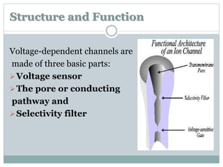
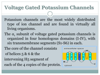
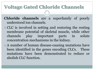
Ion Channels 
FARAZA JAVED 
MPHIL PHARMACOLOGY
Ion Channels 
Ion channels are pore-forming membrane proteins 
whose function is establishing a resting membrane 
potential, shaping action potentials and other 
electrical signals by gating the flow of ions across 
the cell membrane, controlling the flow of ions 
across membranes, and regulating cell volume.
They are often described as narrow, water-filled 
tunnels that accept only specific type of ions. This 
characteristic is called selective permeability. 
Ion channels are integral membrane proteins, 
formed as assemblies of several proteins. Such 
"multi-subunit" assemblies usually make a circular 
arrangement of identical or homologous proteins 
closely packed around a water-filled pore through 
the plane of the lipid bilayer membrane.
Ion channels are different from other transporter 
proteins: 
The rate of ion transport through the channel is 
very high (often 106 ions per second or above). 
Ions pass through channels down their 
electrochemical gradient, which is a function of ion 
concentration and membrane potential, 
"downhill", without the input of metabolic energy 
(e.g. Adenosine triphosphate, active transport 
mechanisms, co-transport mechanisms).
History 
By the late 1800s, the chemical mechanism 
underlying nerve and muscle tissue messaging was 
a mystery. 
Ludimar Hermann was able to conclude that nerve 
and muscle cells were capable of exhibiting a "self-propagating 
wave of negative charge which 
advances in steps along the tissue ".
Julius Bernstein made the first real theoretical 
contribution, for he postulated the ionic theory, 
the Nernst equation, and the assumption of a semi-permeable 
membrane surrounding nerve and 
muscle cells could all help explain the mounting 
electrophysiological evidence of the previous 
statement.
Sidney Ringer used a solution of water and ran it 
through the vessels of an isolated heart from a frog 
in the 1880s and discovered that in order for the 
heart to continue beating salts needed to be 
present in the water. 
Specifically, sodium, calcium, and potassium salts 
were needed and they had to be in special 
concentrations relative to each other.
In 1937, John Z. Young 
use squid neuron to study 
ion current. 
He made important experiments possible for the 
first time, including the first intracellular recordings 
of the nerve cell action potential.
The next improvement in instrumentation took place 
in the late 1940s by Kenneth Cole. This involved 
placing a glass electrode inside the cell in order to 
"voltage clamp" the interior of the cell. 
 Voltage clamping made it possible to distinguish the 
voltage effects caused by influx of sodium or efflux of 
potassium.
 Later in the 1960s and 70s, many other small 
molecules and peptides would be discovered to 
help gate these channels, including glutamate, 
GABA, glycine, serotonin, dopamine, and 
norepinephrine.
In 1970s, the existence of ion channels was 
confirmed by the invention of ‘patch clamp’ 
technique by Erwin Neher and Bert Sakmann who 
won a Nobel Prize for it. 
In 2003, the Nobel Prize was awarded to American 
scientists, Roderick MacKinon and Peter Agre for 
their x-ray crystallographic structure studies on 
ion channels.
Since the discovery of ion channels, the etiology of 
many diseases has been traced back to 
channelopathies. Many toxins produced by snakes, 
fish, spiders and other insects paralyze the ion 
channels. Many physiological mechanisms have 
been studied at molecular level and have 
confirmed the involvement of ion channels. The 
ion channels are the recent target sites for 
pharmaceutical biosynthesis of new drugs.
Types of Ion Channels 
2 major types: 
Voltage gated ion channels 
Ligand gated ion channels
Voltage Gated Ion Channels
Structure and Function 
Voltage-dependent channels are 
made of three basic parts: 
Voltage sensor 
The pore or conducting 
pathway and 
Selectivity filter
Voltage gated ion channels consist of a highly 
processed 
 α subunit, associated with 
 auxiliary β subunits. 
The pore-forming α subunit is sufficient for functional 
expression, but the kinetics and voltage dependence 
of channel gating are modified by the β subunits.
The α subunits are organized in four homologous 
domains (I-IV) each with six transmembrane 
segments (S1-S6) - 24 transmembrane segments in 
total. The pore forming segments are formed by S5 
and S6.
Each of these segments 
coils is called a 
transmembrane 
domain, and within 
a transmembrane domain 
the side chains necessarily 
face outward where they 
readily interact with the 
lipids of the membrane are known as Polypeptide 
chain.
For clarity in the figure, the alpha-helices are shown 
spread out and in a row. 
In an actual membrane, the 
alpha-helices are 
not in a line, but clustered. 
This is shown in top view in 
the figure. 
At the center of the four 
domains is the channel 
through which the ions movement take place.
Voltage 
Gated 
Channels 
Domains TM 
Segment 
s 
Sub- 
Units 
Pore 
Forming 
Regions 
Voltage 
Sensor 
V.G Na+ 4 6 1 S5-S6 S4 
V.G Ca2+ 4 6 4 S5-S6 S4 
V.G Cl- 4 6 4 S5-S6 S4 
V.G K+ 4 6 1 S5-S6 S4 
Channel Structure
In this diagram, a single transmembrane domain is 
shown as the voltage sensor that operates the gate. 
The S4 segment of voltage gated 
channel is the voltage sensor 
that is responsible for changing 
conformation as the voltage 
changes. All voltage gated 
channels have this S4 segment.
Changes in the membrane potential modulate the 
channel's opening or closing, as changing the 
membrane potential changes the relative amounts of 
positive and negative charges on the inside and 
outside of the membrane. Like charges repel, so the 
positively charged S4 segment 
will be pushed away from 
a positive intracellular fluid 
towards the negative extra 
cellular fluid, changing the 
protein's conformation and opening the channel.
At a typical resting membrane potential (for example, -70 
mV) the channel is closed. Then should any 
factor depolarize the 
membrane potential 
sufficiently (for example, 
to -50 mV), the voltage 
sensor moves outward 
and the gate opens. 
The channel can also 
close (deactivate) by negative voltages that restore the 
down position of S4 and close the gate.
Types of V.G Ion Channels 
4 major types: 
V.G Sodium Channels 
V.G Calcium Channels 
V.G Potassium Channels 
V.G Chloride Channels
Voltage Gated Sodium Channels 
The founding member of the ion channel superfamily 
in terms of its discovery as a protein is the voltage 
gated sodium channel. These channels are 
responsible for the rapid influx of sodium ions that 
underlies the rising phase of the action potential in 
nerve, muscle, and endocrine cells. 
Sodium channel composed of one principal alpha 
subunit and one or two auxiliary beta subunits.
The a subunits of sodium channels are composed of 
four homologous domains that each contain six 
transmembrane segments. 
Different distinct neurotoxin binding sites have been 
identified within the Na channel protein, with 
different effects on ion permeation and gating 
resulting in either inhibition or enhancement of Na 
currents.
Channelopathies 
Extensive research has been done and is continued on 
ion channels. Ion channels are a favorite site for 
invention of new drugs. 
Moreover many genetic disorders are found to be 
caused by defective channel proteins. In addition to 
that, many toxins and venoms produced by spiders, 
snakes, scorpion, bees, fish, snails and others act by 
incapacitating ion channels.
Paramyotonia Congenita (PMC) 
Paramyotonia Congenita (PMC) is one of the periodic 
paralyses caused by mutations in (alpha-1 subunit) 
the sodium channel. PMC causes muscle stiffness 
(myotonia) which is made worse by chilling or 
activity. 
Mechanism: In Paramyotonia the sodium channels 
fail to regulate the flow of ions properly. At first this 
imbalance causes the muscle fiber to contract 
uncontrollably, but as the imbalance worsens the 
muscle stops responding to nerve signals and 
becomes weak or paralyzed.
This can clearly be seen in an EMG measurement of 
muscle from a PMC patient. 
Treatment: 
Tocainide is a new 
antiarrhythmic agent 
which seems to 
reduce effectively 
sodium conductance.
Brugada syndrome 
The Brugada syndrome is a genetic disease that is 
characterized by abnormal electrocardiogram (ECG) 
findings and an increased risk of sudden cardiac 
death. It is the major cause of sudden death in 
young’s and termed as sudden unexplained/ adult 
death syndrome (SUDS) or (SADS).
Genetics: 
The cases of Brugada syndrome have been shown to be 
associated with mutations (loss of function 
mutation) in the gene (named SCN5A of alpha 
subunit) that encodes for the sodium ion channel in 
the cell membranes of the muscle cells of the heart 
(the myocytes).
Treatment: The cause of death in Brugada syndrome 
is ventricular fibrillation. These arrhythmias appear 
with no warning. While there is no exact treatment 
modality, treatment lies in termination of this 
lethal arrhythmia before it causes death. 
This is done via implantation of an implantable 
cardioverter-defibrillator(ICD), which continuously 
monitors the heart rhythm and will defibrillate an 
individual if ventricular fibrillation is noted.
Voltage Gated Calcium Channels 
Voltage-gated calcium channels mediate calcium 
influx in response to membrane depolarization and 
regulate intracellular processes such as contraction, 
secretion, neurotransmission. 
Like sodium channels, the α1 subunit of voltage gated 
calcium channels is organized in four homologous 
domains (I-IV), with six transmembrane segments 
(S1-S6) in each. 
An intracellular β subunit and a transmembrane, 
disulfide-linked α2β subunit complex are 
components of most types of calcium channels.
A γ subunit has also been found in skeletal muscle 
calcium channels, and related subunits are expressed 
in heart and brain. 
There are several different kinds of high-voltage-gated 
calcium channels (VGCCs). 
N-type channel (Most often 
found throughout the brain 
and peripheral nervous 
system).
P/Q-type channel (Purkinje neurons in the 
cerebellum) 
L-type (Skeletal muscle, smooth muscle, bone 
(osteoblasts), ventricular myocytes (responsible for 
prolonged action potential in cardiac cell), dendrites 
and dendritic spines of cortical neurones). 
L-type channels are responsible for excitation-contraction 
coupling of skeletal, smooth, and cardiac 
muscle and for hormone secretion in endocrine cells.
Malignant Hyperthermia 
MH is a life-threatening clinical syndrome of 
hypermetabolism involving the skeletal muscle. It is 
triggered in susceptible individuals primarily by the 
volatile inhalational anesthetic agents (Halothane) 
and the muscle relaxant succinylcholine.
Genetics: 
The defect is typically located on chromosome 
19 (involving the ryanodine receptor) located in the 
N-terminus of the protein, which interacts with L-type 
calcium channels. This region is important for 
allowing Ca2+ passage through the protein following 
opening.
Treatment: During an episode of malignant 
hyperthermia, wrapping the patient in a cooling 
blanket can help reduce fever and the risk of serious 
complications. 
The current treatment of choice is the intravenous 
administration of Dantrolene, the only known 
antidote, and supportive therapy directed at 
correcting hyperthermia.
Migraine 
Calcium channel blockers are effective second-line 
agents. They are a viable alternative in patients who 
cannot tolerate β-blockers. 
Mechanism: Results of recent studies suggest that 
cerebral blood flow during the initial phase of 
migraine is decreased and this decrease probably 
leads to ischemia and hypoxia. Cellular hypoxia, in 
turn, can cause an increase in the flow of calcium, 
resulting in calcium overload and cellular 
dysfunction.
Nimodipine, a calcium-channel blocker that exhibits 
selective effects on cerebral vessels, seems to offer 
protection against the cerebral ischemia and hypoxia 
presumed to be operative during migraine attacks.
Analgesic Activity of CCB 
Ziconotide (Prialt) is the synthetic form of the N-type 
Ca2+ channel blocker in the final stages of 
clinical development, a peptide toxin derived from 
a marine cone snail. 
In humans, spinal infusion of Prialt produces 
significant pain relief in patients with intractable 
pain associated with cancer, AIDS and in some 
neuropathic pain conditions.
Voltage Gated Potassium Channels 
Potassium channels are the most widely distributed 
type of ion channel and are found in virtually all 
living organisms. 
The α1 subunit of voltage gated potassium channels is 
organized in four homologous domains (I-IV), with 
six transmembrane segments (S1-S6) in each. 
The core of the channel consists 
of helices 5 & 6 & the 
intervening H5 segment of 
each of the 4 copies of the protein.
Mutation studies showed that the H5 segment is 
essential for K+ selectivity. 
Potassium channels act to set or reset the resting 
potential in many cells. In excitable cells, such 
as neurons, the delayed counter flow of potassium 
ions shapes the action potential. 
They also regulate cellular processes such as the 
secretion of hormones (e.g., insulin release 
from beta-cells in the pancreas) so their malfunction 
can lead to diseases.
Congenital Hyperinsulinism 
Congenital hyperinsulinism is a condition that causes 
individuals to have abnormally high levels of insulin. 
People with this condition have frequent episodes of 
low blood sugar (hypoglycemia). These conditions 
are present at birth but milder forms may not be 
detected until adult years.
Mechanism: 
In approximately half of people with congenital 
hyperinsulinism, the cause is unknown. Mutations in 
genes that regulate the release (secretion) of insulin, 
which is produced by beta cells in the pancreas is the 
proposed cause.
Treatment: 
Diazoxide and octreotide are the primary 
medications used in long-term treatment of CHI.
Multiple sclerosis 
Multiple sclerosis (MS) is an inflammatory disease of 
CNS characterized by demyelination of axons. 
Uregualtion of V.G Potassium channels increase the 
autoimmune disease process of MS. 
The idea that neurologic function might be improved if 
conduction could be restored in CNS demyelinated 
axons led to the testing of potassium channel 
blockers as a symptomatic treatment.
To date, only 2 broad-spectrum K+ channel blockers, 
4-aminopyridine (4-AP) and 3,4-diaminopyridine 
(3,4-DAP), have been tested in MS patients. 
Although both 4-AP and 3,4-DAP produce clear 
neurologic benefits, their use has been limited due to 
toxicity.
Epilepsy 
 Instead of blocking excitatory ion channels, another 
potentially antiexcitable strategy is to enhance the 
activity of Kv channels. The past decade has seen 
increased interest in Kv channel opening as 
antiepileptic mechanism with focus on Kv channels. 
The leading compound in the pipeline is 
Retigabine which activates neuronal Kv channels. 
Retigabine has successfully completed Phase III trial 
and is presently awaiting approval as an 
antiepileptic.
Voltage Gated Chloride Channels 
Chloride channels are a superfamily of poorly 
understood ion channels. 
CLC is involved in setting and restoring the resting 
membrane potential of skeletal muscle, while other 
channels play important parts in solute 
concentration mechanisms in the kidney. 
A number of human disease-causing mutations have 
been identified in the genes encoding CLCs. These 
mutations have been demonstrated to reduce or 
abolish CLC function.
Cystic Fibrosis 
Cystic fibrosis transmembrane conductance 
regulator (CFTR) is a membrane protein that is 
encoded by the CFTR gene. Mutations of the CFTR 
gene affecting chloride ion channel function lead to 
dysregulation of epithelial fluid transport in the lung, 
pancreas and other organs, resulting in cystic 
fibrosis.
Treatment: 
Respiratory therapy is any treatment that slows 
down lung damage and improves breathing. 
 FDA has just approved a new drug called Ivacaftor 
that will almost certainly be a godsend for 4% of 
cystic fibrosis (CF) sufferers.
References 
 Richard W. Tsien and Curtis F. Barrett. A Brief History 
of Calcium Channel Discovery. Madame Curie 
Bioscience Database. Austin (TX): Landes Bioscience; 
2000. 
 Carafoli E. Calcium signaling: a tale for all seasons. Proc 
Natl Acad Sci USA. 2002;99:1115–1122. 
Hunter JV, Moss AJ. Seizures and Arrythmias: Differing 
phenotypes of a common channelopathy. 
Neurology.2009;72(3):208–9. 
 Grillner S. The motor infrastructure: from ion channel to 
 neuronal networks. Nat Rcv Neurosci 2003;4:573–86.
 Francisco Bezanilla. Voltage-Gated Ion Channels. 
Nanobioscience, Vol. 4, No. 1, March 2005. 
 Susan I.V. Judge, Christopher T. Bever Jr. Potassium 
channel blockers in multiple sclerosis: Neuronal Kv 
channels and effects of symptomatic treatment. 
Pharmacology & Therapeutics 111 (2006) 224 – 259. 
 William A. Catteral. From Ionic Currents to Molecular 
Review Mechanisms: The Structure and Function of 
Voltage-Gated Sodium Channels. Neuron, Vol. 26, 13–25, 
April, 2000. 
 Paul Linsdell. Mechanism of chloride permeation in the 
cystic fibrosis transmembrane conductance regulator 
chloride channel. Exp Physiol (2006). pp 123–129.
 http://genetics.thetech.org/original. 
 Dinarello CA, Porat R. Fever and hyperthermia. In: Fauci A, 
Kasper D, Longo DL, et al, eds. Harrison's Principles of 
Internal Medicine. 17th ed. [online version]. New York, NY: 
McGraw Hill;2008:chap 17. 
 Felix Luessi, MD; Volker Siffrin; Frauke Zipp. 
Neurodegeneration in Multiple Sclerosis: Novel Treatment 
Strategies: Therapeutic Approaches to Neuronal 
Degeneration in MS. Medscape. 2013. 
 Yu-Qing Cao. Voltage-gated calcium channels and pain. 
Pain. 126 (2006) 5–9.
 http://physrev.physiology.org 
 http://www.britannica.com/ion-channel 
 Susan I. V. Judge, Jennifer M. Lee, Christopher T. Bever Jr, 
Paul M. Hoffman. Voltage-gated potassium channels in 
multiple sclerosis: Overview and new implications for 
treatment of central nervous system inflammation and 
degeneration. JRRD. (2006). 43:1. p111-122. 
 William A. Catterall. Structure and Function of Voltage- 
Gated Ion Channels. Annu. Rev. Biochem. 1995. 64:493- 
531.

Ion channels, types and their importace in managment of diseases

  • 1.
    Ion Channels FARAZAJAVED MPHIL PHARMACOLOGY
  • 2.
    Ion Channels Ionchannels are pore-forming membrane proteins whose function is establishing a resting membrane potential, shaping action potentials and other electrical signals by gating the flow of ions across the cell membrane, controlling the flow of ions across membranes, and regulating cell volume.
  • 3.
    They are oftendescribed as narrow, water-filled tunnels that accept only specific type of ions. This characteristic is called selective permeability. Ion channels are integral membrane proteins, formed as assemblies of several proteins. Such "multi-subunit" assemblies usually make a circular arrangement of identical or homologous proteins closely packed around a water-filled pore through the plane of the lipid bilayer membrane.
  • 4.
    Ion channels aredifferent from other transporter proteins: The rate of ion transport through the channel is very high (often 106 ions per second or above). Ions pass through channels down their electrochemical gradient, which is a function of ion concentration and membrane potential, "downhill", without the input of metabolic energy (e.g. Adenosine triphosphate, active transport mechanisms, co-transport mechanisms).
  • 5.
    History By thelate 1800s, the chemical mechanism underlying nerve and muscle tissue messaging was a mystery. Ludimar Hermann was able to conclude that nerve and muscle cells were capable of exhibiting a "self-propagating wave of negative charge which advances in steps along the tissue ".
  • 6.
    Julius Bernstein madethe first real theoretical contribution, for he postulated the ionic theory, the Nernst equation, and the assumption of a semi-permeable membrane surrounding nerve and muscle cells could all help explain the mounting electrophysiological evidence of the previous statement.
  • 7.
    Sidney Ringer useda solution of water and ran it through the vessels of an isolated heart from a frog in the 1880s and discovered that in order for the heart to continue beating salts needed to be present in the water. Specifically, sodium, calcium, and potassium salts were needed and they had to be in special concentrations relative to each other.
  • 8.
    In 1937, JohnZ. Young use squid neuron to study ion current. He made important experiments possible for the first time, including the first intracellular recordings of the nerve cell action potential.
  • 9.
    The next improvementin instrumentation took place in the late 1940s by Kenneth Cole. This involved placing a glass electrode inside the cell in order to "voltage clamp" the interior of the cell.  Voltage clamping made it possible to distinguish the voltage effects caused by influx of sodium or efflux of potassium.
  • 10.
     Later inthe 1960s and 70s, many other small molecules and peptides would be discovered to help gate these channels, including glutamate, GABA, glycine, serotonin, dopamine, and norepinephrine.
  • 11.
    In 1970s, theexistence of ion channels was confirmed by the invention of ‘patch clamp’ technique by Erwin Neher and Bert Sakmann who won a Nobel Prize for it. In 2003, the Nobel Prize was awarded to American scientists, Roderick MacKinon and Peter Agre for their x-ray crystallographic structure studies on ion channels.
  • 12.
    Since the discoveryof ion channels, the etiology of many diseases has been traced back to channelopathies. Many toxins produced by snakes, fish, spiders and other insects paralyze the ion channels. Many physiological mechanisms have been studied at molecular level and have confirmed the involvement of ion channels. The ion channels are the recent target sites for pharmaceutical biosynthesis of new drugs.
  • 13.
    Types of IonChannels 2 major types: Voltage gated ion channels Ligand gated ion channels
  • 14.
  • 15.
    Structure and Function Voltage-dependent channels are made of three basic parts: Voltage sensor The pore or conducting pathway and Selectivity filter
  • 16.
    Voltage gated ionchannels consist of a highly processed  α subunit, associated with  auxiliary β subunits. The pore-forming α subunit is sufficient for functional expression, but the kinetics and voltage dependence of channel gating are modified by the β subunits.
  • 17.
    The α subunitsare organized in four homologous domains (I-IV) each with six transmembrane segments (S1-S6) - 24 transmembrane segments in total. The pore forming segments are formed by S5 and S6.
  • 18.
    Each of thesesegments coils is called a transmembrane domain, and within a transmembrane domain the side chains necessarily face outward where they readily interact with the lipids of the membrane are known as Polypeptide chain.
  • 19.
    For clarity inthe figure, the alpha-helices are shown spread out and in a row. In an actual membrane, the alpha-helices are not in a line, but clustered. This is shown in top view in the figure. At the center of the four domains is the channel through which the ions movement take place.
  • 20.
    Voltage Gated Channels Domains TM Segment s Sub- Units Pore Forming Regions Voltage Sensor V.G Na+ 4 6 1 S5-S6 S4 V.G Ca2+ 4 6 4 S5-S6 S4 V.G Cl- 4 6 4 S5-S6 S4 V.G K+ 4 6 1 S5-S6 S4 Channel Structure
  • 21.
    In this diagram,a single transmembrane domain is shown as the voltage sensor that operates the gate. The S4 segment of voltage gated channel is the voltage sensor that is responsible for changing conformation as the voltage changes. All voltage gated channels have this S4 segment.
  • 22.
    Changes in themembrane potential modulate the channel's opening or closing, as changing the membrane potential changes the relative amounts of positive and negative charges on the inside and outside of the membrane. Like charges repel, so the positively charged S4 segment will be pushed away from a positive intracellular fluid towards the negative extra cellular fluid, changing the protein's conformation and opening the channel.
  • 23.
    At a typicalresting membrane potential (for example, -70 mV) the channel is closed. Then should any factor depolarize the membrane potential sufficiently (for example, to -50 mV), the voltage sensor moves outward and the gate opens. The channel can also close (deactivate) by negative voltages that restore the down position of S4 and close the gate.
  • 24.
    Types of V.GIon Channels 4 major types: V.G Sodium Channels V.G Calcium Channels V.G Potassium Channels V.G Chloride Channels
  • 25.
    Voltage Gated SodiumChannels The founding member of the ion channel superfamily in terms of its discovery as a protein is the voltage gated sodium channel. These channels are responsible for the rapid influx of sodium ions that underlies the rising phase of the action potential in nerve, muscle, and endocrine cells. Sodium channel composed of one principal alpha subunit and one or two auxiliary beta subunits.
  • 26.
    The a subunitsof sodium channels are composed of four homologous domains that each contain six transmembrane segments. Different distinct neurotoxin binding sites have been identified within the Na channel protein, with different effects on ion permeation and gating resulting in either inhibition or enhancement of Na currents.
  • 27.
    Channelopathies Extensive researchhas been done and is continued on ion channels. Ion channels are a favorite site for invention of new drugs. Moreover many genetic disorders are found to be caused by defective channel proteins. In addition to that, many toxins and venoms produced by spiders, snakes, scorpion, bees, fish, snails and others act by incapacitating ion channels.
  • 28.
    Paramyotonia Congenita (PMC) Paramyotonia Congenita (PMC) is one of the periodic paralyses caused by mutations in (alpha-1 subunit) the sodium channel. PMC causes muscle stiffness (myotonia) which is made worse by chilling or activity. Mechanism: In Paramyotonia the sodium channels fail to regulate the flow of ions properly. At first this imbalance causes the muscle fiber to contract uncontrollably, but as the imbalance worsens the muscle stops responding to nerve signals and becomes weak or paralyzed.
  • 29.
    This can clearlybe seen in an EMG measurement of muscle from a PMC patient. Treatment: Tocainide is a new antiarrhythmic agent which seems to reduce effectively sodium conductance.
  • 30.
    Brugada syndrome TheBrugada syndrome is a genetic disease that is characterized by abnormal electrocardiogram (ECG) findings and an increased risk of sudden cardiac death. It is the major cause of sudden death in young’s and termed as sudden unexplained/ adult death syndrome (SUDS) or (SADS).
  • 31.
    Genetics: The casesof Brugada syndrome have been shown to be associated with mutations (loss of function mutation) in the gene (named SCN5A of alpha subunit) that encodes for the sodium ion channel in the cell membranes of the muscle cells of the heart (the myocytes).
  • 32.
    Treatment: The causeof death in Brugada syndrome is ventricular fibrillation. These arrhythmias appear with no warning. While there is no exact treatment modality, treatment lies in termination of this lethal arrhythmia before it causes death. This is done via implantation of an implantable cardioverter-defibrillator(ICD), which continuously monitors the heart rhythm and will defibrillate an individual if ventricular fibrillation is noted.
  • 33.
    Voltage Gated CalciumChannels Voltage-gated calcium channels mediate calcium influx in response to membrane depolarization and regulate intracellular processes such as contraction, secretion, neurotransmission. Like sodium channels, the α1 subunit of voltage gated calcium channels is organized in four homologous domains (I-IV), with six transmembrane segments (S1-S6) in each. An intracellular β subunit and a transmembrane, disulfide-linked α2β subunit complex are components of most types of calcium channels.
  • 34.
    A γ subunithas also been found in skeletal muscle calcium channels, and related subunits are expressed in heart and brain. There are several different kinds of high-voltage-gated calcium channels (VGCCs). N-type channel (Most often found throughout the brain and peripheral nervous system).
  • 35.
    P/Q-type channel (Purkinjeneurons in the cerebellum) L-type (Skeletal muscle, smooth muscle, bone (osteoblasts), ventricular myocytes (responsible for prolonged action potential in cardiac cell), dendrites and dendritic spines of cortical neurones). L-type channels are responsible for excitation-contraction coupling of skeletal, smooth, and cardiac muscle and for hormone secretion in endocrine cells.
  • 36.
    Malignant Hyperthermia MHis a life-threatening clinical syndrome of hypermetabolism involving the skeletal muscle. It is triggered in susceptible individuals primarily by the volatile inhalational anesthetic agents (Halothane) and the muscle relaxant succinylcholine.
  • 37.
    Genetics: The defectis typically located on chromosome 19 (involving the ryanodine receptor) located in the N-terminus of the protein, which interacts with L-type calcium channels. This region is important for allowing Ca2+ passage through the protein following opening.
  • 39.
    Treatment: During anepisode of malignant hyperthermia, wrapping the patient in a cooling blanket can help reduce fever and the risk of serious complications. The current treatment of choice is the intravenous administration of Dantrolene, the only known antidote, and supportive therapy directed at correcting hyperthermia.
  • 40.
    Migraine Calcium channelblockers are effective second-line agents. They are a viable alternative in patients who cannot tolerate β-blockers. Mechanism: Results of recent studies suggest that cerebral blood flow during the initial phase of migraine is decreased and this decrease probably leads to ischemia and hypoxia. Cellular hypoxia, in turn, can cause an increase in the flow of calcium, resulting in calcium overload and cellular dysfunction.
  • 41.
    Nimodipine, a calcium-channelblocker that exhibits selective effects on cerebral vessels, seems to offer protection against the cerebral ischemia and hypoxia presumed to be operative during migraine attacks.
  • 42.
    Analgesic Activity ofCCB Ziconotide (Prialt) is the synthetic form of the N-type Ca2+ channel blocker in the final stages of clinical development, a peptide toxin derived from a marine cone snail. In humans, spinal infusion of Prialt produces significant pain relief in patients with intractable pain associated with cancer, AIDS and in some neuropathic pain conditions.
  • 43.
    Voltage Gated PotassiumChannels Potassium channels are the most widely distributed type of ion channel and are found in virtually all living organisms. The α1 subunit of voltage gated potassium channels is organized in four homologous domains (I-IV), with six transmembrane segments (S1-S6) in each. The core of the channel consists of helices 5 & 6 & the intervening H5 segment of each of the 4 copies of the protein.
  • 44.
    Mutation studies showedthat the H5 segment is essential for K+ selectivity. Potassium channels act to set or reset the resting potential in many cells. In excitable cells, such as neurons, the delayed counter flow of potassium ions shapes the action potential. They also regulate cellular processes such as the secretion of hormones (e.g., insulin release from beta-cells in the pancreas) so their malfunction can lead to diseases.
  • 45.
    Congenital Hyperinsulinism Congenitalhyperinsulinism is a condition that causes individuals to have abnormally high levels of insulin. People with this condition have frequent episodes of low blood sugar (hypoglycemia). These conditions are present at birth but milder forms may not be detected until adult years.
  • 46.
    Mechanism: In approximatelyhalf of people with congenital hyperinsulinism, the cause is unknown. Mutations in genes that regulate the release (secretion) of insulin, which is produced by beta cells in the pancreas is the proposed cause.
  • 47.
    Treatment: Diazoxide andoctreotide are the primary medications used in long-term treatment of CHI.
  • 48.
    Multiple sclerosis Multiplesclerosis (MS) is an inflammatory disease of CNS characterized by demyelination of axons. Uregualtion of V.G Potassium channels increase the autoimmune disease process of MS. The idea that neurologic function might be improved if conduction could be restored in CNS demyelinated axons led to the testing of potassium channel blockers as a symptomatic treatment.
  • 49.
    To date, only2 broad-spectrum K+ channel blockers, 4-aminopyridine (4-AP) and 3,4-diaminopyridine (3,4-DAP), have been tested in MS patients. Although both 4-AP and 3,4-DAP produce clear neurologic benefits, their use has been limited due to toxicity.
  • 50.
    Epilepsy  Insteadof blocking excitatory ion channels, another potentially antiexcitable strategy is to enhance the activity of Kv channels. The past decade has seen increased interest in Kv channel opening as antiepileptic mechanism with focus on Kv channels. The leading compound in the pipeline is Retigabine which activates neuronal Kv channels. Retigabine has successfully completed Phase III trial and is presently awaiting approval as an antiepileptic.
  • 51.
    Voltage Gated ChlorideChannels Chloride channels are a superfamily of poorly understood ion channels. CLC is involved in setting and restoring the resting membrane potential of skeletal muscle, while other channels play important parts in solute concentration mechanisms in the kidney. A number of human disease-causing mutations have been identified in the genes encoding CLCs. These mutations have been demonstrated to reduce or abolish CLC function.
  • 52.
    Cystic Fibrosis Cysticfibrosis transmembrane conductance regulator (CFTR) is a membrane protein that is encoded by the CFTR gene. Mutations of the CFTR gene affecting chloride ion channel function lead to dysregulation of epithelial fluid transport in the lung, pancreas and other organs, resulting in cystic fibrosis.
  • 53.
    Treatment: Respiratory therapyis any treatment that slows down lung damage and improves breathing.  FDA has just approved a new drug called Ivacaftor that will almost certainly be a godsend for 4% of cystic fibrosis (CF) sufferers.
  • 54.
    References  RichardW. Tsien and Curtis F. Barrett. A Brief History of Calcium Channel Discovery. Madame Curie Bioscience Database. Austin (TX): Landes Bioscience; 2000.  Carafoli E. Calcium signaling: a tale for all seasons. Proc Natl Acad Sci USA. 2002;99:1115–1122. Hunter JV, Moss AJ. Seizures and Arrythmias: Differing phenotypes of a common channelopathy. Neurology.2009;72(3):208–9.  Grillner S. The motor infrastructure: from ion channel to  neuronal networks. Nat Rcv Neurosci 2003;4:573–86.
  • 55.
     Francisco Bezanilla.Voltage-Gated Ion Channels. Nanobioscience, Vol. 4, No. 1, March 2005.  Susan I.V. Judge, Christopher T. Bever Jr. Potassium channel blockers in multiple sclerosis: Neuronal Kv channels and effects of symptomatic treatment. Pharmacology & Therapeutics 111 (2006) 224 – 259.  William A. Catteral. From Ionic Currents to Molecular Review Mechanisms: The Structure and Function of Voltage-Gated Sodium Channels. Neuron, Vol. 26, 13–25, April, 2000.  Paul Linsdell. Mechanism of chloride permeation in the cystic fibrosis transmembrane conductance regulator chloride channel. Exp Physiol (2006). pp 123–129.
  • 56.
     http://genetics.thetech.org/original. Dinarello CA, Porat R. Fever and hyperthermia. In: Fauci A, Kasper D, Longo DL, et al, eds. Harrison's Principles of Internal Medicine. 17th ed. [online version]. New York, NY: McGraw Hill;2008:chap 17.  Felix Luessi, MD; Volker Siffrin; Frauke Zipp. Neurodegeneration in Multiple Sclerosis: Novel Treatment Strategies: Therapeutic Approaches to Neuronal Degeneration in MS. Medscape. 2013.  Yu-Qing Cao. Voltage-gated calcium channels and pain. Pain. 126 (2006) 5–9.
  • 57.
     http://physrev.physiology.org http://www.britannica.com/ion-channel  Susan I. V. Judge, Jennifer M. Lee, Christopher T. Bever Jr, Paul M. Hoffman. Voltage-gated potassium channels in multiple sclerosis: Overview and new implications for treatment of central nervous system inflammation and degeneration. JRRD. (2006). 43:1. p111-122.  William A. Catterall. Structure and Function of Voltage- Gated Ion Channels. Annu. Rev. Biochem. 1995. 64:493- 531.