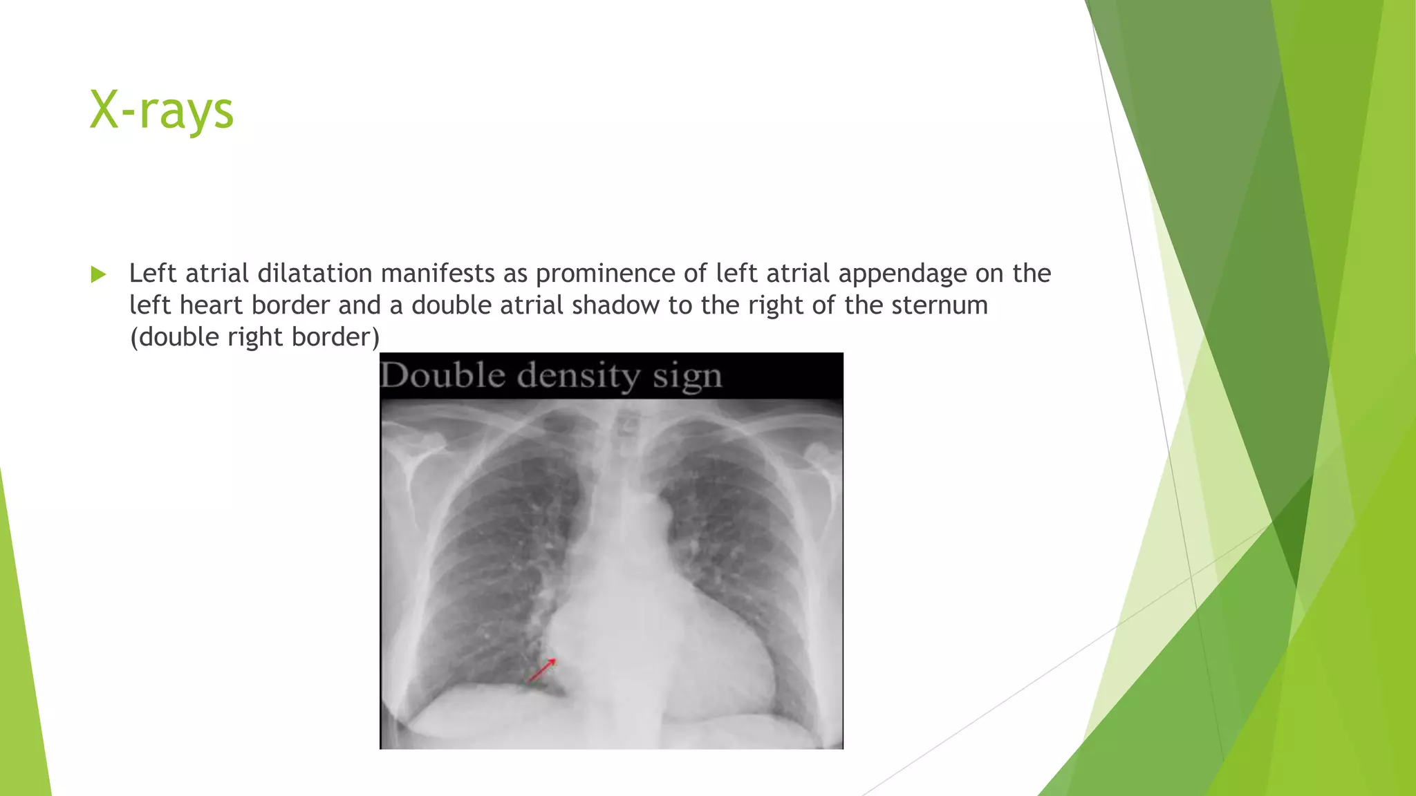
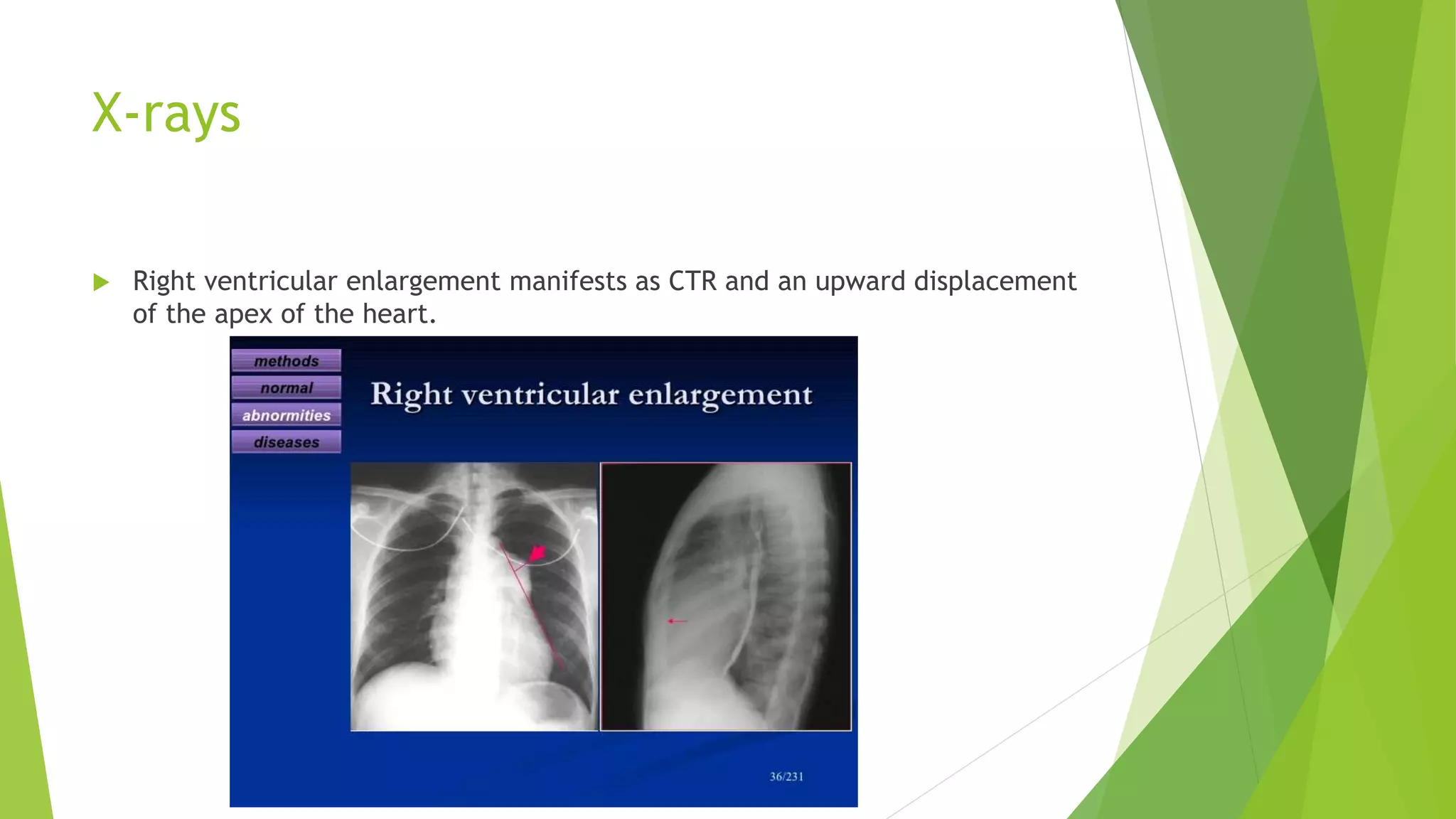
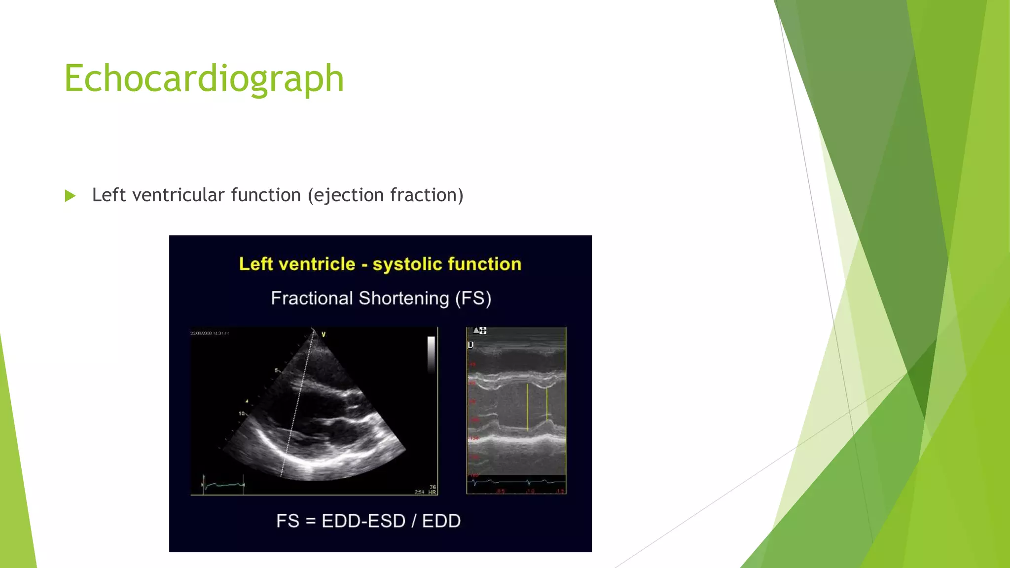
Investigation in cardiac
diseases
CVS
Methods
 ECG(electrocardiograph)
 Chest X-rays
 Echocardiograph
 Ambulatory ECG
 Thallium 201 scanning (radionuclide scanning)
 Technetium-99-labeled sestamibi (radionuclide scanning)
 Cardiac catheterization
 Coronary angiography
ECG
 Ecg is useful to detect the following abnormalities
 Cardiac arrhythmia
 Conduction defects
 Myocardial infarction or ischemia
 Myocardia hypotrophy
 Electrolyte imbalance
 Toxicity of certain drugs
X-rays
 Heart size can be measured only from PA view of chest x-rays because in AP
view cardiac shadow is large.
 The maximum transverse diameter of the heart is compared with the
maximum transvers diameter of the chest is measured from inside of the ribs,
this is called cardiothoracic ratio (CTR)
 It should be less than 50 %.
 Cardiomegaly and pericardial effusion cause an increase in cardiothoracic
ratio.
X-rays
 Pericardial effusion
produce globular
shadow
X-rays
 Left atrial dilatation manifests as prominence of left atrial appendage on the
left heart border and a double atrial shadow to the right of the sternum
(double right border)
X-rays
 Left ventricular enlargement manifests as increased CTR and increased
convexity of the left heart border.
X-rays
 Right atrial enlargement manifests as projection of right border of heart into
the right lower lung field
X-rays
 Right ventricular enlargement manifests as CTR and an upward displacement
of the apex of the heart.
X-rays
 Enlargement of pulmonary artery manifests as a prominent bulge on the left
heart border below the aortic knuckle.
X-rays
 Lung field may indicate pulmonary hypertension by enlargement of the hilar
vessels
 Enlarged right lower lob artery
 Kerly’s B lines and pleural effusion may be present in cardiac failure
Echocardiograph
 It is sensitive method to determination of
 Size of all four chambers of heart
Echocardiograph
 Left ventricular function (ejection fraction)
Echocardiography
 Regional wall motion abnormalities due to myocardial infarction
 Complication of myocardial infarction such as papillary muscles dysfunction
mitral regurgitation
 Structural valve abnormalities stenosis and regurgitation
 Cardiac output
 Ventricular hypertrophy
 Pericardial effusion
 Atrial and ventricular septic defects and other congenital defects.
Types of echocardiography
 Two-dimensional real time echocardiography
 M-mod echocardiograph
 Doppler echocardiograph
Two-dimensional real time
echocardiograph
 Detecting wall motion abnormalities
 Intracardiac masses
 Thrombi and tumors
 Endocarditic vegetations
 Congenital heart disease
M-mod echocardiography
 Measurement of size of chambers of heart
 Calculation of ejection fraction and accurate timing of cardiac events
Doppler echocardiograph
 Abnormal direction of blood flow
 Aortic mitral or mitral regurgitation
 Pulse-wave Doppler (PW)
 Continuous-wave Doppler (CW)
 Color doppler

Investigation in cardiac diseases cvs

  • 1.
  • 2.
    Methods  ECG(electrocardiograph)  ChestX-rays  Echocardiograph  Ambulatory ECG  Thallium 201 scanning (radionuclide scanning)  Technetium-99-labeled sestamibi (radionuclide scanning)  Cardiac catheterization  Coronary angiography
  • 3.
    ECG  Ecg isuseful to detect the following abnormalities  Cardiac arrhythmia  Conduction defects  Myocardial infarction or ischemia  Myocardia hypotrophy  Electrolyte imbalance  Toxicity of certain drugs
  • 4.
    X-rays  Heart sizecan be measured only from PA view of chest x-rays because in AP view cardiac shadow is large.  The maximum transverse diameter of the heart is compared with the maximum transvers diameter of the chest is measured from inside of the ribs, this is called cardiothoracic ratio (CTR)  It should be less than 50 %.  Cardiomegaly and pericardial effusion cause an increase in cardiothoracic ratio.
  • 5.
  • 6.
    X-rays  Left atrialdilatation manifests as prominence of left atrial appendage on the left heart border and a double atrial shadow to the right of the sternum (double right border)
  • 7.
    X-rays  Left ventricularenlargement manifests as increased CTR and increased convexity of the left heart border.
  • 8.
    X-rays  Right atrialenlargement manifests as projection of right border of heart into the right lower lung field
  • 9.
    X-rays  Right ventricularenlargement manifests as CTR and an upward displacement of the apex of the heart.
  • 10.
    X-rays  Enlargement ofpulmonary artery manifests as a prominent bulge on the left heart border below the aortic knuckle.
  • 11.
    X-rays  Lung fieldmay indicate pulmonary hypertension by enlargement of the hilar vessels  Enlarged right lower lob artery  Kerly’s B lines and pleural effusion may be present in cardiac failure
  • 12.
    Echocardiograph  It issensitive method to determination of  Size of all four chambers of heart
  • 13.
    Echocardiograph  Left ventricularfunction (ejection fraction)
  • 14.
    Echocardiography  Regional wallmotion abnormalities due to myocardial infarction  Complication of myocardial infarction such as papillary muscles dysfunction mitral regurgitation  Structural valve abnormalities stenosis and regurgitation  Cardiac output  Ventricular hypertrophy  Pericardial effusion  Atrial and ventricular septic defects and other congenital defects.
  • 15.
    Types of echocardiography Two-dimensional real time echocardiography  M-mod echocardiograph  Doppler echocardiograph
  • 16.
    Two-dimensional real time echocardiograph Detecting wall motion abnormalities  Intracardiac masses  Thrombi and tumors  Endocarditic vegetations  Congenital heart disease
  • 17.
    M-mod echocardiography  Measurementof size of chambers of heart  Calculation of ejection fraction and accurate timing of cardiac events
  • 18.
    Doppler echocardiograph  Abnormaldirection of blood flow  Aortic mitral or mitral regurgitation  Pulse-wave Doppler (PW)  Continuous-wave Doppler (CW)  Color doppler