![Lesson 1
Cardiovascular testing
Tsegaye Melaku (BPharm, MSc)
[Assistant Professor of Clinical Pharmacy]
tsegayemlk@yahoo.com or tsegaye.melaku@ju.edu.et +251913765609December, 2019
Pharmacotherapy of Cardiovascular Disorders](https://image.slidesharecdn.com/1-cardiovasculartesting-200123121032/85/Cardiovascular-testing-1-320.jpg)


![B. Carotid arterial pulse
Diminished pulsations (indicate):
– In ↓ SV, atherosclerotic narrowing of carotid artery,
– Obstruction to LV outflow, AS or HCM.
Very forceful/hyperdynamic/"bounding“ pulsations:
– In ↑ stroke volume, Chronic AR,
– High CO [hyperthyroidism, marked anemia]
29](https://image.slidesharecdn.com/1-cardiovasculartesting-200123121032/85/Cardiovascular-testing-29-320.jpg)

![E. Heart Sounds
– Typical "lub-dub" sound of the normal heart
– S1:precedes ventricular contraction
» Due to closure of the mitral & tricuspid valves
– S2: follows ventricular contraction
» Due to closure of the aortic & pulmonic valves
– Others [S3/S4/Murmur/gallop]:presence of underlying heart
disease
31](https://image.slidesharecdn.com/1-cardiovasculartesting-200123121032/85/Cardiovascular-testing-31-320.jpg)


![ Murmurs
– Auditory vibrations [turbulent blood flow] within the heart
chambers or across the valves.
– Based on timing & duration within cardiac cycle (systolic,
diastolic, or continuous), intensity (grade 1 to 6, from softest to
loudest), pitch (high or low frequency),
– May be some are “Innocent" or "physiologic"
35](https://image.slidesharecdn.com/1-cardiovasculartesting-200123121032/85/Cardiovascular-testing-35-320.jpg)


![ Troponin [I & T]
– Contractile proteins found only in cardiac myocytes
– Most sensitive, tissue-specific
– Detectable in the blood 2 to 4 hrs of onset of sxs
– Remains detectable for 5 to 10 days
41](https://image.slidesharecdn.com/1-cardiovasculartesting-200123121032/85/Cardiovascular-testing-41-320.jpg)


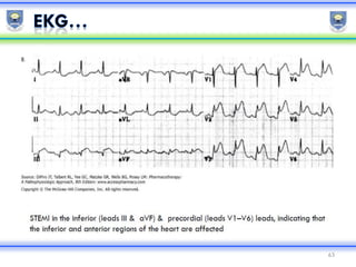


This document provides an overview of cardiovascular testing. It discusses: - Different types of cardiovascular testing including echocardiography, electrocardiography, and cardiac biomarkers. - How these tests are used for cardiovascular disease diagnosis, treatment, and prognosis evaluation. - The importance of obtaining a thorough history and physical exam when evaluating cardiovascular conditions, and assessing elements like jugular venous pressure, heart sounds, and murmurs. - Additional testing modalities like CT, MRI, cardiac catheterization and angiography, and their uses in further evaluating cardiac structure and function.


