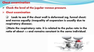
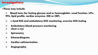
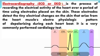
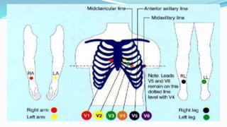
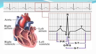
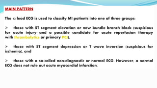
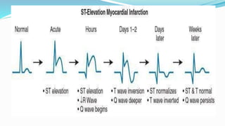
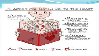
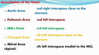
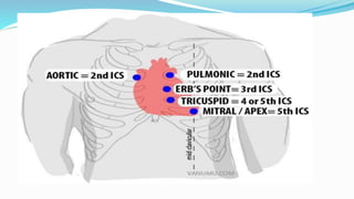
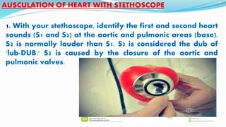
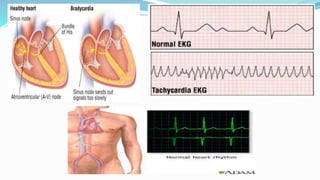
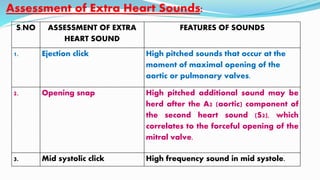
MR. SUDHIR KHUNTIA
INTRODUCTION:-
Assessment of the cardiovascular system is one of the
most important areas of the nurse’s daily patient assessment. Report
your findings as clearly as possible. Charting your results clearly is
essential for others to be able to assess the problem, and good
documentation is also essential for the treatment of the patient as
well as for the nursing care.
PHYSICAL EXAMINATION
General:-
 Build (obesity or wasting); shortness of breath; difficulty in talking; note
whether they look ill.
 Look for pallor, jaundice, sweatiness and clamminess, and for xanthelasma
around the eyes.
Cyanosis
 This is seen below the fingernails and toenails but also in the lips, cheeks,
ears and nose.
 It may increase in the cold and on exertion.
Face
 Malar flush - redness around the cheeks (mitral stenosis).
 Xanthomata - yellowish deposits of lipid around the eyes, palms, or tendons
(hyperlipidaemia).
 Corneal arcus - a ring around the cornea (normal ageing or
hyperlipidaemia).
 Proptosis - forward projection or displacement of the eyeball (Graves'
disease)
Hands
 Finger clubbing.
 Janeway lesions - macules on the back of the hands (infective endocarditis).
 Osler's nodes - tender nodules in the fingertips (infective endocarditis).
Pulse
 Rate: average 72/minute in adults, faster in children and may
slow in old age. Also slower in athletes. Compare with apex rate.
Rhythm:
Respiratory variations are common in healthy individuals (if
there is noticeable quickening in inspiration and slowing in
expiration, this is termed sinus arrhythmia).
•Peripheral pulses:
 Femoral pulses (radial femoral delay in coarctation) and foot
and ankle pulses.
 Listen over the renal and femoral artery for murmurs.
Check blood pressure
 This should be measured in the brachial artery, using a cuff
around the upper arm.
 A large cuff must be used in obese people, because a small cuff
will result in the blood pressure being overestimated.
 Systolic pressure is at the level when first heard (Korotkoff I)
and the diastolic pressure is when silence begins (Korotkoff V).
 In patients with chest pain, or if ever the radial pulses appear
asymmetrical, the pressure should be measured in both arms
because a difference between the two may indicate aortic dissection.
Chest examination
 Check the level of the jugular venous pressure.
 Chest examination:
 Look to see if the chest wall is deformed (eg, funnel chest)
and moves equally (inequality of expansion is usually due to
respiratory disease).
Note the respiratory rate; it is related to the pulse rate in the
ratio of about 1:4 and remains constant in the same individual.
 Ask the patient to breathe out and, using both hands resting lightly on
the side walls of the chest with thumbs meeting in the middle, ask them to
breathe in to assess the expansion of the chest on full inspiration by noting
how far the examiner's thumbs move apart.
 Feel over the anterior chest wall for any thrills associated with cardiac
murmurs.
 Auscultation of the heart -
Examination of other areas
Abdomen - see also separate Abdominal Examination
Palpate the abdomen for hepatomegaly and splenomegaly
(congestive cardiac failure), or spleen alone (infective
endocarditis).
Feel for enlargement of the aorta (aneurysm); feel with the
hands flat either side of the aorta - feel for pulsation and
tenderness.
Investigations
•These may include:
 Blood tests (for fasting glucose and/or haemoglobin, renal function, LFTs,
TFTs, lipid profile, cardiac enzymes, ESR or CRP).
 12-lead ECG and ambulatory ECG monitoring, exercise ECG testing.
 Ambulatory blood pressure monitoring
 chest x-ray
 Spirometry.
 Echocardiogram.
 Cardiac catheterization.
 Angiography.
Electrocardiography (ECG or EKG*) is the process of
recording the electrical activity of the heart over a period of
time using electrodes placed on the skin. These electrodes
detect the tiny electrical changes on the skin that arise from
the heart muscle's electro physiologic pattern
of depolarizing during each heart beat. It is a very
commonly performed cardiology test.
MAIN PATTERN
The 12 lead ECG is used to classify MI patients into one of three groups:
 those with ST segment elevation or new bundle branch block (suspicious
for acute injury and a possible candidate for acute reperfusion therapy
with thrombolytics or primary PCI),
 those with ST segment depression or T wave inversion (suspicious for
ischemia), and
 those with a so-called non-diagnostic or normal ECG. However, a normal
ECG does not rule out acute myocardial infarction.
CARDIAC ASSESSMENT
GUIDE BY: PRESENTED BY:
MRS. SHEFALI CHARAN LECTURER MR. JEEVAN LAL
M.Sc. NURSING M. Sc. NURSING FINAL YEAR
(MEDICAL SURGICAL NURSING)
Health History:-
1. Current Health Status
-chest pain
- shortness of breath
- swelling of ankles or feet
- heart palpitations
- fatigue
2. Past Health History
-Congenital heart disease
- Rheumatic fever
- Heart murmur
- High blood pressure, high cholesterol, diabetes mellitus
- Confusion
- Fatigue
3. Family History
4. Personal Habits
Techniques of Examination
 The patient should be supine with upper body elevated
at a 15-30E angle.
The room must be quiet, warm, and have good lighting.
You should stand to the right of the patient being
examined. Inspection and Palpation of the Heart
The finger pads are more sensitive in detecting pulsations.
Inspect and Palpate for:
Pulsations- these are more visible when patients are thin. A
thick chest wall or increased AP diameter can obscure them.
Pulsations may indicate increased blood volume or
pressure.
Lift or heaves- these are forceful cardiac contractions that
cause a slight to vigorous movement of sternum and ribs.
Thrills- these are the vibrations of loud cardiac murmurs.
They feel like the throat of a purring cat. Thrills occur with
turbulent blood flow.
AREA FINDINGS
Aortic (2nd inter
coastal space to
the right of the
sternum)
A pulsation could indicate an aortic aneurysm
A thrill could indicate aortic stenosis
You should inspect and palpate at the following areas:
Pulmonary
(2nd inter
coastal space
to the left of
the sternum)
A pulsation could indicate pulmonary
hypertension
A thrill could indicate pulmonic stenosis
Tricuspid (4-
5th inter
coastal
space,
lower half
of the
sternum)
A sustained systolic lift could indicate right ventricular enlargement.
A systolic thrill could indicate a ventricular septal defect.
Mitral (5th
intercoastal
space at
mid
clavicular
line)
Increased pulsation could indicate increased output, anxiety,
fever, or pregnancy.
A thrill could indicate mitral regurgitation, or mitral stenosis.
Epigastric
(below
xiphoid
process)
Increased aortic pulsation could indicate right
ventricular pulsation of right ventricular
enlargement and aortic regurgitation.
-
Sternoclavicular
(top of sternum
at junction of
clavicles)
Pulsation of aortic arch may be felt in a thin client.
1. Aortic Area
2nd right interspace close to the
sternum.
2. Pulmonic Area 2nd left interspace.
3. ERB's Point 3rd left interspace.
4. Tricuspid Area
5th left interspace close to the
sternum.
5. Mitral Area
(Apical)
5th left interspace medial to the MCL
Auscultation of the Heart
1. With your stethoscope, identify the first and second heart
sounds (S1 and S2) at the aortic and pulmonic areas (base).
S2 is normally louder than S1. S2 is considered the dub of
'lub-DUB.' S2 is caused by the closure of the aortic and
pulmonic valves.
AUSCULATION OF HEART WITH STETHOSCOPE
2. Identify the heart rate.
tachycardia
bradycardia
3. Identify the rhythm.
if it is irregular, try to identify the pattern.
Do early beats appear on a regular rhythm?
Does the irregularity vary consistently with respiration?
Is rhythm totally irregular?
Contd…….
4. Listen to S1 first, then S2 at the previously mentioned
areas using the diaphragm and then the bell.
note its intensity.
are there any splitting sounds check during inspiration
where S2 usually splits at pulmonic and ERB's point.
A thick chest wall or increased AP diameter may make S2
inaudible.
Contd……
ALTERATION IN S1, S2, S3, S4, and murmur
sound
S.N
O
ALTERATION
IN S1
ALTERATION
IN S2
LISTEN FOR
S3
LISTEN FOR
S4
LISTEN FOR MURMUR
1. S1 is
accentuated in
exercise,
anemia,
hyperthyroidis
m and mitral
stenosis
Normal
physiological
splitting of S2 is
best heard at
pulmonic area.
It occurs on
inspiration.
A physiologic
S3 is
frequently
heard in
children and
in pregnant
women.
It occurs
before S1
It is low
pitched and
best heard
with the bell
Heart murmur are heart sound
produced when blood flows across
one of the heart valves that is loud
enough to be heard with a
stethoscope.
2. S1 is diminished in
first degree heart
block
Splitting of S2 can
indicate pulmonic
stenosis, atrial
septal defect,
right ventricular
failure.
It occurs early in
diastole during
rapid ventricular
filling. It is heard
best at the apex
in the left lateral
decubitus
position.
It may be
caused by
coronary
artery disease,
hypertension,
myocardiopat
hy, or aortic
stenosis.
3. S1 split is most
audible in
tricuspid area
A pathologic S3
occurs in people
over the age of
40. Cause is
usually
myocardial
failure
Sounds like
dee-lub-dub(or
Tennessee)
S.NO ASSESSMENT OF EXTRA
HEART SOUND
FEATURES OF SOUNDS
1. Ejection click High pitched sounds that occur at the
moment of maximal opening of the
aortic or pulmonary valves.
2. Opening snap High pitched additional sound may be
herd after the A2 (aortic) component of
the second heart sound (S2), which
correlates to the forceful opening of the
mitral valve.
3. Mid systolic click High frequency sound in mid systole.
Assessment of Extra Heart Sounds:
Cardiac assessment ppt

Cardiac assessment ppt

  • 1.
  • 2.
    INTRODUCTION:- Assessment of thecardiovascular system is one of the most important areas of the nurse’s daily patient assessment. Report your findings as clearly as possible. Charting your results clearly is essential for others to be able to assess the problem, and good documentation is also essential for the treatment of the patient as well as for the nursing care.
  • 5.
    PHYSICAL EXAMINATION General:-  Build(obesity or wasting); shortness of breath; difficulty in talking; note whether they look ill.  Look for pallor, jaundice, sweatiness and clamminess, and for xanthelasma around the eyes. Cyanosis  This is seen below the fingernails and toenails but also in the lips, cheeks, ears and nose.  It may increase in the cold and on exertion.
  • 6.
    Face  Malar flush- redness around the cheeks (mitral stenosis).  Xanthomata - yellowish deposits of lipid around the eyes, palms, or tendons (hyperlipidaemia).  Corneal arcus - a ring around the cornea (normal ageing or hyperlipidaemia).  Proptosis - forward projection or displacement of the eyeball (Graves' disease) Hands  Finger clubbing.  Janeway lesions - macules on the back of the hands (infective endocarditis).  Osler's nodes - tender nodules in the fingertips (infective endocarditis).
  • 7.
    Pulse  Rate: average72/minute in adults, faster in children and may slow in old age. Also slower in athletes. Compare with apex rate. Rhythm: Respiratory variations are common in healthy individuals (if there is noticeable quickening in inspiration and slowing in expiration, this is termed sinus arrhythmia).
  • 8.
    •Peripheral pulses:  Femoralpulses (radial femoral delay in coarctation) and foot and ankle pulses.  Listen over the renal and femoral artery for murmurs.
  • 9.
    Check blood pressure This should be measured in the brachial artery, using a cuff around the upper arm.  A large cuff must be used in obese people, because a small cuff will result in the blood pressure being overestimated.  Systolic pressure is at the level when first heard (Korotkoff I) and the diastolic pressure is when silence begins (Korotkoff V).  In patients with chest pain, or if ever the radial pulses appear asymmetrical, the pressure should be measured in both arms because a difference between the two may indicate aortic dissection.
  • 10.
    Chest examination  Checkthe level of the jugular venous pressure.  Chest examination:  Look to see if the chest wall is deformed (eg, funnel chest) and moves equally (inequality of expansion is usually due to respiratory disease). Note the respiratory rate; it is related to the pulse rate in the ratio of about 1:4 and remains constant in the same individual.
  • 11.
     Ask thepatient to breathe out and, using both hands resting lightly on the side walls of the chest with thumbs meeting in the middle, ask them to breathe in to assess the expansion of the chest on full inspiration by noting how far the examiner's thumbs move apart.  Feel over the anterior chest wall for any thrills associated with cardiac murmurs.  Auscultation of the heart -
  • 12.
    Examination of otherareas Abdomen - see also separate Abdominal Examination Palpate the abdomen for hepatomegaly and splenomegaly (congestive cardiac failure), or spleen alone (infective endocarditis). Feel for enlargement of the aorta (aneurysm); feel with the hands flat either side of the aorta - feel for pulsation and tenderness.
  • 13.
    Investigations •These may include: Blood tests (for fasting glucose and/or haemoglobin, renal function, LFTs, TFTs, lipid profile, cardiac enzymes, ESR or CRP).  12-lead ECG and ambulatory ECG monitoring, exercise ECG testing.  Ambulatory blood pressure monitoring  chest x-ray  Spirometry.  Echocardiogram.  Cardiac catheterization.  Angiography.
  • 15.
    Electrocardiography (ECG orEKG*) is the process of recording the electrical activity of the heart over a period of time using electrodes placed on the skin. These electrodes detect the tiny electrical changes on the skin that arise from the heart muscle's electro physiologic pattern of depolarizing during each heart beat. It is a very commonly performed cardiology test.
  • 19.
    MAIN PATTERN The 12lead ECG is used to classify MI patients into one of three groups:  those with ST segment elevation or new bundle branch block (suspicious for acute injury and a possible candidate for acute reperfusion therapy with thrombolytics or primary PCI),  those with ST segment depression or T wave inversion (suspicious for ischemia), and  those with a so-called non-diagnostic or normal ECG. However, a normal ECG does not rule out acute myocardial infarction.
  • 22.
    CARDIAC ASSESSMENT GUIDE BY:PRESENTED BY: MRS. SHEFALI CHARAN LECTURER MR. JEEVAN LAL M.Sc. NURSING M. Sc. NURSING FINAL YEAR (MEDICAL SURGICAL NURSING)
  • 24.
    Health History:- 1. CurrentHealth Status -chest pain - shortness of breath - swelling of ankles or feet - heart palpitations - fatigue
  • 25.
    2. Past HealthHistory -Congenital heart disease - Rheumatic fever - Heart murmur - High blood pressure, high cholesterol, diabetes mellitus - Confusion - Fatigue
  • 26.
    3. Family History 4.Personal Habits
  • 28.
    Techniques of Examination The patient should be supine with upper body elevated at a 15-30E angle. The room must be quiet, warm, and have good lighting. You should stand to the right of the patient being examined. Inspection and Palpation of the Heart The finger pads are more sensitive in detecting pulsations.
  • 30.
    Inspect and Palpatefor: Pulsations- these are more visible when patients are thin. A thick chest wall or increased AP diameter can obscure them. Pulsations may indicate increased blood volume or pressure. Lift or heaves- these are forceful cardiac contractions that cause a slight to vigorous movement of sternum and ribs. Thrills- these are the vibrations of loud cardiac murmurs. They feel like the throat of a purring cat. Thrills occur with turbulent blood flow.
  • 32.
    AREA FINDINGS Aortic (2ndinter coastal space to the right of the sternum) A pulsation could indicate an aortic aneurysm A thrill could indicate aortic stenosis You should inspect and palpate at the following areas:
  • 33.
    Pulmonary (2nd inter coastal space tothe left of the sternum) A pulsation could indicate pulmonary hypertension A thrill could indicate pulmonic stenosis
  • 34.
    Tricuspid (4- 5th inter coastal space, lowerhalf of the sternum) A sustained systolic lift could indicate right ventricular enlargement. A systolic thrill could indicate a ventricular septal defect.
  • 35.
    Mitral (5th intercoastal space at mid clavicular line) Increasedpulsation could indicate increased output, anxiety, fever, or pregnancy. A thrill could indicate mitral regurgitation, or mitral stenosis.
  • 36.
    Epigastric (below xiphoid process) Increased aortic pulsationcould indicate right ventricular pulsation of right ventricular enlargement and aortic regurgitation. -
  • 37.
    Sternoclavicular (top of sternum atjunction of clavicles) Pulsation of aortic arch may be felt in a thin client.
  • 38.
    1. Aortic Area 2ndright interspace close to the sternum. 2. Pulmonic Area 2nd left interspace. 3. ERB's Point 3rd left interspace. 4. Tricuspid Area 5th left interspace close to the sternum. 5. Mitral Area (Apical) 5th left interspace medial to the MCL Auscultation of the Heart
  • 40.
    1. With yourstethoscope, identify the first and second heart sounds (S1 and S2) at the aortic and pulmonic areas (base). S2 is normally louder than S1. S2 is considered the dub of 'lub-DUB.' S2 is caused by the closure of the aortic and pulmonic valves. AUSCULATION OF HEART WITH STETHOSCOPE
  • 41.
    2. Identify theheart rate. tachycardia bradycardia 3. Identify the rhythm. if it is irregular, try to identify the pattern. Do early beats appear on a regular rhythm? Does the irregularity vary consistently with respiration? Is rhythm totally irregular? Contd…….
  • 43.
    4. Listen toS1 first, then S2 at the previously mentioned areas using the diaphragm and then the bell. note its intensity. are there any splitting sounds check during inspiration where S2 usually splits at pulmonic and ERB's point. A thick chest wall or increased AP diameter may make S2 inaudible. Contd……
  • 44.
    ALTERATION IN S1,S2, S3, S4, and murmur sound
  • 45.
    S.N O ALTERATION IN S1 ALTERATION IN S2 LISTENFOR S3 LISTEN FOR S4 LISTEN FOR MURMUR 1. S1 is accentuated in exercise, anemia, hyperthyroidis m and mitral stenosis Normal physiological splitting of S2 is best heard at pulmonic area. It occurs on inspiration. A physiologic S3 is frequently heard in children and in pregnant women. It occurs before S1 It is low pitched and best heard with the bell Heart murmur are heart sound produced when blood flows across one of the heart valves that is loud enough to be heard with a stethoscope.
  • 46.
    2. S1 isdiminished in first degree heart block Splitting of S2 can indicate pulmonic stenosis, atrial septal defect, right ventricular failure. It occurs early in diastole during rapid ventricular filling. It is heard best at the apex in the left lateral decubitus position. It may be caused by coronary artery disease, hypertension, myocardiopat hy, or aortic stenosis. 3. S1 split is most audible in tricuspid area A pathologic S3 occurs in people over the age of 40. Cause is usually myocardial failure Sounds like dee-lub-dub(or Tennessee)
  • 47.
    S.NO ASSESSMENT OFEXTRA HEART SOUND FEATURES OF SOUNDS 1. Ejection click High pitched sounds that occur at the moment of maximal opening of the aortic or pulmonary valves. 2. Opening snap High pitched additional sound may be herd after the A2 (aortic) component of the second heart sound (S2), which correlates to the forceful opening of the mitral valve. 3. Mid systolic click High frequency sound in mid systole. Assessment of Extra Heart Sounds: