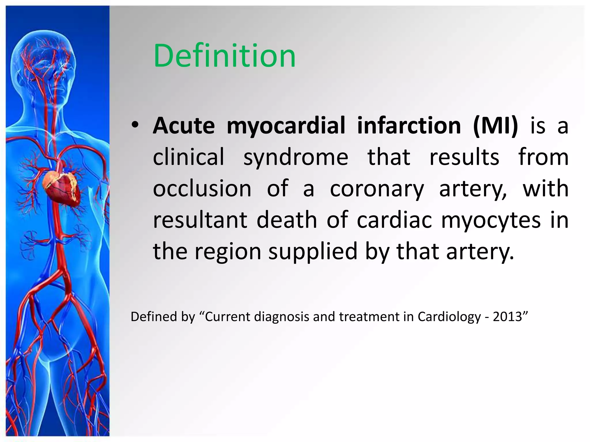
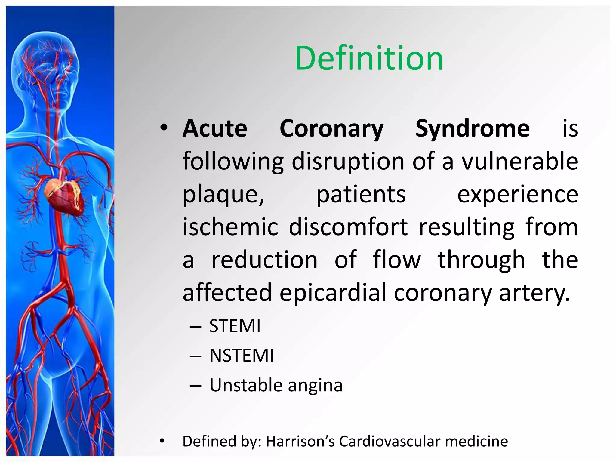
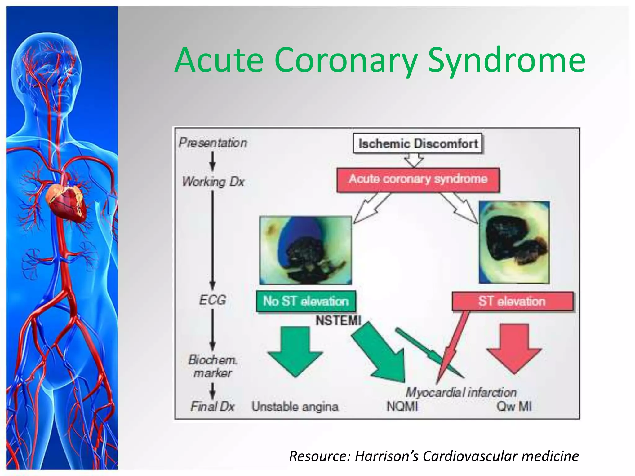
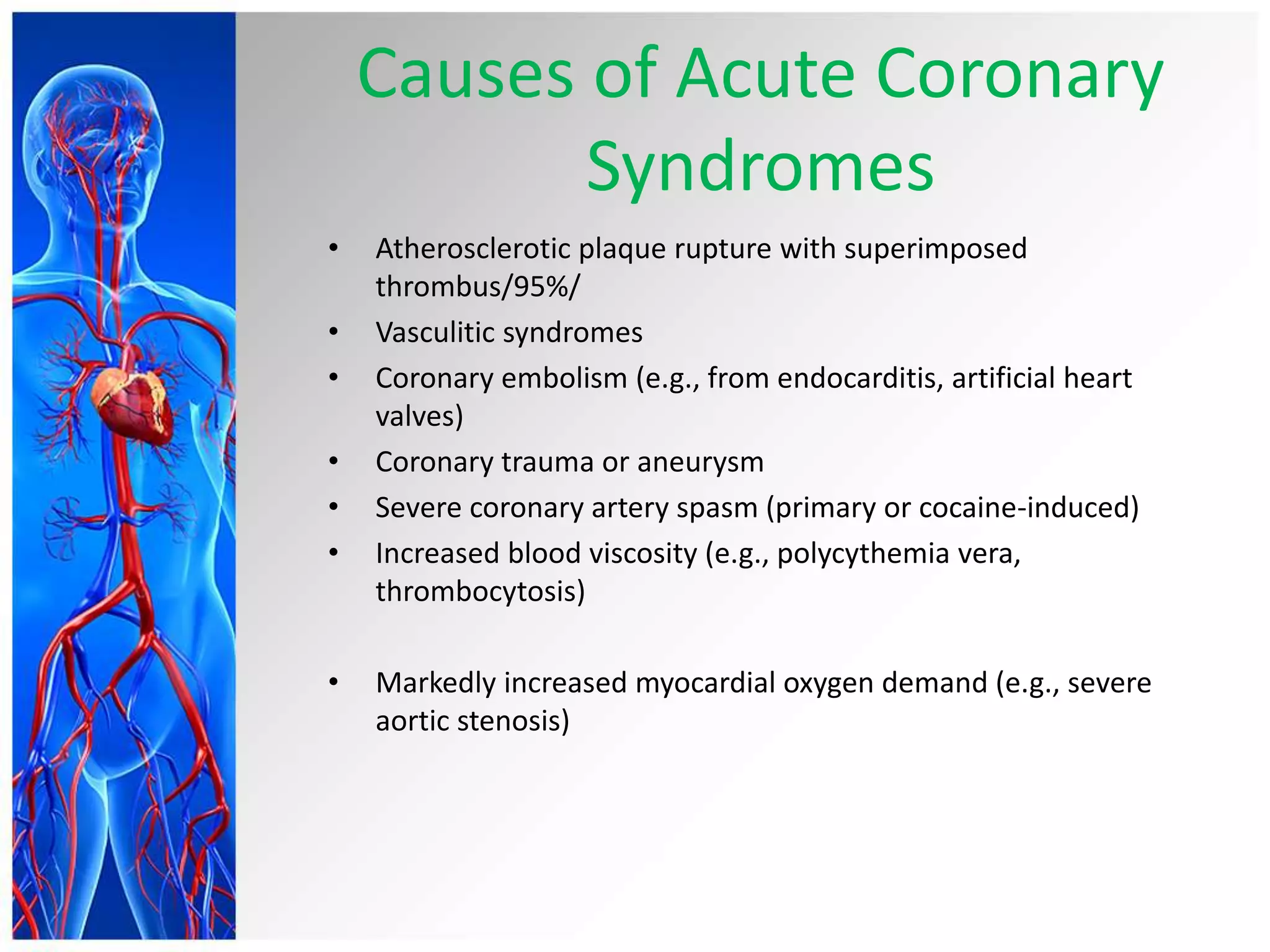
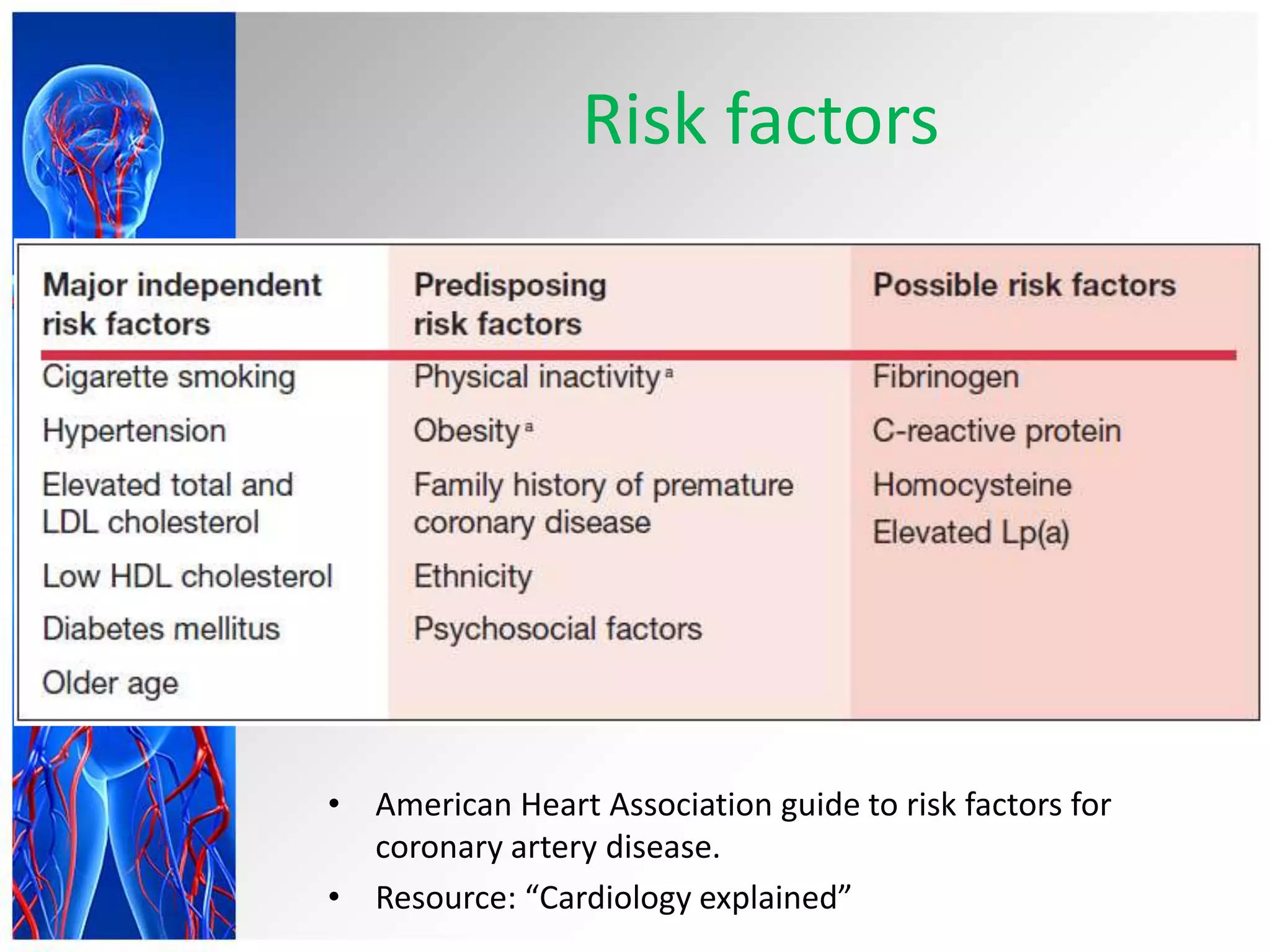
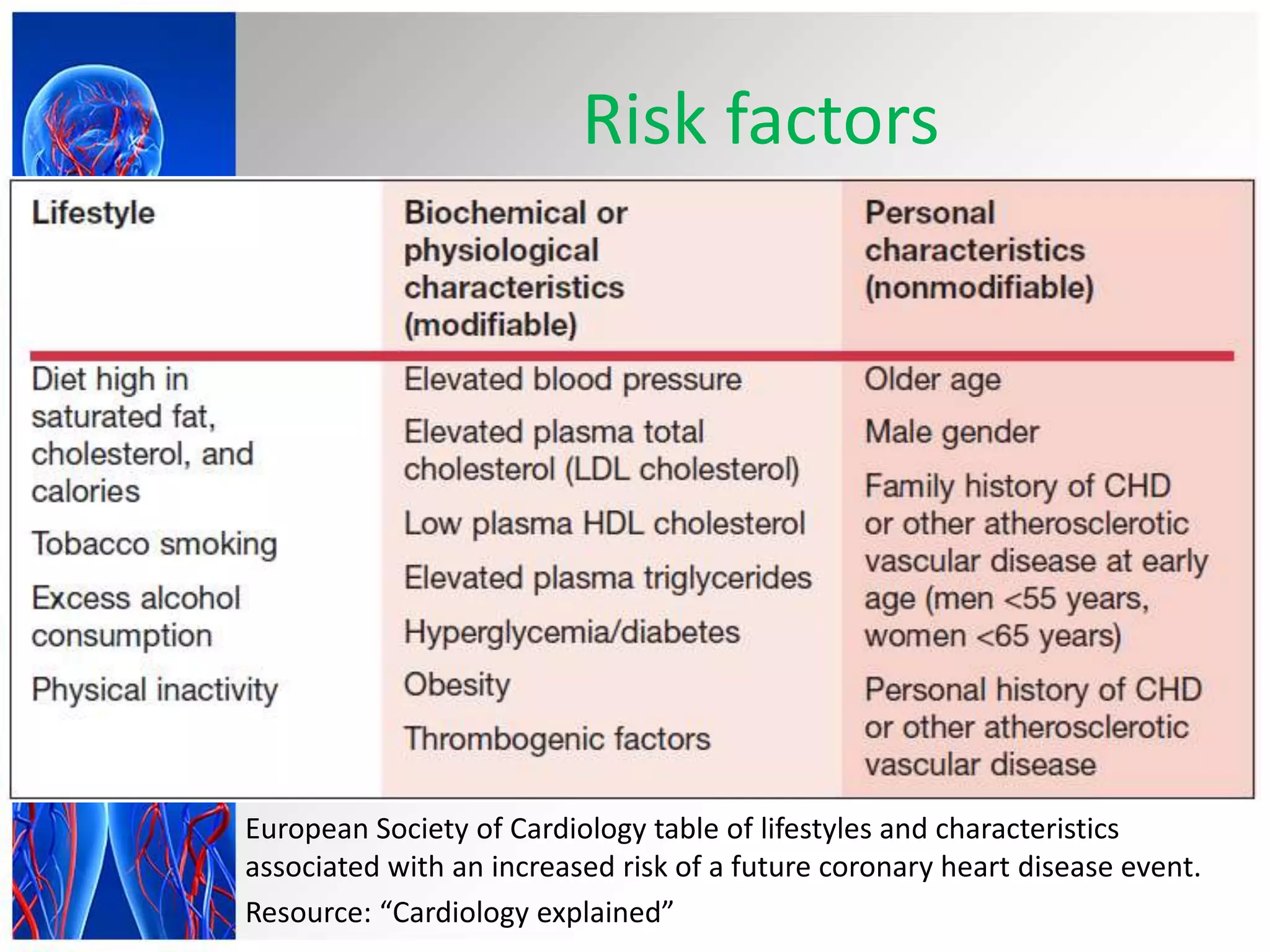
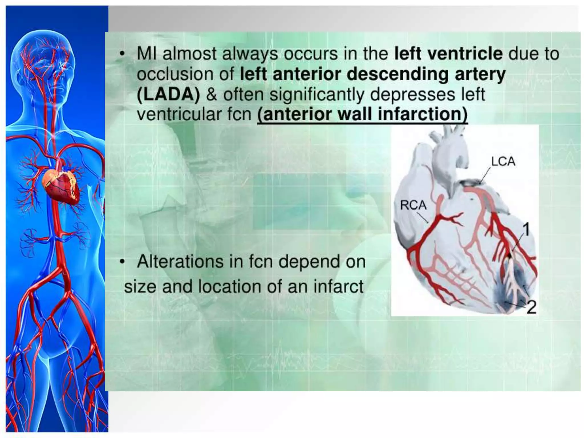
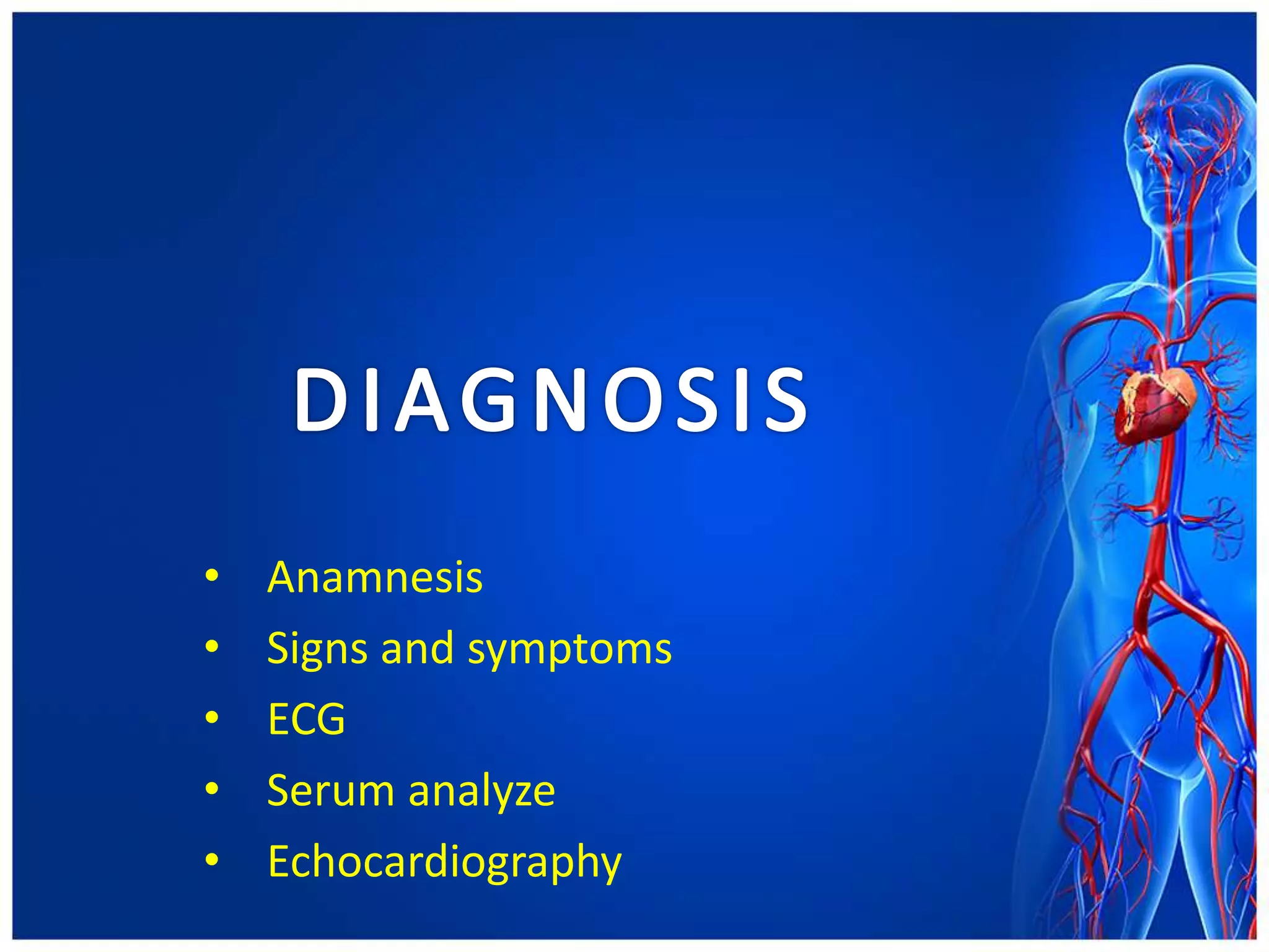
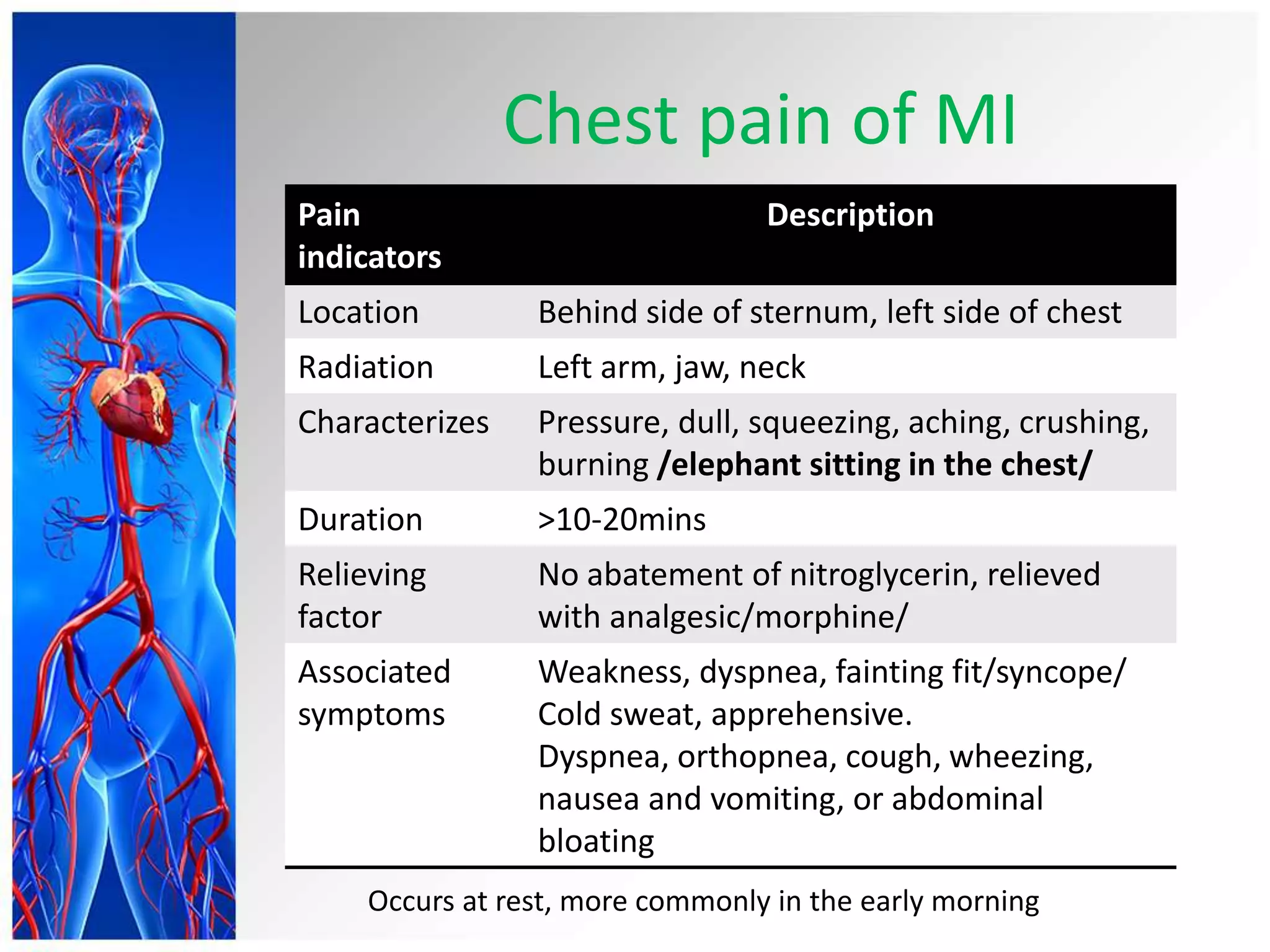
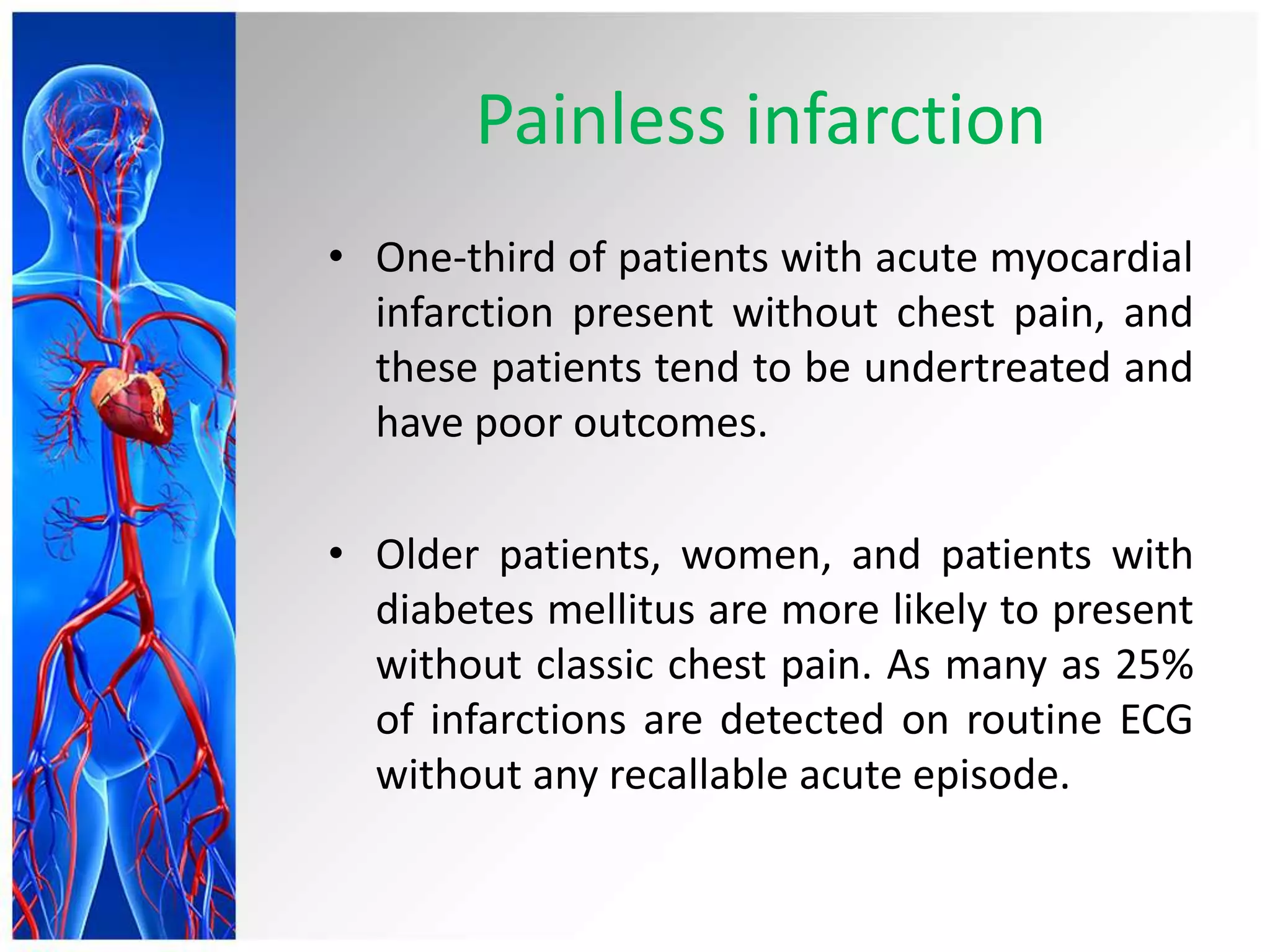
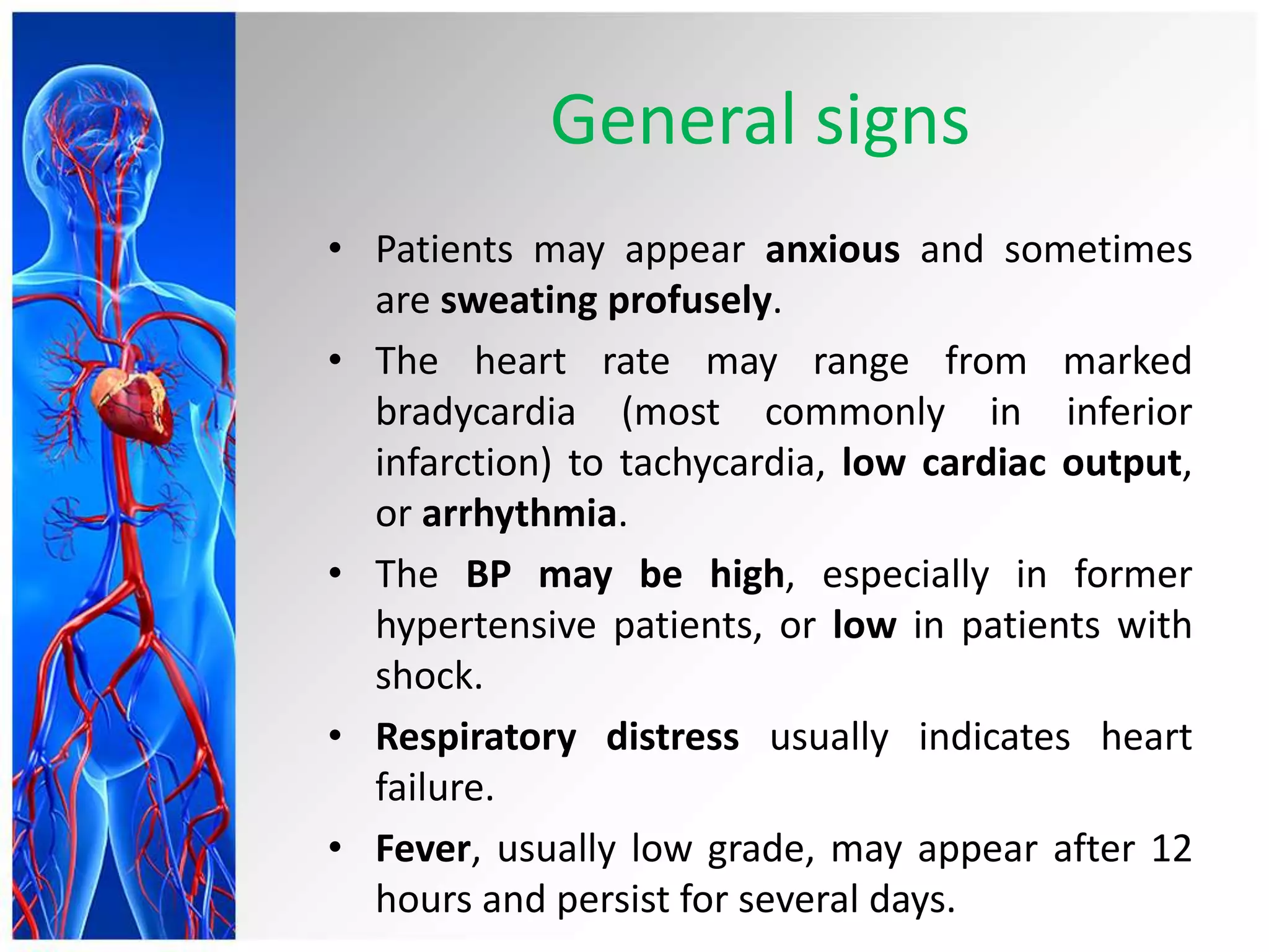
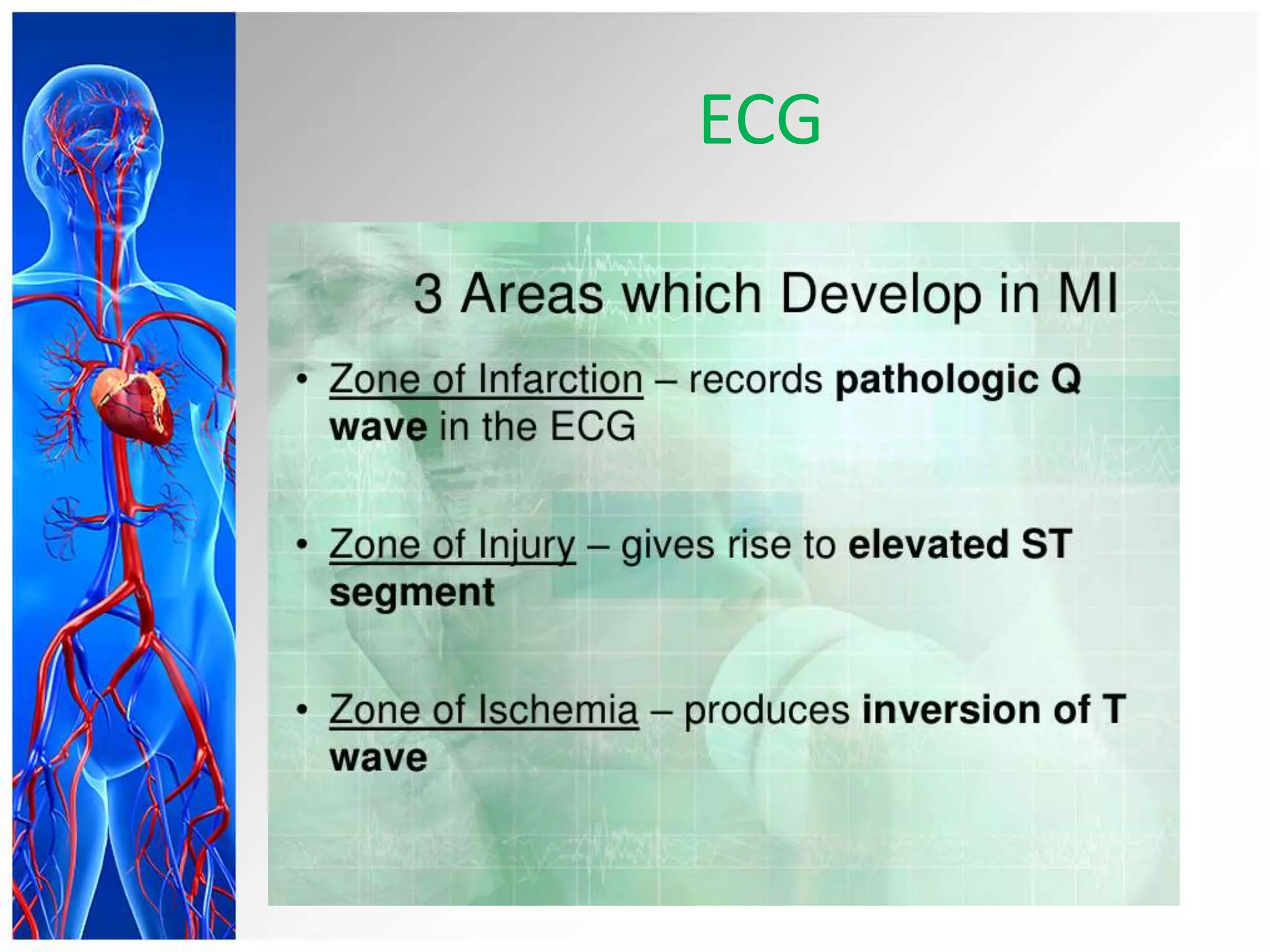
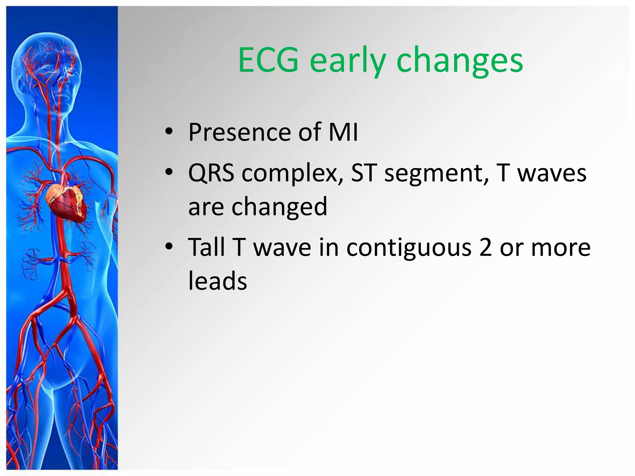
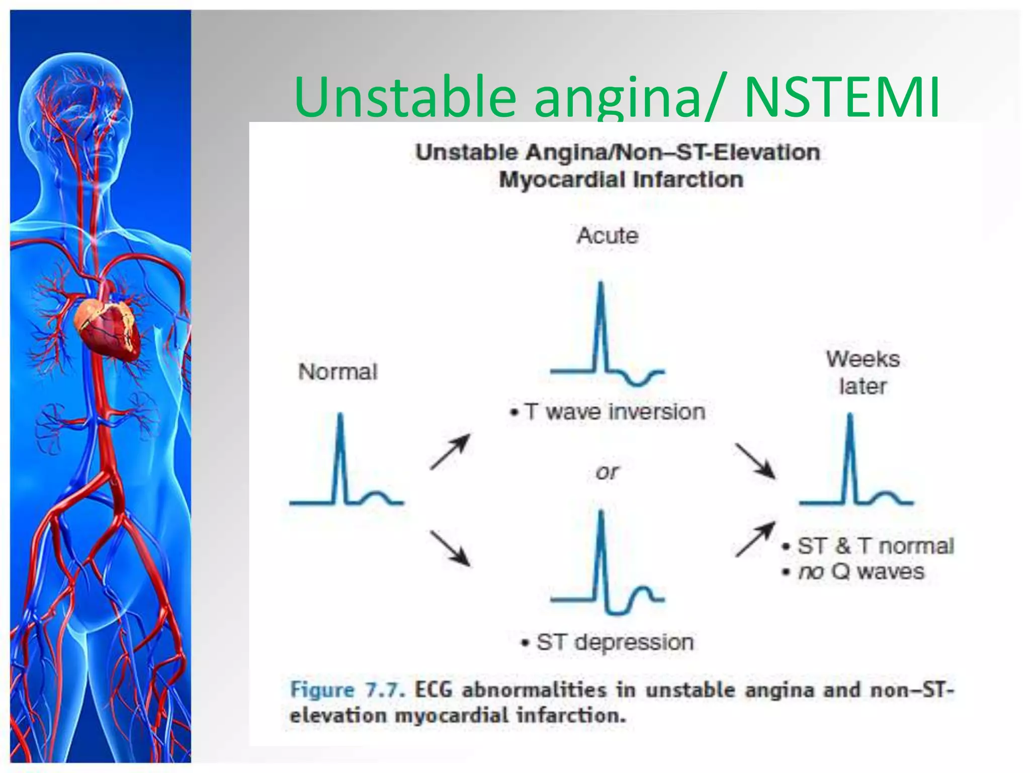
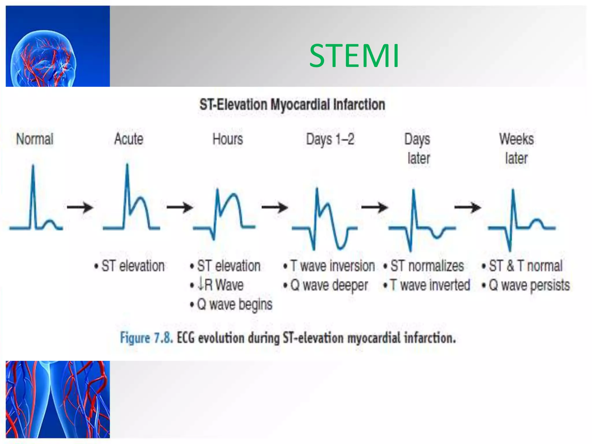
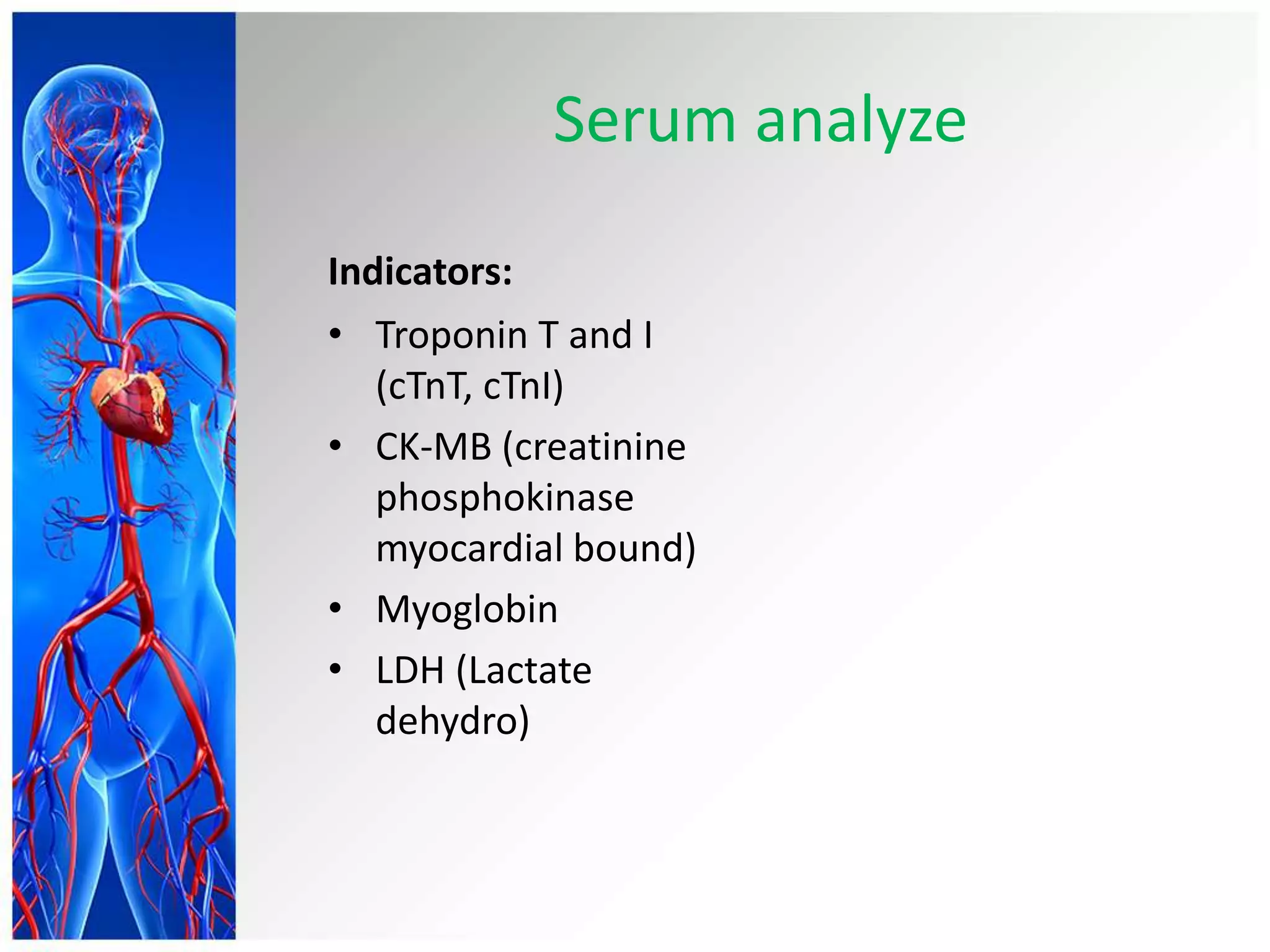
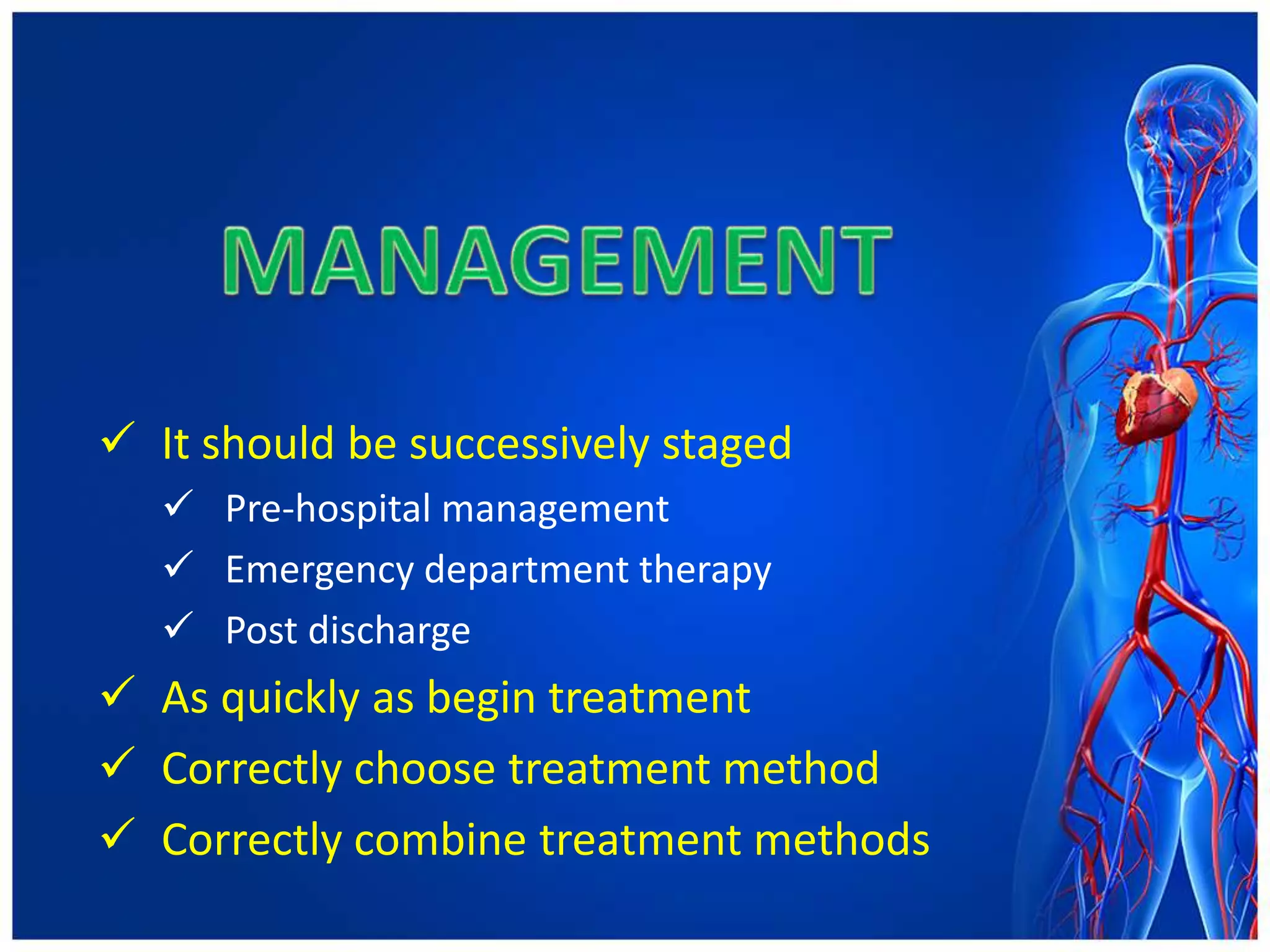
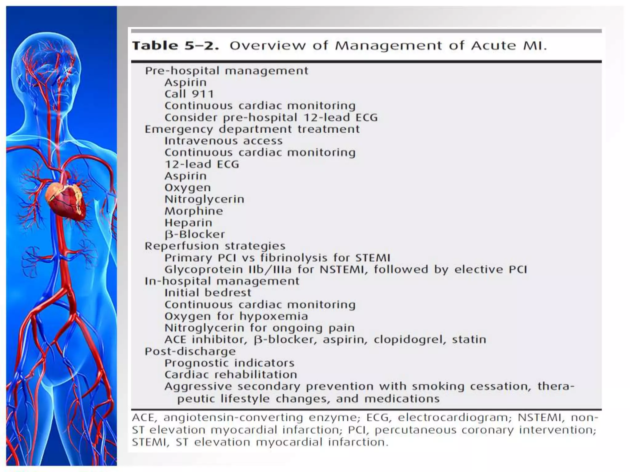
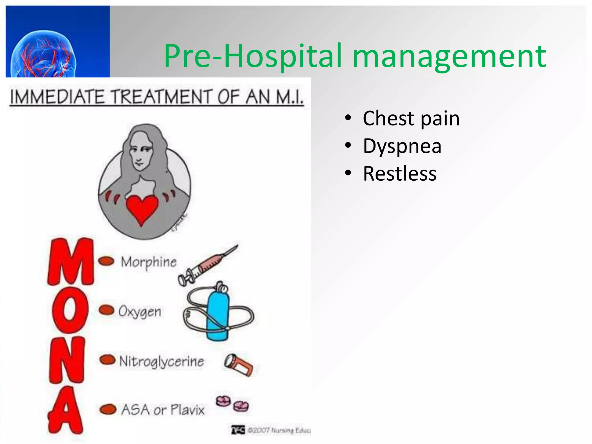
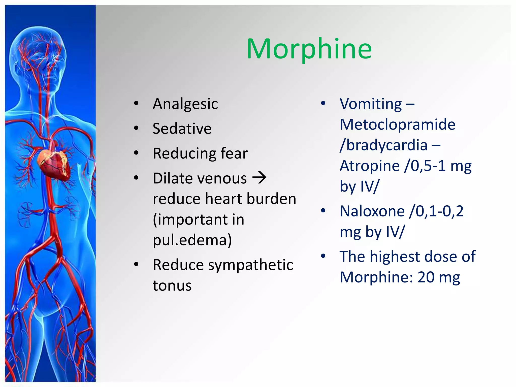
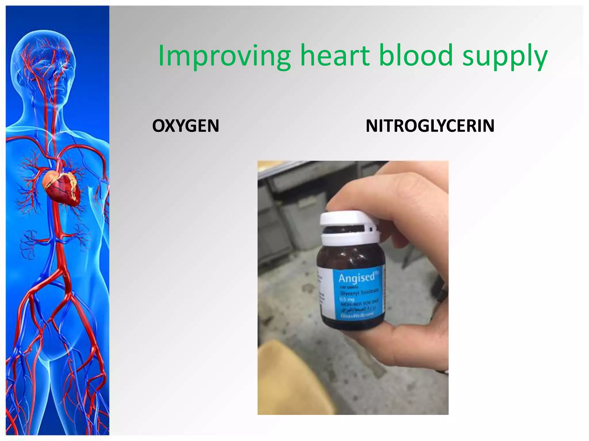
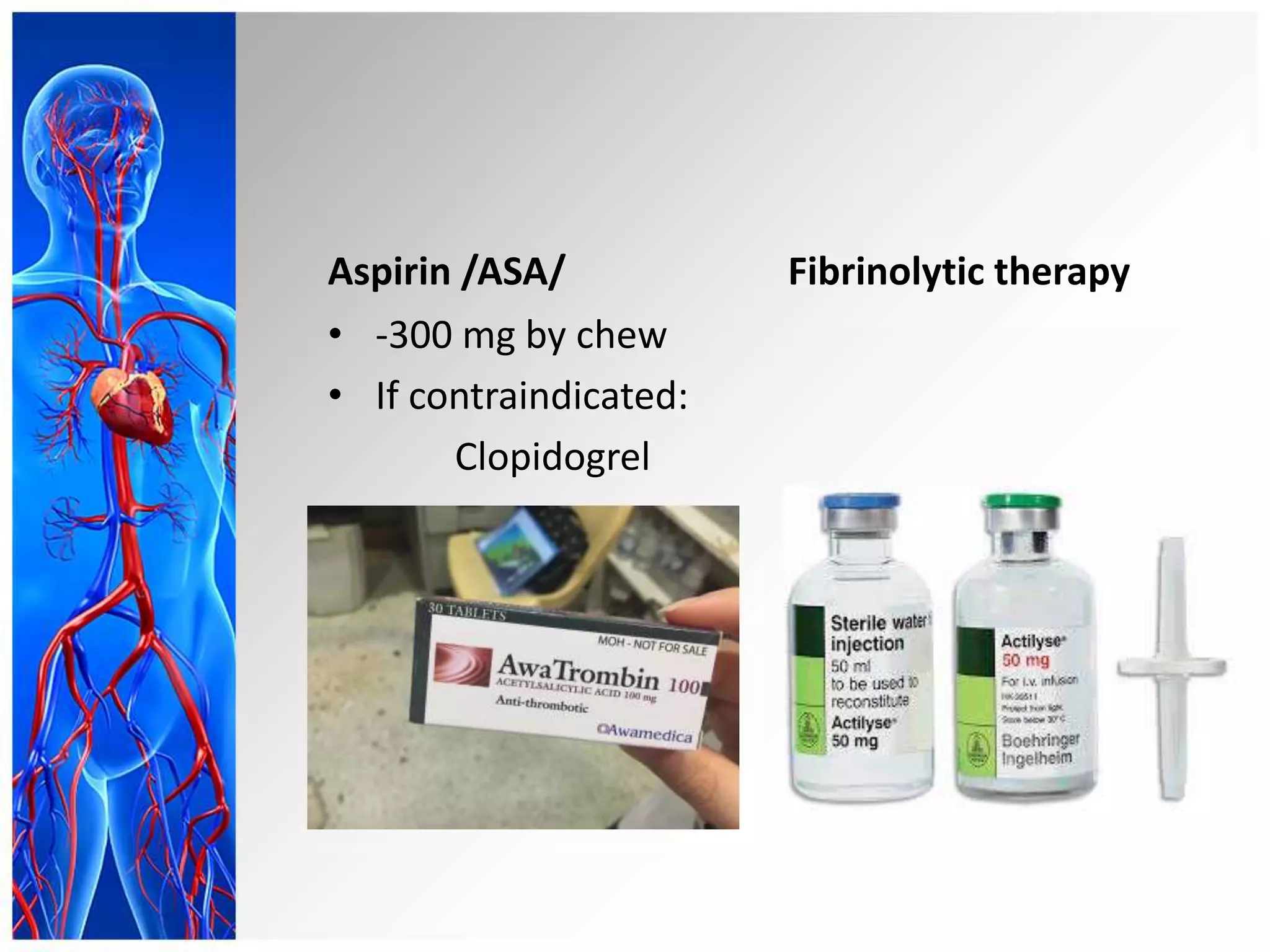
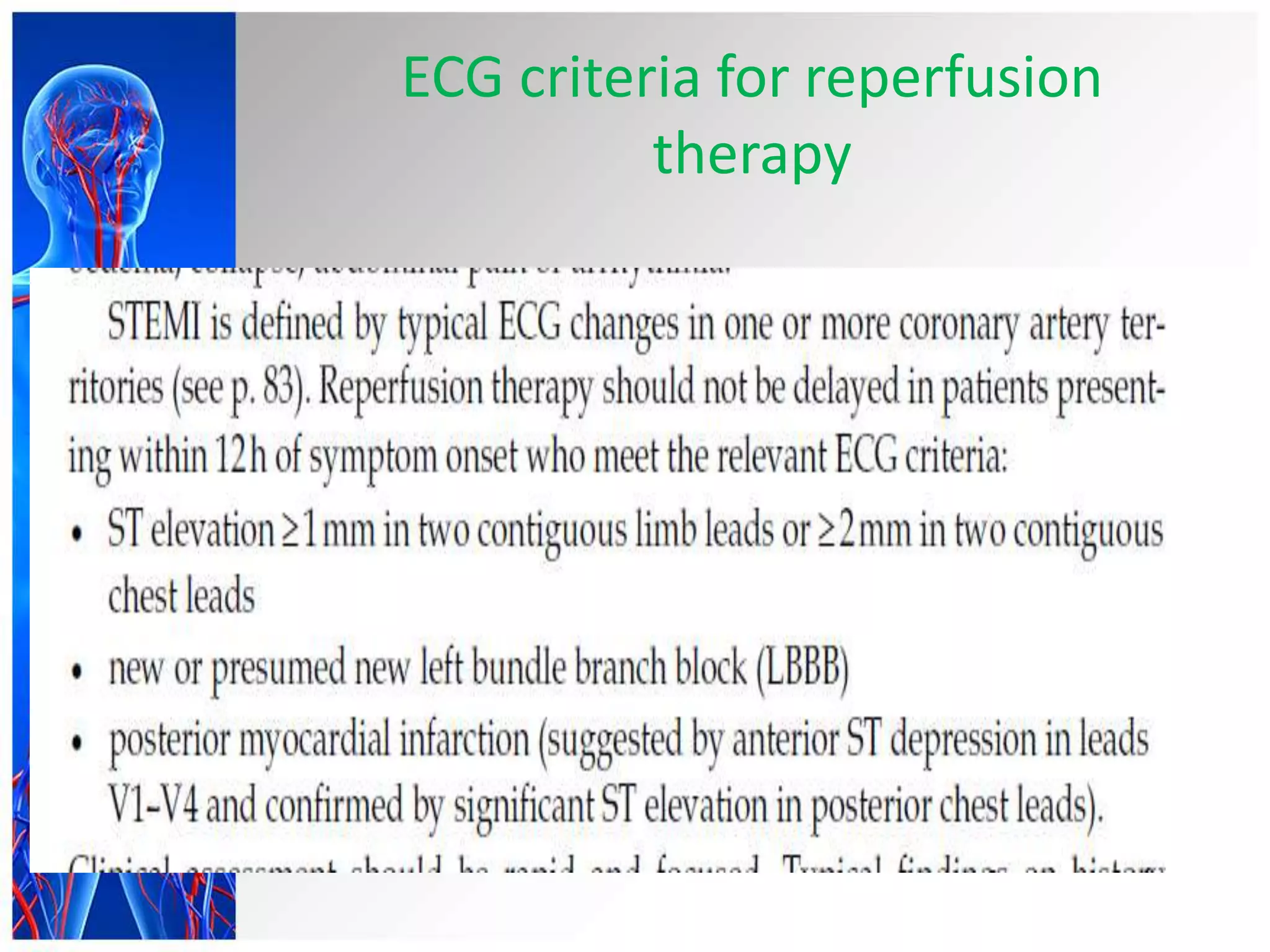
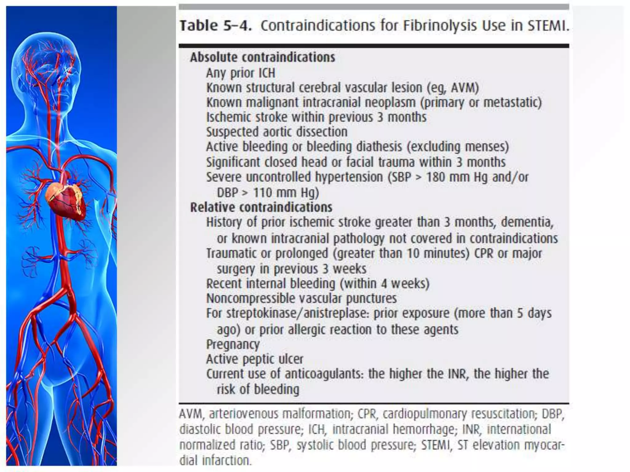
Acute
Myocardial
Infarction
.Done By :- marwa Abbass
Outline
• Introduction
• Etiology and risk factors
• Pathogenesis
• Classification
• Diagnosis
• Management
Definition
• Acute myocardial infarction (MI) is a
clinical syndrome that results from
occlusion of a coronary artery, with
resultant death of cardiac myocytes in
the region supplied by that artery.
Defined by “Current diagnosis and treatment in Cardiology - 2013”
Definition
• Acute Coronary Syndrome is
following disruption of a vulnerable
plaque, patients experience
ischemic discomfort resulting from
a reduction of flow through the
affected epicardial coronary artery.
– STEMI
– NSTEMI
– Unstable angina
• Defined by: Harrison’s Cardiovascular medicine
Acute Coronary Syndrome
Resource: Harrison’s Cardiovascular medicine
Causes of Acute Coronary
Syndromes
• Atherosclerotic plaque rupture with superimposed
thrombus/95%/
• Vasculitic syndromes
• Coronary embolism (e.g., from endocarditis, artificial heart
valves)
• Coronary trauma or aneurysm
• Severe coronary artery spasm (primary or cocaine-induced)
• Increased blood viscosity (e.g., polycythemia vera,
thrombocytosis)
• Markedly increased myocardial oxygen demand (e.g., severe
aortic stenosis)
Risk factors
• American Heart Association guide to risk factors for
coronary artery disease.
• Resource: “Cardiology explained”
Risk factors
European Society of Cardiology table of lifestyles and characteristics
associated with an increased risk of a future coronary heart disease event.
Resource: “Cardiology explained”
Main
• STEMI
• NSTEMI/Unstable angina
By necrotic area:
By progress:
 Initial period: (<6 hours)
 Acute period: (6h – 7 days)
 Recovering period: (7-28 days)
 Convalescence period: (≥29 days)
• Anamnesis
• Signs and symptoms
• ECG
• Serum analyze
• Echocardiography
Chest pain of MI
Pain
indicators
Description
Location Behind side of sternum, left side of chest
Radiation Left arm, jaw, neck
Characterizes Pressure, dull, squeezing, aching, crushing,
burning /elephant sitting in the chest/
Duration >10-20mins
Relieving
factor
No abatement of nitroglycerin, relieved
with analgesic/morphine/
Associated
symptoms
Weakness, dyspnea, fainting fit/syncope/
Cold sweat, apprehensive.
Dyspnea, orthopnea, cough, wheezing,
nausea and vomiting, or abdominal
bloating
Occurs at rest, more commonly in the early morning
Painless infarction
• One-third of patients with acute myocardial
infarction present without chest pain, and
these patients tend to be undertreated and
have poor outcomes.
• Older patients, women, and patients with
diabetes mellitus are more likely to present
without classic chest pain. As many as 25%
of infarctions are detected on routine ECG
without any recallable acute episode.
General signs
• Patients may appear anxious and sometimes
are sweating profusely.
• The heart rate may range from marked
bradycardia (most commonly in inferior
infarction) to tachycardia, low cardiac output,
or arrhythmia.
• The BP may be high, especially in former
hypertensive patients, or low in patients with
shock.
• Respiratory distress usually indicates heart
failure.
• Fever, usually low grade, may appear after 12
hours and persist for several days.
ECG
ECG early changes
• Presence of MI
• QRS complex, ST segment, T waves
are changed
• Tall T wave in contiguous 2 or more
leads
Unstable angina/ NSTEMI
STEMI
Serum analyze
Indicators:
• Troponin T and I
(cTnT, cTnI)
• CK-MB (creatinine
phosphokinase
myocardial bound)
• Myoglobin
• LDH (Lactate
dehydro)
Criteria of MI
• Troponin or CK-MB increased with one
of these:
– Chest pain
– Positive ischemic change in ECG (ST
segment and T wave’s changes)
– Pathological Q wave presence in ECG newly
– Patient had CABG
– Detected pathological changes in autopsy
Assessment of risk
/patient with MI/
Assessment of risk
• Total score is TIMI /Thrombolysis in
myocardial infarction/ assessment’s
score
• Total scores:
– 2-1: Low risk
– 4-3: Moderate risk
– 7-5: High risk
• TIMI is important for deciding esp.
NSTEMI prognosis
 It should be successively staged
 Pre-hospital management
 Emergency department therapy
 Post discharge
 As quickly as begin treatment
 Correctly choose treatment method
 Correctly combine treatment methods
Pre-Hospital management
• Chest pain
• Dyspnea
• Restless
Morphine
• Analgesic
• Sedative
• Reducing fear
• Dilate venous 
reduce heart burden
(important in
pul.edema)
• Reduce sympathetic
tonus
• Vomiting –
Metoclopramide
/bradycardia –
Atropine /0,5-1 mg
by IV/
• Naloxone /0,1-0,2
mg by IV/
• The highest dose of
Morphine: 20 mg
Improving heart blood supply
OXYGEN NITROGLYCERIN
Aspirin /ASA/
• -300 mg by chew
• If contraindicated:
Clopidogrel
Fibrinolytic therapy
ECG criteria for reperfusion
therapy
Reference
• “Davidson’s Foundations
• “Cardiology explained” Euan A Ashley and Josef Niebauer
• “Current diagnosis and treatment in Cardiology” Michael
H. Crawford, 3rd edition
• “Current Medical diagnosis and treatment – 2013” Maxine
A. Papadakis, Stephen J. McPhee
• “Harrison’s Cardiovascular medicine” Joseph Loscalzo, 17th
edition
• “Pathophysiology of heart” Leonard S. Lilly, 5th edition
• “Pathophysiology concepts of Altered Health States” Carol
Mattson Porth, 7th edition

Myocardia linfarction

  • 1.
  • 2.
    Outline • Introduction • Etiologyand risk factors • Pathogenesis • Classification • Diagnosis • Management
  • 3.
    Definition • Acute myocardialinfarction (MI) is a clinical syndrome that results from occlusion of a coronary artery, with resultant death of cardiac myocytes in the region supplied by that artery. Defined by “Current diagnosis and treatment in Cardiology - 2013”
  • 4.
    Definition • Acute CoronarySyndrome is following disruption of a vulnerable plaque, patients experience ischemic discomfort resulting from a reduction of flow through the affected epicardial coronary artery. – STEMI – NSTEMI – Unstable angina • Defined by: Harrison’s Cardiovascular medicine
  • 5.
    Acute Coronary Syndrome Resource:Harrison’s Cardiovascular medicine
  • 6.
    Causes of AcuteCoronary Syndromes • Atherosclerotic plaque rupture with superimposed thrombus/95%/ • Vasculitic syndromes • Coronary embolism (e.g., from endocarditis, artificial heart valves) • Coronary trauma or aneurysm • Severe coronary artery spasm (primary or cocaine-induced) • Increased blood viscosity (e.g., polycythemia vera, thrombocytosis) • Markedly increased myocardial oxygen demand (e.g., severe aortic stenosis)
  • 7.
    Risk factors • AmericanHeart Association guide to risk factors for coronary artery disease. • Resource: “Cardiology explained”
  • 8.
    Risk factors European Societyof Cardiology table of lifestyles and characteristics associated with an increased risk of a future coronary heart disease event. Resource: “Cardiology explained”
  • 10.
  • 11.
  • 12.
    By progress:  Initialperiod: (<6 hours)  Acute period: (6h – 7 days)  Recovering period: (7-28 days)  Convalescence period: (≥29 days)
  • 13.
    • Anamnesis • Signsand symptoms • ECG • Serum analyze • Echocardiography
  • 14.
    Chest pain ofMI Pain indicators Description Location Behind side of sternum, left side of chest Radiation Left arm, jaw, neck Characterizes Pressure, dull, squeezing, aching, crushing, burning /elephant sitting in the chest/ Duration >10-20mins Relieving factor No abatement of nitroglycerin, relieved with analgesic/morphine/ Associated symptoms Weakness, dyspnea, fainting fit/syncope/ Cold sweat, apprehensive. Dyspnea, orthopnea, cough, wheezing, nausea and vomiting, or abdominal bloating Occurs at rest, more commonly in the early morning
  • 15.
    Painless infarction • One-thirdof patients with acute myocardial infarction present without chest pain, and these patients tend to be undertreated and have poor outcomes. • Older patients, women, and patients with diabetes mellitus are more likely to present without classic chest pain. As many as 25% of infarctions are detected on routine ECG without any recallable acute episode.
  • 16.
    General signs • Patientsmay appear anxious and sometimes are sweating profusely. • The heart rate may range from marked bradycardia (most commonly in inferior infarction) to tachycardia, low cardiac output, or arrhythmia. • The BP may be high, especially in former hypertensive patients, or low in patients with shock. • Respiratory distress usually indicates heart failure. • Fever, usually low grade, may appear after 12 hours and persist for several days.
  • 17.
  • 18.
    ECG early changes •Presence of MI • QRS complex, ST segment, T waves are changed • Tall T wave in contiguous 2 or more leads
  • 19.
  • 20.
  • 21.
    Serum analyze Indicators: • TroponinT and I (cTnT, cTnI) • CK-MB (creatinine phosphokinase myocardial bound) • Myoglobin • LDH (Lactate dehydro)
  • 22.
    Criteria of MI •Troponin or CK-MB increased with one of these: – Chest pain – Positive ischemic change in ECG (ST segment and T wave’s changes) – Pathological Q wave presence in ECG newly – Patient had CABG – Detected pathological changes in autopsy
  • 23.
  • 24.
    Assessment of risk •Total score is TIMI /Thrombolysis in myocardial infarction/ assessment’s score • Total scores: – 2-1: Low risk – 4-3: Moderate risk – 7-5: High risk • TIMI is important for deciding esp. NSTEMI prognosis
  • 25.
     It shouldbe successively staged  Pre-hospital management  Emergency department therapy  Post discharge  As quickly as begin treatment  Correctly choose treatment method  Correctly combine treatment methods
  • 27.
    Pre-Hospital management • Chestpain • Dyspnea • Restless
  • 28.
    Morphine • Analgesic • Sedative •Reducing fear • Dilate venous  reduce heart burden (important in pul.edema) • Reduce sympathetic tonus • Vomiting – Metoclopramide /bradycardia – Atropine /0,5-1 mg by IV/ • Naloxone /0,1-0,2 mg by IV/ • The highest dose of Morphine: 20 mg
  • 29.
    Improving heart bloodsupply OXYGEN NITROGLYCERIN
  • 30.
    Aspirin /ASA/ • -300mg by chew • If contraindicated: Clopidogrel Fibrinolytic therapy
  • 31.
    ECG criteria forreperfusion therapy
  • 33.
    Reference • “Davidson’s Foundations •“Cardiology explained” Euan A Ashley and Josef Niebauer • “Current diagnosis and treatment in Cardiology” Michael H. Crawford, 3rd edition • “Current Medical diagnosis and treatment – 2013” Maxine A. Papadakis, Stephen J. McPhee • “Harrison’s Cardiovascular medicine” Joseph Loscalzo, 17th edition • “Pathophysiology of heart” Leonard S. Lilly, 5th edition • “Pathophysiology concepts of Altered Health States” Carol Mattson Porth, 7th edition

Editor's Notes

  • #6 The flow reduction may be caused by a completely occlusive thrombus (right) or subtotally occlusive thrombus (left). Patients with ischemic discomfort may present with or without ST-segment elevation. Of patients with ST-segment elevation, the majority (wide red arrow) ultimately develop a Q-wave on the ECG (QwMI), while a minority (thin red arrow) do not develop Q-wave and in older literature where said to have sustained a non-Q-wave MI (NQMI). Patients who present without ST-segment elevation are suffering from either unstable angina or a non-ST-segment elevation MI (NSTEMI) (wide green arrows), a distinction that is ultimately made on the presence or absence of a serum cardiac marker such as CK-MB or a cardiac troponin detected in the blood. The majority of patients presenting with NSTEMI do not develop a Q-wave on the ECG; a minority develop a QwMI (thin green arrow).