SlideShare a Scribd company logo
1 of 15
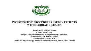
INVESTIGATIVE PROCEDURES USED IN PATIENTS
WITH CARDIAC DISEASES
Submitted by – Iffat Parveen
Class – Bpt 4th year
Subject – Physiotherapy in Cardiopulmonary Conditions
Submitted to – Dr. Jamal Ali Moiz
Submitted on – 19-01-2020
Centre for physiotherapy and Rehabilitation Sciences, Jamia Millia Islamia
• Cardiovascular diseases are diagnosed using an array of laboratory tests and imaging studies. The primary
part of diagnosis is medical and family histories of the patient, risk factors, physical examination and
coordination of these findings with the results from tests and procedures.
• Some of the common tests used to diagnose cardiovascular diseases include:
1. Blood test
2. Chest X-ray
3. ECG
4. Stress testing
5. ECHO
6. CT
7. MRI
Blood Tests - Haematology and clinical chemistry
• When the muscle has been damaged, as in a heart attack, body releases substances in blood. Blood tests can
measure the substances and show if, and how much of, heart muscle has been damaged.
• Blood tests are also done to measure the level of other substances in blood, such as blood fats (e.g. cholesterol
and triglycerides), vitamins and minerals. Blood sample is taken from a vein in your arm. A laboratory then
tests it and sends the results to the doctor, who will explain the results.
• As anaemia can unmask angina or exacerbate heart failure, a full blood count is useful and helps guide the
safe use of antiplatelet therapies and anticoagulants.
• Measurement of the erythrocyte sedimentation rate and serology are indicated if connective tissue disease is
suspected.
• Urea and electrolytes are measured and liver function tests performed prior to starting therapies that may
impact on renal function or cause hepatotoxicity.
• Blood glucose and a lipid profile help identify patients with diabetes mellitus and assess cardiovascular risk.
C-reactive protein (CRP) and other protein markers like Apolipoprotein A1 and B are used to detect
inflammation that may lead to heart diseases
• In patients with acute chest pain, cardiac troponin is measured to determine whether there is myocardial injury
or infarction. Cardiac Troponin-T is one of the markers of heart attack.
Chest X-Ray (CXR)
A chest x-ray aides in the differentiation between respiratory and
cardiac causes of dyspnoea. In those with heart failure, common
findings include cardiomegaly, interstitial oedema, pulmonary
oedema and pleural effusions. Evidence of surgery (eg CABG,
valve repair, ICD implantation) is also detected on CXR.
• This is a test that shows the shape and size of the heart lungs
and major blood vessels. This test is seldom used in diagnosis
of heart diseases as it does not provide added information over
echocardiography and other imaging studies.
• The maximum width of the heart divided by the maximum
width of the thorax on a posteroanterior chest X-ray (the
cardiothoracic ratio) should normally be <0.5. An increased
cardiothoracic ratio is common in valvular heart disease and
heart failure. In heart failure this is often accompanied by
distension of the upper lobe pulmonary veins, diffuse
shadowing within the lungs due to pulmonary oedema, and
Kerley B lines (horizontal, engorged lymphatics at the
periphery of the lower lobes).
• A widened mediastinum may indicate a thoracic aneurysm.
Fig: Chest X-ray in heart failure. This shows
cardiomegaly with patchy alveolar shadowing of
pulmonary oedema and Kerley B lines (engorged
lymphatics, arrow) at the periphery of both lungs.
Electrocardiography (ECG)
• This is a simple and a painless test that records the heart’s electrical activity. The patient is strapped to the
instrument with several patches or leads placed over his or her chest, wrists and ankles. A small portable
machine records the activities of the heart on a strip of graph paper.
• It is a simple test that identifies heart rate, conduction disturbances, myocardial ischaemia and possible
structural defects.ECG aids in the diagnosis of underlying causes of heart disease such as coronary artery
disease or arrhythmias.
• Tall p wave – p pulmonale(right atrial enlargement), Widened p wave – p mitrale( left atrial enlargement)
• PR interval – normal range (0.12-0.20 sec). If > 0.20 sec– first degree heart block, if < 0.12 sec – Wolfe
parckinson wide syndrome
• QT interval – normal range(0.36-0.44 sec).If prolonged,indicative of hypokalemia,hypocalcemia.
• QRS complex duration normal is < 0.11 sec. If > 0.11 sec – bundle branch block
• ST segment elevation or depression may represent ischaemia or infarction.
• Large voltage QRS complexes, downward sloping ST segments and T wave inversion may represent chamber
hypertrophy.
• Rhythm disturbances such as atrial arrhythmias, heart block and intraventricular septal conduction delays are
common in heart failure secondary to cardiac remodelling and may also exacerbate heart failure.
Ambulatory ECG monitoring
Continuous ECG recording over 24–48 hours can be used
to identify symptomatic or asymptomatic rhythm
disturbances in patients with palpitation or syncope. If
symptoms are less frequent, it may be necessary to use
patient-activated recorders that record the heart rhythm
only when the patient is symptomatic; the device is
activated by the patient
Fig: Electrocardiography (ECG). A 12-lead ECG
lead placement. B Normal PQRST complex.C
Acute anterior myocardial infarction. Note the ST
elevation in leads V1–V6 and aVL, and
‘reciprocal’ ST depression in leads II, III and aVF.
Fig:Printout from a 24-hour ambulatory electrocardiogram
recording, showing complete heart block. Arrows indicate
visible P waves. At times, these are masked by the QRS
complex or T wave.
Exercise Stress Testing
• For this test, the patient is made to work hard e.g. run on a
treadmill or exercise while the leads of ECG are placed over their
body. The test detects the effects of the exercise on the heart.
• Exercise stress testing may identify myocardial ischaemia,
haemodynamic/ electrical instability, or other exertion-related signs
or symptoms.
- Note: cardiac “stress” may also be induced using medications, when
an individual is unable to perform the exercise test as required.
• An exercise ECG is useful in the diagnosis and functional
assessment of patients with suspected coronary artery disease.
Down-sloping ST segment depression, particularly when it occurs
during minor exertion.
• In patients with athereosclerosis and coronary heart diseases
the arteries that are narrowed by plaques cannot supply
adequate blood to the heart muscles while it is beating faster.
This may lead to shortness of breath and chest pain. The ECG
pattern, arrhythmias etc. also show the possibility of a
coronary artery disease.
Echocardiography
• Another noninvasive method of evaluating the heart uses the physical properties of sound & uses sound
waves to create a moving picture of the heart. In this test, a probe is rolled over the chest and the machine
creates the image of the heart on the monitor. This provides information on the blood flow,
structure,functions, valves and chambers of the heart. Motion mode (M-mode) and two-dimensional (2-0)
images are highly reliable methods of evaluation.
- Stress echocardiography
• Stress echo assesses patients with suspected or known myocardial ischaemia. Exercise or medication is
used to stress the heart. Cardiac function is then evaluated using echocardiography pre and immediately
post stress. Myocardial response may be described as
• hypokinetic (decreased), dyskinetic (impaired) or akinetic (absent).
• This test is valuable in assessment of
viable/ischaemic myocardium in known
CVD being considered for revascularisation.
Radionuclide studies - Nuclear cardiac stress test
• This test is sometimes called an ‘exercise thallium scan’, a ‘dual isotope treadmill’ or an ‘exercise nuclear
scan’.
• A radioactive substance called a ‘tracer’(Technetium-99) is injected intravenously into bloodstream and
detected using a gamma camera to assess left ventricular function.
• The doctor uses this picture to see how much blood flows to your heart muscle and how well your heart
pumps blood when you are resting and doing physical activity. This test also helps the doctor to see if heart
muscle is damaged. Thallium and sestamibi are taken up by myocardial cells and indicate myocardial
perfusion at rest and exercise.
Computed Tomography
• A computed tomography (CT) scan of the chest provides a series of cross-sectional x-ray images. CT helps to
detect the calcium deposits or calcifications in the walls of the coronary arteries. These are early markers of
atherosclerosis and coronary heart disease. This is not a routine test in coronary heart disease
• CT scan has been found to be a useful first-line diagnostic tool in the evaluation of mediastinal disease,
including mediastinal masses, staging of mediastinal cancers, and identification of cysts (Lillington, 1987).
• It provides identification of anatomical abnormalities such as aneurysms or valve dysfunction, as well as
providing information about pulmonary vein anatomy. Cardiac CT also provides information about patency of
grafts following CABG.
• Computed tomography (CT), with its superior temporal resolution of the coronary arteries, is particularly
useful for investigating symptomatic patients at low to intermediate risk of coronary artery disease. It can
reduce the need for invasive investigation in patients with a low probability of occlusive coronary disease
who require valve surgery.
Coronary Angiography and Cardiac Catheterization
It is an invasive test, with a small but serious risk to the patient.
• Coronary angiography investigates integrity of coronary arteries by insertion of a catheter into the coronary
vasculature and the use of dye to produce the image. Coronary angiography detects blockages in the large
coronary arteries.The presence, location and extent of vessel narrowing is identified on the image and likely
sources of symptoms (“culprit lesions”) may be identified.
• The cardiac catheterization laboratory requires an x-ray generator tube and image intensifier/cine camera.
Data are stored using 35mm file (Reagan, Boxt, and Katz, 1994). The procedure for cardiac catheterization of
the left side of the heart requires the threading of a catheter, guided by fluoroscopy, through the femoral artery
or brachial artcry 10 thc aorta. Direct measurements of chamber pressures, blood flow, and oxygen saturation
can be obtained.
• A fine catheter is introduced under local anaesthetic via a peripheral artery (usually the radial or femoral) and
advanced to the heart under X-ray guidance. Although measurements of intracardiac pressures, and therefore
estimates of valvular and cardiac function, are possible, the primary application of this technique is imaging
of the coronary circulation using contrast
• Cardiac ventriculography is when dye is injected
into the ventricle and the entire chamber becomes
outlined. Several cardiac cycles are recorded on
cine. It provides valuable information about
global and segmental wall motion, valve motion,
and the presence of abnormal anatomy
(Grossman, 1986).
• When selective coronary arteriography is
performed, radiopaque contrast material is
injected into the left main or right coronary artery.
Cine recordings are made after the injection, and
injections are repeated until the entire coronary
tree is visualized.
Cardiac Magnetic Resonance Imaging
(MRI)
• Cardiac MRI uses high intensity magnetic fields and radiofrequency and a computer to produce images with
high resolution. This gives a 3D image of the moving as well as still pictures of the heart.
• The image provides accurate information about cardiac volumes, muscle mass, contractility, tissue scarring
and ejection fraction. Location and size of myocardial infarction can be described with precision and may
provide useful information regarding patency of bypass grafts.
• The image can identify regional wall motion abnormalities (RWMA) and wall dyssynchrony, valve sclerosis,
stenosis or regurgitation and provide information regarding myocardial fibrosis and assists in the diagnosis of
amyloid cardiomyopathy, myocarditis and cardiac sarcoid.
• Magnetic resonance imaging (MRI) provides superior
spatial resolution and is the imaging modality of choice
for investigating the aetiology of heart muscle diseases
(cardiomyopathy).
Thankyou
References:
• Donna Frownfelter & Elizabeth Dean Principle and practice of Cardiopulmonary Physical
Therapy, 3rd edition,
• J Alastair Innes, Anna R Dover, Karen Fairhurst Macleod’s Clinical examination 14th edition.
• Atul Luthra. ECG made easy, 4tg edition.Jaypee.
• www.heartonline.org.au/resources
• https://www.news-medical.net/health/Cardiovascular-Disease-Diagnosis.aspx
• https://www.heart.org/en/health-topics/heart-attack/diagnosing-a-heart-attack/noninvasive-tests-
and-procedures
• https://healthywa.wa.gov.au/Articles/A_E/Common-medical-tests-to-diagnose-heart-conditions

More Related Content

What's hot (20)

Cardiac stress test
Cardiac stress testCardiac stress test
Cardiac stress test
 
Breathing ex
Breathing exBreathing ex
Breathing ex
 
EET (Exercise Tolerance Test )
EET (Exercise Tolerance Test )EET (Exercise Tolerance Test )
EET (Exercise Tolerance Test )
 
Abnormal breathing pattern
Abnormal breathing patternAbnormal breathing pattern
Abnormal breathing pattern
 
Cardiac rehabilitation
Cardiac rehabilitation Cardiac rehabilitation
Cardiac rehabilitation
 
Cardiac tamponade
Cardiac tamponadeCardiac tamponade
Cardiac tamponade
 
Myocardial infarction
Myocardial infarctionMyocardial infarction
Myocardial infarction
 
Heart valve
Heart valveHeart valve
Heart valve
 
Diagnostic tests in cardiology
Diagnostic tests in cardiologyDiagnostic tests in cardiology
Diagnostic tests in cardiology
 
Cardiac surgeries
Cardiac surgeriesCardiac surgeries
Cardiac surgeries
 
Myocarditis
MyocarditisMyocarditis
Myocarditis
 
2. investigation of cardiovascular system )2(
2. investigation of cardiovascular system )2(2. investigation of cardiovascular system )2(
2. investigation of cardiovascular system )2(
 
Cardiac Stress Testing Seminar
Cardiac Stress Testing Seminar Cardiac Stress Testing Seminar
Cardiac Stress Testing Seminar
 
coronary artery disease
coronary artery diseasecoronary artery disease
coronary artery disease
 
Lobectomy
LobectomyLobectomy
Lobectomy
 
Cardiac rehabilitation
Cardiac rehabilitationCardiac rehabilitation
Cardiac rehabilitation
 
Cardiac assessment
Cardiac assessmentCardiac assessment
Cardiac assessment
 
exercise tolerance test.pptx
exercise tolerance test.pptxexercise tolerance test.pptx
exercise tolerance test.pptx
 
Breathing patterns
Breathing patternsBreathing patterns
Breathing patterns
 
Incentive spirometry ppt
Incentive spirometry pptIncentive spirometry ppt
Incentive spirometry ppt
 

Similar to Investigative procedures in cardiac diseases.

Investigation of cardiovascular diseases
Investigation of cardiovascular diseasesInvestigation of cardiovascular diseases
Investigation of cardiovascular diseasesmushfiq1970
 
Cardiac diagnostics and laboratory tests
Cardiac diagnostics and laboratory tests Cardiac diagnostics and laboratory tests
Cardiac diagnostics and laboratory tests Dr. Rima Das
 
Diagnostic measures in cardiology
Diagnostic measures in cardiologyDiagnostic measures in cardiology
Diagnostic measures in cardiologySUDESHNA BANERJEE
 
Preoperative management
Preoperative managementPreoperative management
Preoperative managementAbeer Nakera
 
invasive non invasive procedures.pdf for bsc nursing students
invasive non invasive procedures.pdf for bsc nursing studentsinvasive non invasive procedures.pdf for bsc nursing students
invasive non invasive procedures.pdf for bsc nursing studentsshanmukhadevi
 
Pedia Cardiac Imaging by Caffey.pptx
Pedia Cardiac Imaging by Caffey.pptxPedia Cardiac Imaging by Caffey.pptx
Pedia Cardiac Imaging by Caffey.pptxFreceGaming1
 
The role of Echocardiography In coronary artery disease and Acute Myocardial...
The role of Echocardiography In  coronary artery disease and Acute Myocardial...The role of Echocardiography In  coronary artery disease and Acute Myocardial...
The role of Echocardiography In coronary artery disease and Acute Myocardial...Nizam Uddin
 
Diagnostic procedures for coronary heart disease
Diagnostic procedures for coronary heart diseaseDiagnostic procedures for coronary heart disease
Diagnostic procedures for coronary heart diseasetasha0203
 
non invasive and invasive cardiac monitoring.pptx
non invasive and invasive cardiac monitoring.pptxnon invasive and invasive cardiac monitoring.pptx
non invasive and invasive cardiac monitoring.pptxshafina27
 
1. Cardiovascular 2022 copy.pdf
1.  Cardiovascular 2022 copy.pdf1.  Cardiovascular 2022 copy.pdf
1. Cardiovascular 2022 copy.pdfShougAlmutairi
 
Cardiovascular Diagnostic Procedures by Nadia Abdulallah
Cardiovascular Diagnostic Procedures by Nadia AbdulallahCardiovascular Diagnostic Procedures by Nadia Abdulallah
Cardiovascular Diagnostic Procedures by Nadia AbdulallahNadia Abdulallah
 
CAD.pdf
CAD.pdfCAD.pdf
CAD.pdfDinu85
 
CSA cardiovascular /emergency /cardiovascular
CSA  cardiovascular /emergency /cardiovascularCSA  cardiovascular /emergency /cardiovascular
CSA cardiovascular /emergency /cardiovascularSaraRmk
 
Cardiovascular assessment and diagnostic investigations ppt slideshare
Cardiovascular assessment and diagnostic investigations ppt slideshareCardiovascular assessment and diagnostic investigations ppt slideshare
Cardiovascular assessment and diagnostic investigations ppt slidesharesonam
 

Similar to Investigative procedures in cardiac diseases. (20)

Investigation of cardiovascular diseases
Investigation of cardiovascular diseasesInvestigation of cardiovascular diseases
Investigation of cardiovascular diseases
 
Cardiac diagnostics and laboratory tests
Cardiac diagnostics and laboratory tests Cardiac diagnostics and laboratory tests
Cardiac diagnostics and laboratory tests
 
Diagnostic measures in cardiology
Diagnostic measures in cardiologyDiagnostic measures in cardiology
Diagnostic measures in cardiology
 
Preoperative management
Preoperative managementPreoperative management
Preoperative management
 
invasive non invasive procedures.pdf for bsc nursing students
invasive non invasive procedures.pdf for bsc nursing studentsinvasive non invasive procedures.pdf for bsc nursing students
invasive non invasive procedures.pdf for bsc nursing students
 
Cardiovascular testing 1
Cardiovascular testing 1Cardiovascular testing 1
Cardiovascular testing 1
 
Cvs6 cvs
Cvs6 cvsCvs6 cvs
Cvs6 cvs
 
Pedia Cardiac Imaging by Caffey.pptx
Pedia Cardiac Imaging by Caffey.pptxPedia Cardiac Imaging by Caffey.pptx
Pedia Cardiac Imaging by Caffey.pptx
 
Stemi in the young
Stemi in the youngStemi in the young
Stemi in the young
 
Cardiology 101 back to the basics
Cardiology 101 back to the basicsCardiology 101 back to the basics
Cardiology 101 back to the basics
 
The role of Echocardiography In coronary artery disease and Acute Myocardial...
The role of Echocardiography In  coronary artery disease and Acute Myocardial...The role of Echocardiography In  coronary artery disease and Acute Myocardial...
The role of Echocardiography In coronary artery disease and Acute Myocardial...
 
Diagnostic procedures for coronary heart disease
Diagnostic procedures for coronary heart diseaseDiagnostic procedures for coronary heart disease
Diagnostic procedures for coronary heart disease
 
non invasive and invasive cardiac monitoring.pptx
non invasive and invasive cardiac monitoring.pptxnon invasive and invasive cardiac monitoring.pptx
non invasive and invasive cardiac monitoring.pptx
 
Myocardial infraction
Myocardial infractionMyocardial infraction
Myocardial infraction
 
1. Cardiovascular 2022 copy.pdf
1.  Cardiovascular 2022 copy.pdf1.  Cardiovascular 2022 copy.pdf
1. Cardiovascular 2022 copy.pdf
 
Cardiovascular Diagnostic Procedures by Nadia Abdulallah
Cardiovascular Diagnostic Procedures by Nadia AbdulallahCardiovascular Diagnostic Procedures by Nadia Abdulallah
Cardiovascular Diagnostic Procedures by Nadia Abdulallah
 
CAD.pdf
CAD.pdfCAD.pdf
CAD.pdf
 
CSA cardiovascular /emergency /cardiovascular
CSA  cardiovascular /emergency /cardiovascularCSA  cardiovascular /emergency /cardiovascular
CSA cardiovascular /emergency /cardiovascular
 
Cardiovascular assessment and diagnostic investigations ppt slideshare
Cardiovascular assessment and diagnostic investigations ppt slideshareCardiovascular assessment and diagnostic investigations ppt slideshare
Cardiovascular assessment and diagnostic investigations ppt slideshare
 
CARDIO docx
CARDIO docxCARDIO docx
CARDIO docx
 

More from BPT4thyearJamiaMilli (20)

Humidification
Humidification Humidification
Humidification
 
Physiotherapy assessment of cardiac conditions
Physiotherapy assessment of cardiac conditionsPhysiotherapy assessment of cardiac conditions
Physiotherapy assessment of cardiac conditions
 
M mrc scale
M mrc scaleM mrc scale
M mrc scale
 
Monitoring system in icu
Monitoring system in icuMonitoring system in icu
Monitoring system in icu
 
Pft interpretation
Pft interpretationPft interpretation
Pft interpretation
 
Abg interpretation
Abg interpretation Abg interpretation
Abg interpretation
 
Cardiac auscultation
Cardiac auscultationCardiac auscultation
Cardiac auscultation
 
cases of ecg interpretation
 cases of ecg interpretation cases of ecg interpretation
cases of ecg interpretation
 
Cardiac axis
Cardiac axisCardiac axis
Cardiac axis
 
Chest auscultation
Chest auscultationChest auscultation
Chest auscultation
 
Placement of ecg leads during exercise (cardio ppt)
Placement of ecg leads during exercise (cardio ppt)Placement of ecg leads during exercise (cardio ppt)
Placement of ecg leads during exercise (cardio ppt)
 
Pt assessment
Pt assessment Pt assessment
Pt assessment
 
Acapella
AcapellaAcapella
Acapella
 
Pulmonary rehabilitation strength training
Pulmonary rehabilitation strength trainingPulmonary rehabilitation strength training
Pulmonary rehabilitation strength training
 
Cardiopulmonary sgrq questionnaire
Cardiopulmonary  sgrq questionnaireCardiopulmonary  sgrq questionnaire
Cardiopulmonary sgrq questionnaire
 
Nyha
NyhaNyha
Nyha
 
Pt assessment of cardiac surgery conditions
 Pt assessment of cardiac surgery conditions Pt assessment of cardiac surgery conditions
Pt assessment of cardiac surgery conditions
 
Cardiac arrhythmia.
Cardiac arrhythmia.Cardiac arrhythmia.
Cardiac arrhythmia.
 
Ecg placement resting
Ecg placement restingEcg placement resting
Ecg placement resting
 
Cardiopulmonary resucitation
Cardiopulmonary resucitationCardiopulmonary resucitation
Cardiopulmonary resucitation
 

Recently uploaded

Enzyme, Pharmaceutical Aids, Miscellaneous Last Part of Chapter no 5th.pdf
Enzyme, Pharmaceutical Aids, Miscellaneous Last Part of Chapter no 5th.pdfEnzyme, Pharmaceutical Aids, Miscellaneous Last Part of Chapter no 5th.pdf
Enzyme, Pharmaceutical Aids, Miscellaneous Last Part of Chapter no 5th.pdfSumit Tiwari
 
भारत-रोम व्यापार.pptx, Indo-Roman Trade,
भारत-रोम व्यापार.pptx, Indo-Roman Trade,भारत-रोम व्यापार.pptx, Indo-Roman Trade,
भारत-रोम व्यापार.pptx, Indo-Roman Trade,Virag Sontakke
 
ECONOMIC CONTEXT - LONG FORM TV DRAMA - PPT
ECONOMIC CONTEXT - LONG FORM TV DRAMA - PPTECONOMIC CONTEXT - LONG FORM TV DRAMA - PPT
ECONOMIC CONTEXT - LONG FORM TV DRAMA - PPTiammrhaywood
 
EPANDING THE CONTENT OF AN OUTLINE using notes.pptx
EPANDING THE CONTENT OF AN OUTLINE using notes.pptxEPANDING THE CONTENT OF AN OUTLINE using notes.pptx
EPANDING THE CONTENT OF AN OUTLINE using notes.pptxRaymartEstabillo3
 
Sanyam Choudhary Chemistry practical.pdf
Sanyam Choudhary Chemistry practical.pdfSanyam Choudhary Chemistry practical.pdf
Sanyam Choudhary Chemistry practical.pdfsanyamsingh5019
 
Paris 2024 Olympic Geographies - an activity
Paris 2024 Olympic Geographies - an activityParis 2024 Olympic Geographies - an activity
Paris 2024 Olympic Geographies - an activityGeoBlogs
 
Science 7 - LAND and SEA BREEZE and its Characteristics
Science 7 - LAND and SEA BREEZE and its CharacteristicsScience 7 - LAND and SEA BREEZE and its Characteristics
Science 7 - LAND and SEA BREEZE and its CharacteristicsKarinaGenton
 
Mastering the Unannounced Regulatory Inspection
Mastering the Unannounced Regulatory InspectionMastering the Unannounced Regulatory Inspection
Mastering the Unannounced Regulatory InspectionSafetyChain Software
 
How to Configure Email Server in Odoo 17
How to Configure Email Server in Odoo 17How to Configure Email Server in Odoo 17
How to Configure Email Server in Odoo 17Celine George
 
Computed Fields and api Depends in the Odoo 17
Computed Fields and api Depends in the Odoo 17Computed Fields and api Depends in the Odoo 17
Computed Fields and api Depends in the Odoo 17Celine George
 
The Most Excellent Way | 1 Corinthians 13
The Most Excellent Way | 1 Corinthians 13The Most Excellent Way | 1 Corinthians 13
The Most Excellent Way | 1 Corinthians 13Steve Thomason
 
Proudly South Africa powerpoint Thorisha.pptx
Proudly South Africa powerpoint Thorisha.pptxProudly South Africa powerpoint Thorisha.pptx
Proudly South Africa powerpoint Thorisha.pptxthorishapillay1
 
CARE OF CHILD IN INCUBATOR..........pptx
CARE OF CHILD IN INCUBATOR..........pptxCARE OF CHILD IN INCUBATOR..........pptx
CARE OF CHILD IN INCUBATOR..........pptxGaneshChakor2
 
Biting mechanism of poisonous snakes.pdf
Biting mechanism of poisonous snakes.pdfBiting mechanism of poisonous snakes.pdf
Biting mechanism of poisonous snakes.pdfadityarao40181
 
Crayon Activity Handout For the Crayon A
Crayon Activity Handout For the Crayon ACrayon Activity Handout For the Crayon A
Crayon Activity Handout For the Crayon AUnboundStockton
 
ENGLISH5 QUARTER4 MODULE1 WEEK1-3 How Visual and Multimedia Elements.pptx
ENGLISH5 QUARTER4 MODULE1 WEEK1-3 How Visual and Multimedia Elements.pptxENGLISH5 QUARTER4 MODULE1 WEEK1-3 How Visual and Multimedia Elements.pptx
ENGLISH5 QUARTER4 MODULE1 WEEK1-3 How Visual and Multimedia Elements.pptxAnaBeatriceAblay2
 
POINT- BIOCHEMISTRY SEM 2 ENZYMES UNIT 5.pptx
POINT- BIOCHEMISTRY SEM 2 ENZYMES UNIT 5.pptxPOINT- BIOCHEMISTRY SEM 2 ENZYMES UNIT 5.pptx
POINT- BIOCHEMISTRY SEM 2 ENZYMES UNIT 5.pptxSayali Powar
 

Recently uploaded (20)

Enzyme, Pharmaceutical Aids, Miscellaneous Last Part of Chapter no 5th.pdf
Enzyme, Pharmaceutical Aids, Miscellaneous Last Part of Chapter no 5th.pdfEnzyme, Pharmaceutical Aids, Miscellaneous Last Part of Chapter no 5th.pdf
Enzyme, Pharmaceutical Aids, Miscellaneous Last Part of Chapter no 5th.pdf
 
भारत-रोम व्यापार.pptx, Indo-Roman Trade,
भारत-रोम व्यापार.pptx, Indo-Roman Trade,भारत-रोम व्यापार.pptx, Indo-Roman Trade,
भारत-रोम व्यापार.pptx, Indo-Roman Trade,
 
Staff of Color (SOC) Retention Efforts DDSD
Staff of Color (SOC) Retention Efforts DDSDStaff of Color (SOC) Retention Efforts DDSD
Staff of Color (SOC) Retention Efforts DDSD
 
ECONOMIC CONTEXT - LONG FORM TV DRAMA - PPT
ECONOMIC CONTEXT - LONG FORM TV DRAMA - PPTECONOMIC CONTEXT - LONG FORM TV DRAMA - PPT
ECONOMIC CONTEXT - LONG FORM TV DRAMA - PPT
 
EPANDING THE CONTENT OF AN OUTLINE using notes.pptx
EPANDING THE CONTENT OF AN OUTLINE using notes.pptxEPANDING THE CONTENT OF AN OUTLINE using notes.pptx
EPANDING THE CONTENT OF AN OUTLINE using notes.pptx
 
Sanyam Choudhary Chemistry practical.pdf
Sanyam Choudhary Chemistry practical.pdfSanyam Choudhary Chemistry practical.pdf
Sanyam Choudhary Chemistry practical.pdf
 
Paris 2024 Olympic Geographies - an activity
Paris 2024 Olympic Geographies - an activityParis 2024 Olympic Geographies - an activity
Paris 2024 Olympic Geographies - an activity
 
Model Call Girl in Bikash Puri Delhi reach out to us at 🔝9953056974🔝
Model Call Girl in Bikash Puri  Delhi reach out to us at 🔝9953056974🔝Model Call Girl in Bikash Puri  Delhi reach out to us at 🔝9953056974🔝
Model Call Girl in Bikash Puri Delhi reach out to us at 🔝9953056974🔝
 
Model Call Girl in Tilak Nagar Delhi reach out to us at 🔝9953056974🔝
Model Call Girl in Tilak Nagar Delhi reach out to us at 🔝9953056974🔝Model Call Girl in Tilak Nagar Delhi reach out to us at 🔝9953056974🔝
Model Call Girl in Tilak Nagar Delhi reach out to us at 🔝9953056974🔝
 
Science 7 - LAND and SEA BREEZE and its Characteristics
Science 7 - LAND and SEA BREEZE and its CharacteristicsScience 7 - LAND and SEA BREEZE and its Characteristics
Science 7 - LAND and SEA BREEZE and its Characteristics
 
Mastering the Unannounced Regulatory Inspection
Mastering the Unannounced Regulatory InspectionMastering the Unannounced Regulatory Inspection
Mastering the Unannounced Regulatory Inspection
 
How to Configure Email Server in Odoo 17
How to Configure Email Server in Odoo 17How to Configure Email Server in Odoo 17
How to Configure Email Server in Odoo 17
 
Computed Fields and api Depends in the Odoo 17
Computed Fields and api Depends in the Odoo 17Computed Fields and api Depends in the Odoo 17
Computed Fields and api Depends in the Odoo 17
 
The Most Excellent Way | 1 Corinthians 13
The Most Excellent Way | 1 Corinthians 13The Most Excellent Way | 1 Corinthians 13
The Most Excellent Way | 1 Corinthians 13
 
Proudly South Africa powerpoint Thorisha.pptx
Proudly South Africa powerpoint Thorisha.pptxProudly South Africa powerpoint Thorisha.pptx
Proudly South Africa powerpoint Thorisha.pptx
 
CARE OF CHILD IN INCUBATOR..........pptx
CARE OF CHILD IN INCUBATOR..........pptxCARE OF CHILD IN INCUBATOR..........pptx
CARE OF CHILD IN INCUBATOR..........pptx
 
Biting mechanism of poisonous snakes.pdf
Biting mechanism of poisonous snakes.pdfBiting mechanism of poisonous snakes.pdf
Biting mechanism of poisonous snakes.pdf
 
Crayon Activity Handout For the Crayon A
Crayon Activity Handout For the Crayon ACrayon Activity Handout For the Crayon A
Crayon Activity Handout For the Crayon A
 
ENGLISH5 QUARTER4 MODULE1 WEEK1-3 How Visual and Multimedia Elements.pptx
ENGLISH5 QUARTER4 MODULE1 WEEK1-3 How Visual and Multimedia Elements.pptxENGLISH5 QUARTER4 MODULE1 WEEK1-3 How Visual and Multimedia Elements.pptx
ENGLISH5 QUARTER4 MODULE1 WEEK1-3 How Visual and Multimedia Elements.pptx
 
POINT- BIOCHEMISTRY SEM 2 ENZYMES UNIT 5.pptx
POINT- BIOCHEMISTRY SEM 2 ENZYMES UNIT 5.pptxPOINT- BIOCHEMISTRY SEM 2 ENZYMES UNIT 5.pptx
POINT- BIOCHEMISTRY SEM 2 ENZYMES UNIT 5.pptx
 

Investigative procedures in cardiac diseases.

  • 1. INVESTIGATIVE PROCEDURES USED IN PATIENTS WITH CARDIAC DISEASES Submitted by – Iffat Parveen Class – Bpt 4th year Subject – Physiotherapy in Cardiopulmonary Conditions Submitted to – Dr. Jamal Ali Moiz Submitted on – 19-01-2020 Centre for physiotherapy and Rehabilitation Sciences, Jamia Millia Islamia
  • 2. • Cardiovascular diseases are diagnosed using an array of laboratory tests and imaging studies. The primary part of diagnosis is medical and family histories of the patient, risk factors, physical examination and coordination of these findings with the results from tests and procedures. • Some of the common tests used to diagnose cardiovascular diseases include: 1. Blood test 2. Chest X-ray 3. ECG 4. Stress testing 5. ECHO 6. CT 7. MRI
  • 3. Blood Tests - Haematology and clinical chemistry • When the muscle has been damaged, as in a heart attack, body releases substances in blood. Blood tests can measure the substances and show if, and how much of, heart muscle has been damaged. • Blood tests are also done to measure the level of other substances in blood, such as blood fats (e.g. cholesterol and triglycerides), vitamins and minerals. Blood sample is taken from a vein in your arm. A laboratory then tests it and sends the results to the doctor, who will explain the results. • As anaemia can unmask angina or exacerbate heart failure, a full blood count is useful and helps guide the safe use of antiplatelet therapies and anticoagulants. • Measurement of the erythrocyte sedimentation rate and serology are indicated if connective tissue disease is suspected. • Urea and electrolytes are measured and liver function tests performed prior to starting therapies that may impact on renal function or cause hepatotoxicity. • Blood glucose and a lipid profile help identify patients with diabetes mellitus and assess cardiovascular risk. C-reactive protein (CRP) and other protein markers like Apolipoprotein A1 and B are used to detect inflammation that may lead to heart diseases • In patients with acute chest pain, cardiac troponin is measured to determine whether there is myocardial injury or infarction. Cardiac Troponin-T is one of the markers of heart attack.
  • 4. Chest X-Ray (CXR) A chest x-ray aides in the differentiation between respiratory and cardiac causes of dyspnoea. In those with heart failure, common findings include cardiomegaly, interstitial oedema, pulmonary oedema and pleural effusions. Evidence of surgery (eg CABG, valve repair, ICD implantation) is also detected on CXR. • This is a test that shows the shape and size of the heart lungs and major blood vessels. This test is seldom used in diagnosis of heart diseases as it does not provide added information over echocardiography and other imaging studies. • The maximum width of the heart divided by the maximum width of the thorax on a posteroanterior chest X-ray (the cardiothoracic ratio) should normally be <0.5. An increased cardiothoracic ratio is common in valvular heart disease and heart failure. In heart failure this is often accompanied by distension of the upper lobe pulmonary veins, diffuse shadowing within the lungs due to pulmonary oedema, and Kerley B lines (horizontal, engorged lymphatics at the periphery of the lower lobes). • A widened mediastinum may indicate a thoracic aneurysm. Fig: Chest X-ray in heart failure. This shows cardiomegaly with patchy alveolar shadowing of pulmonary oedema and Kerley B lines (engorged lymphatics, arrow) at the periphery of both lungs.
  • 5. Electrocardiography (ECG) • This is a simple and a painless test that records the heart’s electrical activity. The patient is strapped to the instrument with several patches or leads placed over his or her chest, wrists and ankles. A small portable machine records the activities of the heart on a strip of graph paper. • It is a simple test that identifies heart rate, conduction disturbances, myocardial ischaemia and possible structural defects.ECG aids in the diagnosis of underlying causes of heart disease such as coronary artery disease or arrhythmias. • Tall p wave – p pulmonale(right atrial enlargement), Widened p wave – p mitrale( left atrial enlargement) • PR interval – normal range (0.12-0.20 sec). If > 0.20 sec– first degree heart block, if < 0.12 sec – Wolfe parckinson wide syndrome • QT interval – normal range(0.36-0.44 sec).If prolonged,indicative of hypokalemia,hypocalcemia. • QRS complex duration normal is < 0.11 sec. If > 0.11 sec – bundle branch block • ST segment elevation or depression may represent ischaemia or infarction. • Large voltage QRS complexes, downward sloping ST segments and T wave inversion may represent chamber hypertrophy. • Rhythm disturbances such as atrial arrhythmias, heart block and intraventricular septal conduction delays are common in heart failure secondary to cardiac remodelling and may also exacerbate heart failure.
  • 6. Ambulatory ECG monitoring Continuous ECG recording over 24–48 hours can be used to identify symptomatic or asymptomatic rhythm disturbances in patients with palpitation or syncope. If symptoms are less frequent, it may be necessary to use patient-activated recorders that record the heart rhythm only when the patient is symptomatic; the device is activated by the patient Fig: Electrocardiography (ECG). A 12-lead ECG lead placement. B Normal PQRST complex.C Acute anterior myocardial infarction. Note the ST elevation in leads V1–V6 and aVL, and ‘reciprocal’ ST depression in leads II, III and aVF. Fig:Printout from a 24-hour ambulatory electrocardiogram recording, showing complete heart block. Arrows indicate visible P waves. At times, these are masked by the QRS complex or T wave.
  • 7. Exercise Stress Testing • For this test, the patient is made to work hard e.g. run on a treadmill or exercise while the leads of ECG are placed over their body. The test detects the effects of the exercise on the heart. • Exercise stress testing may identify myocardial ischaemia, haemodynamic/ electrical instability, or other exertion-related signs or symptoms. - Note: cardiac “stress” may also be induced using medications, when an individual is unable to perform the exercise test as required. • An exercise ECG is useful in the diagnosis and functional assessment of patients with suspected coronary artery disease. Down-sloping ST segment depression, particularly when it occurs during minor exertion. • In patients with athereosclerosis and coronary heart diseases the arteries that are narrowed by plaques cannot supply adequate blood to the heart muscles while it is beating faster. This may lead to shortness of breath and chest pain. The ECG pattern, arrhythmias etc. also show the possibility of a coronary artery disease.
  • 8. Echocardiography • Another noninvasive method of evaluating the heart uses the physical properties of sound & uses sound waves to create a moving picture of the heart. In this test, a probe is rolled over the chest and the machine creates the image of the heart on the monitor. This provides information on the blood flow, structure,functions, valves and chambers of the heart. Motion mode (M-mode) and two-dimensional (2-0) images are highly reliable methods of evaluation. - Stress echocardiography • Stress echo assesses patients with suspected or known myocardial ischaemia. Exercise or medication is used to stress the heart. Cardiac function is then evaluated using echocardiography pre and immediately post stress. Myocardial response may be described as • hypokinetic (decreased), dyskinetic (impaired) or akinetic (absent). • This test is valuable in assessment of viable/ischaemic myocardium in known CVD being considered for revascularisation.
  • 9. Radionuclide studies - Nuclear cardiac stress test • This test is sometimes called an ‘exercise thallium scan’, a ‘dual isotope treadmill’ or an ‘exercise nuclear scan’. • A radioactive substance called a ‘tracer’(Technetium-99) is injected intravenously into bloodstream and detected using a gamma camera to assess left ventricular function. • The doctor uses this picture to see how much blood flows to your heart muscle and how well your heart pumps blood when you are resting and doing physical activity. This test also helps the doctor to see if heart muscle is damaged. Thallium and sestamibi are taken up by myocardial cells and indicate myocardial perfusion at rest and exercise.
  • 10. Computed Tomography • A computed tomography (CT) scan of the chest provides a series of cross-sectional x-ray images. CT helps to detect the calcium deposits or calcifications in the walls of the coronary arteries. These are early markers of atherosclerosis and coronary heart disease. This is not a routine test in coronary heart disease • CT scan has been found to be a useful first-line diagnostic tool in the evaluation of mediastinal disease, including mediastinal masses, staging of mediastinal cancers, and identification of cysts (Lillington, 1987). • It provides identification of anatomical abnormalities such as aneurysms or valve dysfunction, as well as providing information about pulmonary vein anatomy. Cardiac CT also provides information about patency of grafts following CABG. • Computed tomography (CT), with its superior temporal resolution of the coronary arteries, is particularly useful for investigating symptomatic patients at low to intermediate risk of coronary artery disease. It can reduce the need for invasive investigation in patients with a low probability of occlusive coronary disease who require valve surgery.
  • 11. Coronary Angiography and Cardiac Catheterization It is an invasive test, with a small but serious risk to the patient. • Coronary angiography investigates integrity of coronary arteries by insertion of a catheter into the coronary vasculature and the use of dye to produce the image. Coronary angiography detects blockages in the large coronary arteries.The presence, location and extent of vessel narrowing is identified on the image and likely sources of symptoms (“culprit lesions”) may be identified. • The cardiac catheterization laboratory requires an x-ray generator tube and image intensifier/cine camera. Data are stored using 35mm file (Reagan, Boxt, and Katz, 1994). The procedure for cardiac catheterization of the left side of the heart requires the threading of a catheter, guided by fluoroscopy, through the femoral artery or brachial artcry 10 thc aorta. Direct measurements of chamber pressures, blood flow, and oxygen saturation can be obtained. • A fine catheter is introduced under local anaesthetic via a peripheral artery (usually the radial or femoral) and advanced to the heart under X-ray guidance. Although measurements of intracardiac pressures, and therefore estimates of valvular and cardiac function, are possible, the primary application of this technique is imaging of the coronary circulation using contrast
  • 12. • Cardiac ventriculography is when dye is injected into the ventricle and the entire chamber becomes outlined. Several cardiac cycles are recorded on cine. It provides valuable information about global and segmental wall motion, valve motion, and the presence of abnormal anatomy (Grossman, 1986). • When selective coronary arteriography is performed, radiopaque contrast material is injected into the left main or right coronary artery. Cine recordings are made after the injection, and injections are repeated until the entire coronary tree is visualized.
  • 13. Cardiac Magnetic Resonance Imaging (MRI) • Cardiac MRI uses high intensity magnetic fields and radiofrequency and a computer to produce images with high resolution. This gives a 3D image of the moving as well as still pictures of the heart. • The image provides accurate information about cardiac volumes, muscle mass, contractility, tissue scarring and ejection fraction. Location and size of myocardial infarction can be described with precision and may provide useful information regarding patency of bypass grafts. • The image can identify regional wall motion abnormalities (RWMA) and wall dyssynchrony, valve sclerosis, stenosis or regurgitation and provide information regarding myocardial fibrosis and assists in the diagnosis of amyloid cardiomyopathy, myocarditis and cardiac sarcoid. • Magnetic resonance imaging (MRI) provides superior spatial resolution and is the imaging modality of choice for investigating the aetiology of heart muscle diseases (cardiomyopathy).
  • 15. References: • Donna Frownfelter & Elizabeth Dean Principle and practice of Cardiopulmonary Physical Therapy, 3rd edition, • J Alastair Innes, Anna R Dover, Karen Fairhurst Macleod’s Clinical examination 14th edition. • Atul Luthra. ECG made easy, 4tg edition.Jaypee. • www.heartonline.org.au/resources • https://www.news-medical.net/health/Cardiovascular-Disease-Diagnosis.aspx • https://www.heart.org/en/health-topics/heart-attack/diagnosing-a-heart-attack/noninvasive-tests- and-procedures • https://healthywa.wa.gov.au/Articles/A_E/Common-medical-tests-to-diagnose-heart-conditions