NERVOUS
SYSTEM
Nervous System
โ€ข 
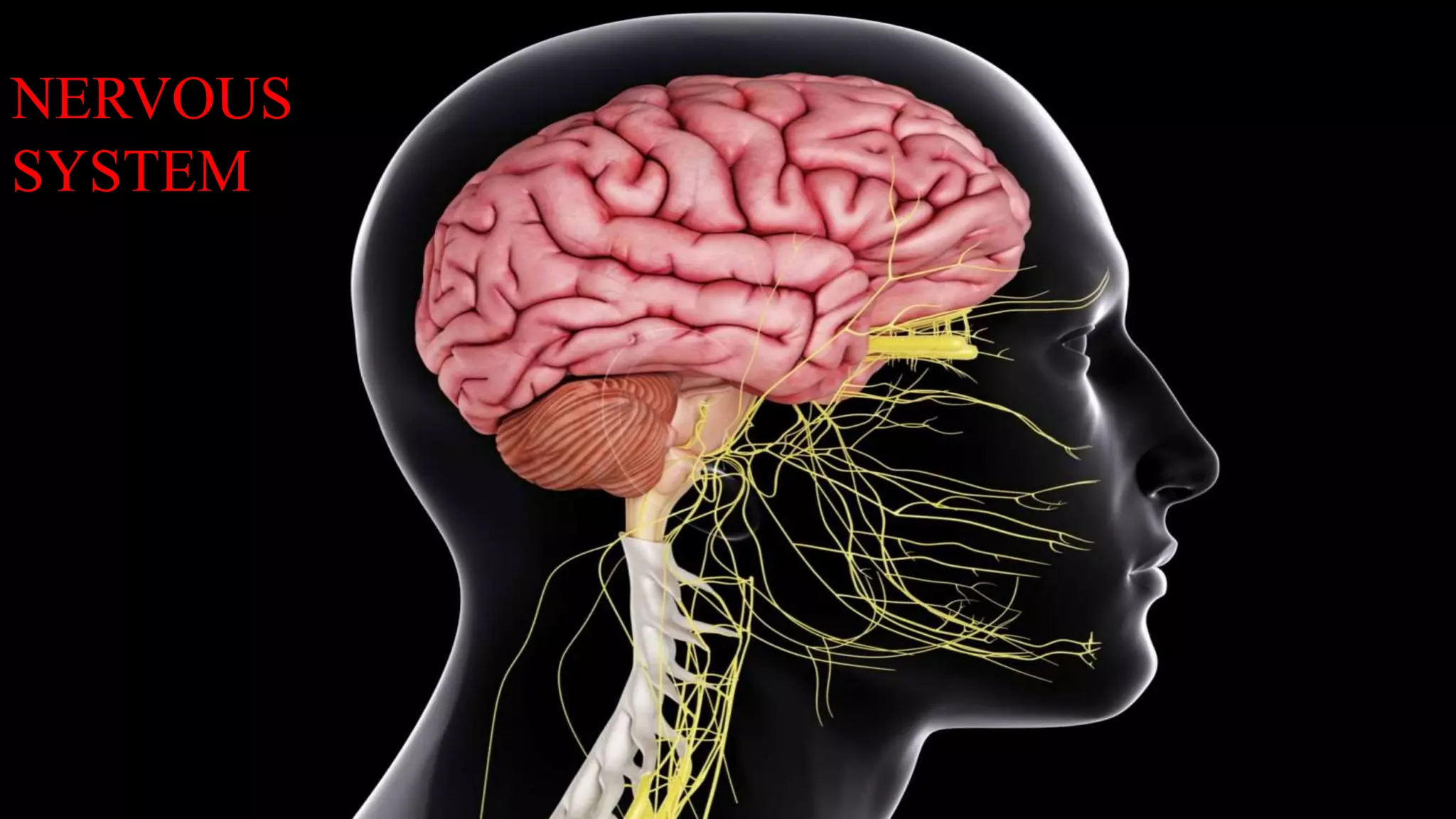
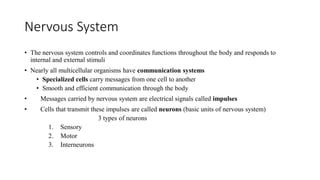
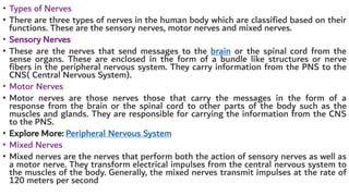
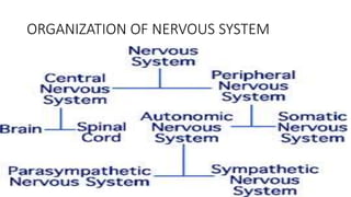
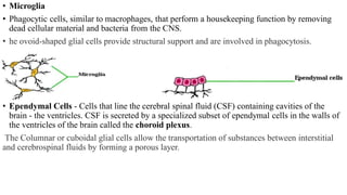
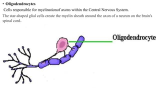
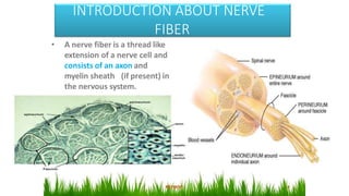
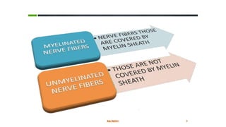
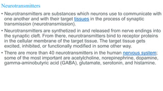
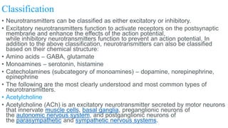
The nervous system controls and coordinates functions throughout the body and responds to
internal and external stimuli
โ€ข Nearly all multicellular organisms have communication systems
โ€ข Specialized cells carry messages from one cell to another
โ€ข Smooth and efficient communication through the body
โ€ข Messages carried by nervous system are electrical signals called impulses
โ€ข Cells that transmit these impulses are called neurons (basic units of nervous system)
3 types of neurons
1. Sensory
2. Motor
3. Interneurons
โ€ข Types of Nerves
โ€ข There are three types of nerves in the human body which are classified based on their
functions. These are the sensory nerves, motor nerves and mixed nerves.
โ€ข Sensory Nerves
โ€ข These are the nerves that send messages to the brain or the spinal cord from the
sense organs. These are enclosed in the form of a bundle like structures or nerve
fibers in the peripheral nervous system. They carry information from the PNS to the
CNS( Central Nervous System).
โ€ข Motor Nerves
โ€ข Motor nerves are those nerves those that carry the messages in the form of a
response from the brain or the spinal cord to other parts of the body such as the
muscles and glands. They are responsible for carrying the information from the CNS
to the PNS.
โ€ข Explore More: Peripheral Nervous System
โ€ข Mixed Nerves
โ€ข Mixed nerves are the nerves that perform both the action of sensory nerves as well as
a motor nerve. They transform electrical impulses from the central nervous system to
the muscles of the body. Generally, the mixed nerves transmit impulses at the rate of
120 meters per second
ORGANIZATION OF NERVOUS SYSTEM
NEURON
โ€ข A nerve cell with its process is called a neuron.
Neuron is the structural & functional unit of nervous
system
โ€ข Neuron is responsible for reception, conduction,
integration & transmission of nerve impulse
โ€ข SPECIAL CHARACTERISTICS OF NEURONS
Amitotic - no mitosis cell division; they cannot
reproduce or regenerate after certain point in life
Longevity - neurons can survive entire lifetime
High metabolic rate - require OXYGEN and
GLUCOSE at all times
PARTS OF NEURON
โ€ข The neuron can be generally divided into three mainfunctional parts: the cell body, the
axon, and the dendrites.
โ€ข The Cell Body (Soma)
๏‚ท In many ways, the cell body is similar to other types of cells. It has a nucleus with at
least one nucleolus and contains many of the typical cytoplasmic organelles.
๏‚ท It lacks centrioles, however. Because centrioles function in cell division, the fact that
neurons lack these organelles is consistent with the amitotic nature of the cell.
๏‚ท Nissl Bodies โ€“
o made from Rough Endoplasmic Reticulum (rER)
o are present in cytoplasm of cell body and extend into dendrites but do not extend
into axon.
o More numerous in motor nerve cells thansensory nerve cells.
o They are chromatophilic substances.
โ€ข Functions:
๏‚ท It contains the nucleus of the cell, and therefore is where most protein synthesis occurs.
๏‚ท Each neuron cell body is a center for receiving and sending nerve impulses.
โ€ข Dendrites are the finger-like cells present on the end of a neuron. They are
short, branching fibers extending from the cell body of the nerve cell. This
fiber increases the surface area available for receiving incoming
information.
โ€ข Dendrites are the receiving or input portions of a neuron. They are usually
short, narrow and highly branched structures.
โ€ข In neurons, these dendrites resemble a tree or branches of a tree,
extending from the cell body of the nerve cell. The cytoplasm of dendrites
contains Nissl bodies, mitochondria and other organelles. Dendrite
provides an increased surface area for the reception of stimuli from the
external world to the other neurons. Dendrite is the shortest fiber, which
stretches out from the cell body of a neuron.
โ€ข Function of Dendrites
โ€ข Dendrites are the structures on the neuron, that function by receiving electrical
messages. The functions of dendrites are to transfer the received information to
the soma of the neuron. Other biological processes of Dendrites are:
โ€ข Dendrites receive the data or signals from another neuron.
โ€ข Dendrite collects and stores all incoming information from axon terminals.
โ€ข Dendrites are the structures of neurons, which conduct electrical impulses
toward the cell body of the nerve cell.
โ€ข Dendrites collect messages through other neurons or the nerves in the human
body. These messages are passed through the nervous systems towards the
brain, where the brain sends the instructions back to different parts of the body
so that a reaction can occur.
1.Axon is a single, long fiber that extends from the other side of the
cell body of neuron.
2.It is covered by a sheath called myelin sheath.
3.Axon is the most important part of the neuron.
4.It carries messages from the cell body of one neuron to another.
โ€ข function of axon
โ€ข An axon, is a long, slender projection of a nerve cell, or neuron, that
typically conducts electrical impulses away from the neuron's cell body.
โ€ข It is a long extension part of the nerve cell, that conducts electrical
impulses towards the brain.
โ€ข The function of the axon is to transmit information to different neurons.
โ€ข Myelin sheath
Definition
Myelin is a dielectric (electrically insulating) material that forms a layer, the myelin sheath, usually
around only the axon of a neuron.
๏‚ท Formation
Myelin is formed in the peripheral nervous system by Schwann cells, which are special supportingcells
that surround the axons. In the central nervous system, supporting cells called glia produce myelin.
โ€ข Composition
Myelin is composed of lipid (about 80%) and protein (about 20%).
๏‚ท Function
Myelin serves as an insulator to prevent spreading of an impulse; thus conduction of impulse is faster
in the myelinated nerve than in the non-myelinated nerve.
Schwann cell
โ€ข Schwann cell or neurilemma cell is a type of neuroglia called oligodendrocytes of PNS (peripheral
nervous system). Schwann cells are named after German physiologist Theodor Schwann, who
discovered them in the 19th century.
โ€ข "The Schwann cells are the cells that produce the myelin sheath around the axon in a neuron."
Neuroglia
โ€ข Neuroglia - Cells that provide metabolic supportand immune protection forneurons. Neurogliaoutnumber
neurons by about10:1 in the Central Nervous System. Neuroglia do not generate or conduct nerve impulses.
However, unlike neurons, glial cells canregenerate if injured
โ€ข Astrocytes - Provide for the energy and other metabolic needs of neurons as well as giving nervous tissue structural
support. When neurons of the brain or the spinal cord are injured and destroyed, they arereplaced with scar tissue
made up of astrocytes (aprocess called gliosis). 0r Astrocytes: The star-shaped glial cells are present between
neurons and blood vessels, and help in the transportation of substances between neurons and blood vessels.
โ€ข Microglia
โ€ข Phagocytic cells, similar to macrophages, that perform a housekeeping function by removing
dead cellular material and bacteria from the CNS.
โ€ข he ovoid-shaped glial cells provide structural support and are involved in phagocytosis.
โ€ข Ependymal Cells - Cells that line the cerebral spinal fluid (CSF) containing cavities of the
brain - the ventricles. CSF is secreted by a specialized subset of ependymal cells in the walls of
the ventricles of the brain called the choroid plexus.
The Columnar or cuboidal glial cells allow the transportation of substances between interstitial
and cerebrospinal fluids by forming a porous layer.
โ€ข Oligodendrocytes
Cells responsible for myelinationof axons within the Central Nervous System.
The star-shaped glial cells create the myelin sheath around the axon of a neuron on the brain's
spinal cord.
INTRODUCTION ABOUT NERVE
FIBER
โ€ข A nerve fiber is a thread like
extension of a nerve cell and
consists of an axon and
myelin sheath (if present) in
the nervous system.
Depending upon STRUCTURE
Depending upon DISTRIBUTION
Somatic nerve fibers
โ€ข These are ฮฑ type motor nerve fibers.
โ€ข The neurotransmitter released at the neuron
endings is acetylcholine(Ach).
โ€ข It always leads to muscles excitation . Inhibition takes
place centrally due to participation of interneurons.
Autonomic nerve fibers
โ€ข They innervate smooth muscles , cardiac
muscles and Glands.
โ€ข Their main work is to maintain homeostasis with
the help of autonomic nervous system.
โ€ข They can lead to either excitation or
inhibition of Effector organs.
Depending upon
diameter and velocity
of conduction (Erlanger
and Grasserโ€™s
classification)
Numerical classification
Properties of Nerve
Fiber
Important Properties
โ€ข Excitability
โ€ข Conductivity
โ€ข Unfatigability
โ€ข Refractive period
โ€ข All or none
response
โ€ข Summation
โ€ข Accommodation
Excitability:
โ€ข nerve fibres are highly excitable tissue
โ€ข respond to various stimuli
โ€ข Capable of generating electrical impulse
Conductivity:
โ€ข action potential is generated in the nerve
fibre, which is propagated along its entire
length to the axon terminal.
Refractive period:
โ€ข during action potential the excitability of a
nerve become reduced
โ€ข that is a new impulse cannot be generated
during a Action Potential
โ€ข Types:
a. Absolute refractory period (ARP)
b. Relative refractory period( RRP)
Unfatiguability :
โ€ข Nerve fibres can not be fatigued even when they are stimulated
continuously.
All or none response:
โ€ข Either all of the action potential is seen or none at all
โ€ข If a stimulus of threshold strength is applied action Potential will be
generated
โ€ข Further increase in strength of stimulus or duration has no effect on
amplitude of action Potential
โ€ข But can affect frequency
Summation:
โ€ข Application of a sub threshold stimulus does not evoke an action
potential. However if sub threshold stimuli are applied in rapid
succession they are added and they produce an action potential.
Accommodation:
โ€ข Application of continuous stimuli may decrease the excitability of
nerve fibre.
โ€ข BIOPHYSICAL BASIS OF ACTION POTENTIAL
โ€ข Action potentials result from the presence in a cell's membrane of
specialtypes of voltage-gated ion channels. A voltage-gated ion
channel is a cluster of proteins embedded in the membrane that has
three key properties:
โšซ It is capable of assuming more than one conformation.
โšซ At least one of the conformations creates a channel through
themembrane that is permeable to specific types of ions.
โšซ The transition between conformations is influenced by the
membranepotential.
โ€ข STAGES OF ACTION POTENTIAL
โ€ข There are six stages of Action Potential. They are:
1. Resting stage
2. Depolarization stage
3. Repolarization stage
4. Spike potential
5. Negative after potential
6. Positive after potential or Hyperpolarization
Resting stage: This is the resting membrane potential before the action potential begins. The
membrane is said to be โ€œPolarizedโ€ during this stage because of the very large membrane
potential that is present.
Depolarization stage: When the negativity of the membrane potentialrises rapidly in positive
direction due to influx (enter) of sodium ions is called depolarization stage.
Repolarization stage: Immediately after depolarization the negativity ofthe membrane potential
re-establishes towards the normal negative membrane due to efflux of potassium ion. This is
called the repolarization membrane.
Spike potential: Initially , the depolarization wave over shoots the zeroline and then sharply falls.
This sharp rise of the depolarization wave and the rapid fall of repolarization wave is called spike
potential.
Negative after potential: At the termination of spike potential , the membrane potential
sometimes fails to reach the normal resting stage. Thisis called negative after potential.
Positive after potential: Sometimes the negativity of membrane potential become more
than its normal level. This is called positive afterpotential.
โ€ขFunction of an action potential
An action potential is part of the process that
occurs during the firing of a neuron. During the
action potential, part of the neural membrane
opens to allow positively charged ions inside the
cell and negatively charged ions out. This
process causes a rapid increase in the positive
charge of the nerve fiber.
Neurotransmitters
โ€ข Neurotransmitters are substances which neurons use to communicate with
one another and with their target tissues in the process of synaptic
transmission (neurotransmission).
โ€ข Neurotransmitters are synthetized in and released from nerve endings into
the synaptic cleft. From there, neurotransmitters bind to receptor proteins
in the cellular membrane of the target tissue. The target tissue gets
excited, inhibited, or functionally modified in some other way.
โ€ข There are more than 40 neurotransmitters in the human nervous system;
some of the most important are acetylcholine, norepinephrine, dopamine,
gamma-aminobutyric acid (GABA), glutamate, serotonin, and histamine.
Mechanism of neurotransmission
โ€ข Neurons communicate with their target tissues at synapses into which they
release chemical substances called neurotransmitters (ligands). As this
communication is mediated with chemical substances, the process is called
chemical neurotransmission and happens within chemical synapses.
โ€ข Each synapse consists of the:
โ€ข Presynaptic membrane โ€“ membrane of the terminal bouton (axon ending) of the
presynaptic nerve fiber
โ€ข Postsynaptic membrane โ€“ membrane of the target cell
โ€ข Synaptic cleft โ€“ a gap between the presynaptic and postsynaptic membranes
โ€ข Inside the terminal bouton of the presynaptic nerve fiber, numerous vesicles that
contain neurotransmitters are produced and stored. When the presynaptic
membrane is depolarized by an action potential, calcium voltage-gated channels
open (found in the membranes of the terminal buttons). This leads to an influx of
calcium ions into the terminal bouton, which changes the state of certain
membrane proteins in the presynaptic membrane, and results in exocytosis of
neurotransmitters from the terminal bouton into the synaptic cleft.
โ€ข After crossing the synaptic cleft, neurotransmitters bind to their receptors on
the postsynaptic membrane. Once the neurotransmitter binds to its receptor, the
ligand-gated channels of the postsynaptic membrane either open or close.
These ligand-gated channels are ion channels, and their opening or closing
alters the permeability of the postsynaptic membrane to calcium, sodium,
potassium, and chloride ions. This leads to a stimulatory or inhibitory response.
โ€ข If a neurotransmitter stimulates the target cell to an action, then it is
an excitatory neurotransmitter acting in an excitatory synapse. On the other
hand, if it inhibits the target cell, it is an inhibitory neurotransmitter acting in an
inhibitory synapse. So, the type of the synapse and the response of the target
tissue depends on the type of neurotransmitter. Excitatory neurotransmitters
cause depolarization of the postsynaptic cells and generate an action potential;
for example acetylcholine stimulates muscle contraction. Inhibitory synapses
cause hyperpolarization of the target cells, leading them farther from the action
potential threshold, thus inhibiting their action; for example GABA inhibits
involuntary movements.
โ€ข The neurotransmitter released into the synaptic cleft acts for a very short
duration, only minutes or even seconds. It is either destroyed by enzymes, such
as acetylcholine esterase, or is reabsorbed into the terminal button of the
presynaptic neuron by reuptake mechanisms and then recycled. The best-
known neurotransmitters responsible for such fast, but short-lived excitatory
action are acetylcholine, norepinephrine, and epinephrine while GABA is the
major inhibitory neurotransmitter
Classification
โ€ข Neurotransmitters can be classified as either excitatory or inhibitory.
โ€ข Excitatory neurotransmitters function to activate receptors on the postsynaptic
membrane and enhance the effects of the action potential,
while inhibitory neurotransmitters function to prevent an action potential. In
addition to the above classification, neurotransmitters can also be classified
based on their chemical structure:
โ€ข Amino acids โ€“ GABA, glutamate
โ€ข Monoamines โ€“ serotonin, histamine
โ€ข Catecholamines (subcategory of monoamines) โ€“ dopamine, norepinephrine,
epinephrine
โ€ข The following are the most clearly understood and most common types of
neurotransmitters.
โ€ข Acetylcholine
โ€ข Acetylcholine (ACh) is an excitatory neurotransmitter secreted by motor neurons
that innervate muscle cells, basal ganglia, preganglionic neurons of
the autonomic nervous system, and postganglionic neurons of
the parasympathetic and sympathetic nervous systems.
โ€ข Its main function is to stimulate muscle contraction. However, the only
exception to this, where acetylcholine is an inhibitory neurotransmitter, is at
the parasympathetic endings of the vagus nerve. These inhibit the heart
muscle through the cardiac plexus.
โ€ข It is also found in sensory neurons and in the autonomic nervous system, and
has a part in scheduling the โ€œdream stateโ€ while an individual is fast asleep.
Acetylcholine plays a vital role in the normal functioning of muscles. For
example, poisonous plants like curare and hemlock cause paralysis of
muscles by blocking the acetylcholine receptor sites of myocytes (muscle
cells). The well-known poison botulin works by preventing vesicles in the
terminal bouton from releasing acetylcholine, thus leading to paralysis of the
effector muscle.
โ€ข Norepinephrine
โ€ข Norepinephrine (NE), also known as noradrenaline (NAd), is an excitatory
neurotransmitter produced by the brainstem, hypothalamus, and adrenal
glands and released into the bloodstream. In the brain it increases the level of
alertness and wakefulness.
โ€ข In the body, it is secreted by most postganglionic sympathetic nerves. It acts to
stimulate the processes in the body. For example, it is very important in the
endogenous production of epinephrine. Norepinephrine has been implicated in
mood disorders such as depression and anxiety, in which case its concentration
in the body is abnormally low. Alternatively, an abnormally high concentration of
it may lead to an impaired sleep cycle.
โ€ข Epinephrine
โ€ข Also known as adrenaline (Ad), epinephrine (Epi) is an excitatory
neurotransmitter produced by the chromaffin cells of the adrenal gland. It
prepares the body for the fight-or-flight response. That means that when a
person is highly stimulated (fear, anger etc.), extra amounts of epinephrine are
released into the bloodstream.
โ€ข This release of epinephrine increases heart rate, blood pressure, and glucose
release from the liver (via glycogenolysis). In this way, the nervous
and endocrine systems prepare the body for dangerous and extreme situations
by increasing nutrient supply to key tissues.
โ€ข Dopamine
โ€ข Dopamine (DA) is a neurotransmitter secreted by the neurons of the substantia
nigra. It is considered a special type of neurotransmitter because its effects are
both excitatory and inhibitory. Which effect depends on the type of receptor that
dopamine binds to.
โ€ข As a part of the extrapyramidal motor system which involves the basal
ganglia, dopamine is important for movement coordination by inhibiting
unnecessary movements. In the pituitary gland, it inhibits the release of
prolactin, and stimulates the secretion of growth hormone.
โ€ข Dopamine deficiency related to the destruction of the substantia nigra
leads to Parkinsonโ€™s disease. Increased activity of dopaminergic neurons
contributes to the pathophysiology of psychotic disorders and
schizophrenia. Drug and alcohol abuse can temporarily increase
dopamine levels in the blood, leading to confusion and the inability to
focus. However, an appropriate secretion of dopamine in the bloodstream
plays a role in the motivation or desire to complete a task.
โ€ข GABA
โ€ข gamma-Aminobutyric acid (GABA) is the most powerful inhibitory
neurotransmitter produced by the neurons of the spinal cord, cerebellum,
basal ganglia, and many areas of the cerebral cortex. It is derived from
glutamate.
โ€ข Functions of GABA are closely related to mood and emotions. It is an inhibitory
neurotransmitter that acts as a brake to excitatory neurotransmitters; thus when
it is abnormally low this can lead to anxiety. It is widely distributed in the brain
and plays a principal role in reducing neuronal excitability throughout the
nervous system.
โ€ข Glutamate
โ€ข Glutamate (Glu) is the most powerful excitatory neurotransmitter of the central
nervous system which ensures homeostasis with the effects of GABA. It is
secreted by neurons of the many of the sensory pathways entering the central
nervous system, as well as the cerebral cortex.
โ€ข Glutamate is the most common neurotransmitter in the central nervous system;
it takes part in the regulation of general excitability of the central nervous
system, learning processes, and memory. Thus, inappropriate glutamate
neurotransmission contributes to developing epilepsy and cognitive and
affective disorders.
โ€ข Serotonin
โ€ข Serotonin (5-hydroxytryptamine, 5-HT) is an inhibitory neurotransmitter that has
been found to be intimately involved in emotion and mood. It is secreted by the
neurons of the brainstem and by neurons that innervate the gastrointestinal
โ€ข It participates in regulation of body temperature, perception of pain,
emotions, and sleep cycle. An insufficient secretion of serotonin may result
in decreased immune system function, as well as a range of emotional
disorders like depression, anger control problems, obsessive-compulsive
disorder, and even suicidal tendencies.
โ€ข Histamine
โ€ข Histamine is an excitatory neurotransmitter produced by neurons of
the hypothalamus, cells of the stomach mucosa, mast cells, and basophils
in the blood. In the central nervous system, it is important for wakefulness,
blood pressure, pain, and sexual behavior. In the stomach, it increases the
acidity.
โ€ข It is involved primarily in the inflammatory response, as well as a range of
other functions such as vasodilation and regulation of the immune
response to foreign bodies. For example, when allergens are introduced
into the bloodstream, histamine assists in the fight against these
microorganisms causing itching of the skin or irritations of the throat, nose,
CNS.pdf

CNS.pdf

  • 1.
  • 2.
    Nervous System โ€ข Thenervous system controls and coordinates functions throughout the body and responds to internal and external stimuli โ€ข Nearly all multicellular organisms have communication systems โ€ข Specialized cells carry messages from one cell to another โ€ข Smooth and efficient communication through the body โ€ข Messages carried by nervous system are electrical signals called impulses โ€ข Cells that transmit these impulses are called neurons (basic units of nervous system) 3 types of neurons 1. Sensory 2. Motor 3. Interneurons
  • 3.
    โ€ข Types ofNerves โ€ข There are three types of nerves in the human body which are classified based on their functions. These are the sensory nerves, motor nerves and mixed nerves. โ€ข Sensory Nerves โ€ข These are the nerves that send messages to the brain or the spinal cord from the sense organs. These are enclosed in the form of a bundle like structures or nerve fibers in the peripheral nervous system. They carry information from the PNS to the CNS( Central Nervous System). โ€ข Motor Nerves โ€ข Motor nerves are those nerves those that carry the messages in the form of a response from the brain or the spinal cord to other parts of the body such as the muscles and glands. They are responsible for carrying the information from the CNS to the PNS. โ€ข Explore More: Peripheral Nervous System โ€ข Mixed Nerves โ€ข Mixed nerves are the nerves that perform both the action of sensory nerves as well as a motor nerve. They transform electrical impulses from the central nervous system to the muscles of the body. Generally, the mixed nerves transmit impulses at the rate of 120 meters per second
  • 4.
  • 5.
    NEURON โ€ข A nervecell with its process is called a neuron. Neuron is the structural & functional unit of nervous system โ€ข Neuron is responsible for reception, conduction, integration & transmission of nerve impulse โ€ข SPECIAL CHARACTERISTICS OF NEURONS Amitotic - no mitosis cell division; they cannot reproduce or regenerate after certain point in life Longevity - neurons can survive entire lifetime High metabolic rate - require OXYGEN and GLUCOSE at all times
  • 6.
    PARTS OF NEURON โ€ขThe neuron can be generally divided into three mainfunctional parts: the cell body, the axon, and the dendrites. โ€ข The Cell Body (Soma) ๏‚ท In many ways, the cell body is similar to other types of cells. It has a nucleus with at least one nucleolus and contains many of the typical cytoplasmic organelles. ๏‚ท It lacks centrioles, however. Because centrioles function in cell division, the fact that neurons lack these organelles is consistent with the amitotic nature of the cell. ๏‚ท Nissl Bodies โ€“ o made from Rough Endoplasmic Reticulum (rER) o are present in cytoplasm of cell body and extend into dendrites but do not extend into axon. o More numerous in motor nerve cells thansensory nerve cells. o They are chromatophilic substances. โ€ข Functions: ๏‚ท It contains the nucleus of the cell, and therefore is where most protein synthesis occurs. ๏‚ท Each neuron cell body is a center for receiving and sending nerve impulses.
  • 7.
    โ€ข Dendrites arethe finger-like cells present on the end of a neuron. They are short, branching fibers extending from the cell body of the nerve cell. This fiber increases the surface area available for receiving incoming information. โ€ข Dendrites are the receiving or input portions of a neuron. They are usually short, narrow and highly branched structures. โ€ข In neurons, these dendrites resemble a tree or branches of a tree, extending from the cell body of the nerve cell. The cytoplasm of dendrites contains Nissl bodies, mitochondria and other organelles. Dendrite provides an increased surface area for the reception of stimuli from the external world to the other neurons. Dendrite is the shortest fiber, which stretches out from the cell body of a neuron.
  • 8.
    โ€ข Function ofDendrites โ€ข Dendrites are the structures on the neuron, that function by receiving electrical messages. The functions of dendrites are to transfer the received information to the soma of the neuron. Other biological processes of Dendrites are: โ€ข Dendrites receive the data or signals from another neuron. โ€ข Dendrite collects and stores all incoming information from axon terminals. โ€ข Dendrites are the structures of neurons, which conduct electrical impulses toward the cell body of the nerve cell. โ€ข Dendrites collect messages through other neurons or the nerves in the human body. These messages are passed through the nervous systems towards the brain, where the brain sends the instructions back to different parts of the body so that a reaction can occur.
  • 9.
    1.Axon is asingle, long fiber that extends from the other side of the cell body of neuron. 2.It is covered by a sheath called myelin sheath. 3.Axon is the most important part of the neuron. 4.It carries messages from the cell body of one neuron to another. โ€ข function of axon โ€ข An axon, is a long, slender projection of a nerve cell, or neuron, that typically conducts electrical impulses away from the neuron's cell body. โ€ข It is a long extension part of the nerve cell, that conducts electrical impulses towards the brain. โ€ข The function of the axon is to transmit information to different neurons.
  • 10.
    โ€ข Myelin sheath Definition Myelinis a dielectric (electrically insulating) material that forms a layer, the myelin sheath, usually around only the axon of a neuron. ๏‚ท Formation Myelin is formed in the peripheral nervous system by Schwann cells, which are special supportingcells that surround the axons. In the central nervous system, supporting cells called glia produce myelin. โ€ข Composition Myelin is composed of lipid (about 80%) and protein (about 20%). ๏‚ท Function Myelin serves as an insulator to prevent spreading of an impulse; thus conduction of impulse is faster in the myelinated nerve than in the non-myelinated nerve. Schwann cell โ€ข Schwann cell or neurilemma cell is a type of neuroglia called oligodendrocytes of PNS (peripheral nervous system). Schwann cells are named after German physiologist Theodor Schwann, who discovered them in the 19th century. โ€ข "The Schwann cells are the cells that produce the myelin sheath around the axon in a neuron."
  • 11.
  • 12.
    โ€ข Neuroglia -Cells that provide metabolic supportand immune protection forneurons. Neurogliaoutnumber neurons by about10:1 in the Central Nervous System. Neuroglia do not generate or conduct nerve impulses. However, unlike neurons, glial cells canregenerate if injured โ€ข Astrocytes - Provide for the energy and other metabolic needs of neurons as well as giving nervous tissue structural support. When neurons of the brain or the spinal cord are injured and destroyed, they arereplaced with scar tissue made up of astrocytes (aprocess called gliosis). 0r Astrocytes: The star-shaped glial cells are present between neurons and blood vessels, and help in the transportation of substances between neurons and blood vessels.
  • 13.
    โ€ข Microglia โ€ข Phagocyticcells, similar to macrophages, that perform a housekeeping function by removing dead cellular material and bacteria from the CNS. โ€ข he ovoid-shaped glial cells provide structural support and are involved in phagocytosis. โ€ข Ependymal Cells - Cells that line the cerebral spinal fluid (CSF) containing cavities of the brain - the ventricles. CSF is secreted by a specialized subset of ependymal cells in the walls of the ventricles of the brain called the choroid plexus. The Columnar or cuboidal glial cells allow the transportation of substances between interstitial and cerebrospinal fluids by forming a porous layer.
  • 14.
    โ€ข Oligodendrocytes Cells responsiblefor myelinationof axons within the Central Nervous System. The star-shaped glial cells create the myelin sheath around the axon of a neuron on the brain's spinal cord.
  • 15.
    INTRODUCTION ABOUT NERVE FIBER โ€ขA nerve fiber is a thread like extension of a nerve cell and consists of an axon and myelin sheath (if present) in the nervous system.
  • 17.
  • 19.
  • 21.
    Somatic nerve fibers โ€ขThese are ฮฑ type motor nerve fibers. โ€ข The neurotransmitter released at the neuron endings is acetylcholine(Ach). โ€ข It always leads to muscles excitation . Inhibition takes place centrally due to participation of interneurons.
  • 22.
    Autonomic nerve fibers โ€ขThey innervate smooth muscles , cardiac muscles and Glands. โ€ข Their main work is to maintain homeostasis with the help of autonomic nervous system. โ€ข They can lead to either excitation or inhibition of Effector organs.
  • 23.
    Depending upon diameter andvelocity of conduction (Erlanger and Grasserโ€™s classification)
  • 24.
  • 25.
  • 26.
    Important Properties โ€ข Excitability โ€ขConductivity โ€ข Unfatigability โ€ข Refractive period โ€ข All or none response โ€ข Summation โ€ข Accommodation
  • 27.
    Excitability: โ€ข nerve fibresare highly excitable tissue โ€ข respond to various stimuli โ€ข Capable of generating electrical impulse Conductivity: โ€ข action potential is generated in the nerve fibre, which is propagated along its entire length to the axon terminal.
  • 28.
    Refractive period: โ€ข duringaction potential the excitability of a nerve become reduced โ€ข that is a new impulse cannot be generated during a Action Potential โ€ข Types: a. Absolute refractory period (ARP) b. Relative refractory period( RRP)
  • 29.
    Unfatiguability : โ€ข Nervefibres can not be fatigued even when they are stimulated continuously. All or none response: โ€ข Either all of the action potential is seen or none at all โ€ข If a stimulus of threshold strength is applied action Potential will be generated โ€ข Further increase in strength of stimulus or duration has no effect on amplitude of action Potential โ€ข But can affect frequency
  • 30.
    Summation: โ€ข Application ofa sub threshold stimulus does not evoke an action potential. However if sub threshold stimuli are applied in rapid succession they are added and they produce an action potential. Accommodation: โ€ข Application of continuous stimuli may decrease the excitability of nerve fibre.
  • 32.
    โ€ข BIOPHYSICAL BASISOF ACTION POTENTIAL โ€ข Action potentials result from the presence in a cell's membrane of specialtypes of voltage-gated ion channels. A voltage-gated ion channel is a cluster of proteins embedded in the membrane that has three key properties: โšซ It is capable of assuming more than one conformation. โšซ At least one of the conformations creates a channel through themembrane that is permeable to specific types of ions. โšซ The transition between conformations is influenced by the membranepotential.
  • 33.
    โ€ข STAGES OFACTION POTENTIAL โ€ข There are six stages of Action Potential. They are: 1. Resting stage 2. Depolarization stage 3. Repolarization stage 4. Spike potential 5. Negative after potential 6. Positive after potential or Hyperpolarization
  • 34.
    Resting stage: Thisis the resting membrane potential before the action potential begins. The membrane is said to be โ€œPolarizedโ€ during this stage because of the very large membrane potential that is present. Depolarization stage: When the negativity of the membrane potentialrises rapidly in positive direction due to influx (enter) of sodium ions is called depolarization stage. Repolarization stage: Immediately after depolarization the negativity ofthe membrane potential re-establishes towards the normal negative membrane due to efflux of potassium ion. This is called the repolarization membrane. Spike potential: Initially , the depolarization wave over shoots the zeroline and then sharply falls. This sharp rise of the depolarization wave and the rapid fall of repolarization wave is called spike potential. Negative after potential: At the termination of spike potential , the membrane potential sometimes fails to reach the normal resting stage. Thisis called negative after potential. Positive after potential: Sometimes the negativity of membrane potential become more than its normal level. This is called positive afterpotential.
  • 35.
    โ€ขFunction of anaction potential An action potential is part of the process that occurs during the firing of a neuron. During the action potential, part of the neural membrane opens to allow positively charged ions inside the cell and negatively charged ions out. This process causes a rapid increase in the positive charge of the nerve fiber.
  • 46.
    Neurotransmitters โ€ข Neurotransmitters aresubstances which neurons use to communicate with one another and with their target tissues in the process of synaptic transmission (neurotransmission). โ€ข Neurotransmitters are synthetized in and released from nerve endings into the synaptic cleft. From there, neurotransmitters bind to receptor proteins in the cellular membrane of the target tissue. The target tissue gets excited, inhibited, or functionally modified in some other way. โ€ข There are more than 40 neurotransmitters in the human nervous system; some of the most important are acetylcholine, norepinephrine, dopamine, gamma-aminobutyric acid (GABA), glutamate, serotonin, and histamine.
  • 47.
    Mechanism of neurotransmission โ€ขNeurons communicate with their target tissues at synapses into which they release chemical substances called neurotransmitters (ligands). As this communication is mediated with chemical substances, the process is called chemical neurotransmission and happens within chemical synapses. โ€ข Each synapse consists of the: โ€ข Presynaptic membrane โ€“ membrane of the terminal bouton (axon ending) of the presynaptic nerve fiber โ€ข Postsynaptic membrane โ€“ membrane of the target cell โ€ข Synaptic cleft โ€“ a gap between the presynaptic and postsynaptic membranes โ€ข Inside the terminal bouton of the presynaptic nerve fiber, numerous vesicles that contain neurotransmitters are produced and stored. When the presynaptic membrane is depolarized by an action potential, calcium voltage-gated channels open (found in the membranes of the terminal buttons). This leads to an influx of calcium ions into the terminal bouton, which changes the state of certain membrane proteins in the presynaptic membrane, and results in exocytosis of neurotransmitters from the terminal bouton into the synaptic cleft.
  • 49.
    โ€ข After crossingthe synaptic cleft, neurotransmitters bind to their receptors on the postsynaptic membrane. Once the neurotransmitter binds to its receptor, the ligand-gated channels of the postsynaptic membrane either open or close. These ligand-gated channels are ion channels, and their opening or closing alters the permeability of the postsynaptic membrane to calcium, sodium, potassium, and chloride ions. This leads to a stimulatory or inhibitory response. โ€ข If a neurotransmitter stimulates the target cell to an action, then it is an excitatory neurotransmitter acting in an excitatory synapse. On the other hand, if it inhibits the target cell, it is an inhibitory neurotransmitter acting in an inhibitory synapse. So, the type of the synapse and the response of the target tissue depends on the type of neurotransmitter. Excitatory neurotransmitters cause depolarization of the postsynaptic cells and generate an action potential; for example acetylcholine stimulates muscle contraction. Inhibitory synapses cause hyperpolarization of the target cells, leading them farther from the action potential threshold, thus inhibiting their action; for example GABA inhibits involuntary movements. โ€ข The neurotransmitter released into the synaptic cleft acts for a very short duration, only minutes or even seconds. It is either destroyed by enzymes, such as acetylcholine esterase, or is reabsorbed into the terminal button of the presynaptic neuron by reuptake mechanisms and then recycled. The best- known neurotransmitters responsible for such fast, but short-lived excitatory action are acetylcholine, norepinephrine, and epinephrine while GABA is the major inhibitory neurotransmitter
  • 51.
    Classification โ€ข Neurotransmitters canbe classified as either excitatory or inhibitory. โ€ข Excitatory neurotransmitters function to activate receptors on the postsynaptic membrane and enhance the effects of the action potential, while inhibitory neurotransmitters function to prevent an action potential. In addition to the above classification, neurotransmitters can also be classified based on their chemical structure: โ€ข Amino acids โ€“ GABA, glutamate โ€ข Monoamines โ€“ serotonin, histamine โ€ข Catecholamines (subcategory of monoamines) โ€“ dopamine, norepinephrine, epinephrine โ€ข The following are the most clearly understood and most common types of neurotransmitters. โ€ข Acetylcholine โ€ข Acetylcholine (ACh) is an excitatory neurotransmitter secreted by motor neurons that innervate muscle cells, basal ganglia, preganglionic neurons of the autonomic nervous system, and postganglionic neurons of the parasympathetic and sympathetic nervous systems.
  • 52.
    โ€ข Its mainfunction is to stimulate muscle contraction. However, the only exception to this, where acetylcholine is an inhibitory neurotransmitter, is at the parasympathetic endings of the vagus nerve. These inhibit the heart muscle through the cardiac plexus. โ€ข It is also found in sensory neurons and in the autonomic nervous system, and has a part in scheduling the โ€œdream stateโ€ while an individual is fast asleep. Acetylcholine plays a vital role in the normal functioning of muscles. For example, poisonous plants like curare and hemlock cause paralysis of muscles by blocking the acetylcholine receptor sites of myocytes (muscle cells). The well-known poison botulin works by preventing vesicles in the terminal bouton from releasing acetylcholine, thus leading to paralysis of the effector muscle. โ€ข Norepinephrine โ€ข Norepinephrine (NE), also known as noradrenaline (NAd), is an excitatory neurotransmitter produced by the brainstem, hypothalamus, and adrenal glands and released into the bloodstream. In the brain it increases the level of alertness and wakefulness.
  • 53.
    โ€ข In thebody, it is secreted by most postganglionic sympathetic nerves. It acts to stimulate the processes in the body. For example, it is very important in the endogenous production of epinephrine. Norepinephrine has been implicated in mood disorders such as depression and anxiety, in which case its concentration in the body is abnormally low. Alternatively, an abnormally high concentration of it may lead to an impaired sleep cycle. โ€ข Epinephrine โ€ข Also known as adrenaline (Ad), epinephrine (Epi) is an excitatory neurotransmitter produced by the chromaffin cells of the adrenal gland. It prepares the body for the fight-or-flight response. That means that when a person is highly stimulated (fear, anger etc.), extra amounts of epinephrine are released into the bloodstream. โ€ข This release of epinephrine increases heart rate, blood pressure, and glucose release from the liver (via glycogenolysis). In this way, the nervous and endocrine systems prepare the body for dangerous and extreme situations by increasing nutrient supply to key tissues. โ€ข Dopamine โ€ข Dopamine (DA) is a neurotransmitter secreted by the neurons of the substantia nigra. It is considered a special type of neurotransmitter because its effects are both excitatory and inhibitory. Which effect depends on the type of receptor that dopamine binds to.
  • 54.
    โ€ข As apart of the extrapyramidal motor system which involves the basal ganglia, dopamine is important for movement coordination by inhibiting unnecessary movements. In the pituitary gland, it inhibits the release of prolactin, and stimulates the secretion of growth hormone. โ€ข Dopamine deficiency related to the destruction of the substantia nigra leads to Parkinsonโ€™s disease. Increased activity of dopaminergic neurons contributes to the pathophysiology of psychotic disorders and schizophrenia. Drug and alcohol abuse can temporarily increase dopamine levels in the blood, leading to confusion and the inability to focus. However, an appropriate secretion of dopamine in the bloodstream plays a role in the motivation or desire to complete a task. โ€ข GABA โ€ข gamma-Aminobutyric acid (GABA) is the most powerful inhibitory neurotransmitter produced by the neurons of the spinal cord, cerebellum, basal ganglia, and many areas of the cerebral cortex. It is derived from glutamate.
  • 55.
    โ€ข Functions ofGABA are closely related to mood and emotions. It is an inhibitory neurotransmitter that acts as a brake to excitatory neurotransmitters; thus when it is abnormally low this can lead to anxiety. It is widely distributed in the brain and plays a principal role in reducing neuronal excitability throughout the nervous system. โ€ข Glutamate โ€ข Glutamate (Glu) is the most powerful excitatory neurotransmitter of the central nervous system which ensures homeostasis with the effects of GABA. It is secreted by neurons of the many of the sensory pathways entering the central nervous system, as well as the cerebral cortex. โ€ข Glutamate is the most common neurotransmitter in the central nervous system; it takes part in the regulation of general excitability of the central nervous system, learning processes, and memory. Thus, inappropriate glutamate neurotransmission contributes to developing epilepsy and cognitive and affective disorders. โ€ข Serotonin โ€ข Serotonin (5-hydroxytryptamine, 5-HT) is an inhibitory neurotransmitter that has been found to be intimately involved in emotion and mood. It is secreted by the neurons of the brainstem and by neurons that innervate the gastrointestinal
  • 56.
    โ€ข It participatesin regulation of body temperature, perception of pain, emotions, and sleep cycle. An insufficient secretion of serotonin may result in decreased immune system function, as well as a range of emotional disorders like depression, anger control problems, obsessive-compulsive disorder, and even suicidal tendencies. โ€ข Histamine โ€ข Histamine is an excitatory neurotransmitter produced by neurons of the hypothalamus, cells of the stomach mucosa, mast cells, and basophils in the blood. In the central nervous system, it is important for wakefulness, blood pressure, pain, and sexual behavior. In the stomach, it increases the acidity. โ€ข It is involved primarily in the inflammatory response, as well as a range of other functions such as vasodilation and regulation of the immune response to foreign bodies. For example, when allergens are introduced into the bloodstream, histamine assists in the fight against these microorganisms causing itching of the skin or irritations of the throat, nose,