SlideShare a Scribd company logo
1 of 20
Human’s Cells
By

Christian Dennis, SharineSinaga, Kelvin
Prihadi
Content
Heart          Intestine

Liver          Stomach

Blood          Kidney

Skin           Ovum

Muscle         Sperm

Neuron
Heart
Scientists consider heart cells a part of the muscle cell family, though with
unique differences in mitochondria, intercalated disks and t-tubes, as well
as in cellular growth.

Mitochondria
Unlike other muscle cells, heart cells are highly resistant to fatigue
because they have more mitochondria organelles than any other cell in the
body.

Intercalated disks
Intercalated disks regulate the passage of positive and negative electrons.
As electric currents repel and attract, it causes electron depolarization,
which regulates heartbeat contractions.

T-Tubules
Plasma membranes that surround each cell and organize them into pairs
to create the striated muscles strands used to build the myocardium
Liver
     Hepatocytes

Hepatocytesare the main cell for protein synthesis. They are also responsible
    for recycling lipoproteins.

     Kupffer Cells

Kupffercells are specialized macrophages. A macrophage is a cell that is a part
     of the immune system.

     StellateCells

Stellatecells are part of the nervous system. They innervate the organ to
      provide a connection between the brain and the liver. It helps with pain
      and sensory information from the liver to provide communication

     Mitochondria

     Mitochondria are located in most human cells, but there is an elevated
     number in liver cells. Mitochondria play a large role in metabolism of
     proteins, carbohydrates and lipids.
Skin
    Melanocytes: The main function of melanocytes is to produce
    melanin, which is responsible for the colour of our skin

    Langerhans Cells arise from bone marrow and migrate to the
    epidermis. Langerhans cells interact with white blood cells
    called "helper T cells" in immune responses and are easily
    damaged by UV radiation

    Merkel Cells: Merkel cells are located in the deepest layer
    (stratum basale) of the epidermis of hairless skin, where they
    are attached to keratinocytes by desmosomes.

    Dermo-epidermal JunctionThe Epidermis and Dermis are
    separated by the Dermo-Epithelial Junction. This junction holds
    the epidermis and dermis together and this is achieved by
    various fibers including collagen and desmosomes.


.
Muscle

    Muscle is composed of thousands of muscle fibers,
    each composed of a single muscle cell. A muscle cell
    contains a series of ultramicroscopic filaments called
    myofibrils. Each myofibril is a muscle cell that
    contains units called sarcomeres.
    Sarcomerescontain thick microfilaments composed of
    the protein myosin. Sarcomeres also contain thin
    microfilaments composed of the protein actin. The
    actin and myosin filaments are arranged parallel to
    one another, with the myosin filaments' molecular
    “heads” protruding toward the actin filaments.

.
Blood


NEUTROPHIL

This granulocyte has very tiny light staining
granules. The nucleus is frequently multi-lobed
with lobes connected by thin strands of nuclear
material. These cells are capable of
phagocytizing foreign cells, toxins, and viruses.
Blood

T

EOSINOPHIL

his granulocyte has large granules (A) which are
acidophilic and appear pink (or red) in a stained
preparation. This micrograph was color
enhanced to illustrate this feature. The nucleus
often has two lobes connected by a band of
nuclear material.
Blood


BASOPHIL

The basophilic granules in this cell are large, stain deep
blue to purple, and are often so numerous they mask the
nucleus. These granules contain histamines (cause
vasodilation) and heparin (anticoagulant).
Blood

LYMPHOCYTE
The lymphocyte is an agranular cell with very clear
cytoplasm which stains pale blue. Its nucleus is very large
for the size of the cell and stains dark purple. (Notice that
the nucleus almost fills the cell leaving a very thin rim of
cytoplasm.) This cell is much smaller than the three
granulocytes (which are all about the same size). These
cells play an important role in our immune response.
Blood

MONOCYTE

This cell is the largest of the
leukocytes and is agranular. The nucleus is most
often "U" or kidney bean shaped; the cytoplasm
is abundant and light blue. These cells leave the
blood streamto become macrophages. As a
monocyte or macrophage, these cells are
phagocytic and defend the body against viruses
and bacteria.
Blood


ERYTHROCYTE

The background cells in
this micrograph are erythrocytes (red blood
cells). These cells are non-nucleated, biconcave
discs that are filled with hemoglobin. The primary
function of these cells is to carry oxygen from the
lungs to the body cells.
Blood

THROMBOCYTES – PLATELETS

Platelets, which are cell fragments. Platelets are important
for proper blood clotting. Each cubic millimeter of blood
should contain 250,000 to 500,000 of these. If the number
is too high, spontaneous clotting may occur. If the number
is too low, clotting may not occur when necessary.
Intestine
Along the whole length of the gut in the glandular
epithelium are goblet cells. These secrete mucus
which lubricates the passage of food along and
protects it from digestive enzymes.
Ovum
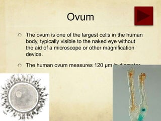
The ovum is one of the largest cells in the human
body, typically visible to the naked eye without
the aid of a microscope or other magnification
device.

The human ovum measures 120 µm in diameter.
Sperm

Motile sperm cells of algae and seedless
plants.Motile sperm cells typically move via
flagella and require water in order to swim toward
the egg for fertilization. These cells cannot swim
backwards due to the nature of their propulsion.

Non-motile sperm cells called spermatia lack
flagella and therefore cannot swim. Spermatia
are produced in a spermatangium.
Kidney

Kidney glomerulus parietal cell

Kidney glomerulus podocyte

Kidney proximal tubule brush border cell

Loop of Henle thin segment cell

Thick ascending limb cell

Kidney distal tubule cell

Kidney collecting duct cellInterstitial kidney cell
Neuron
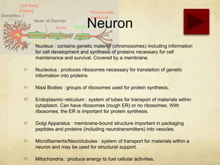
Nucleus : contains genetic material (chromosomes) including information
for cell development and synthesis of proteins necessary for cell
maintenance and survival. Covered by a membrane.

Nucleolus : produces ribosomes necessary for translation of genetic
information into proteins

Nissl Bodies : groups of ribosomes used for protein synthesis.

Endoplasmic reticulum : system of tubes for transport of materials within
cytoplasm. Can have ribosomes (rough ER) or no ribosomes. With
ribosomes, the ER is important for protein synthesis.

Golgi Apparatus : membrane-bound structure important in packaging
peptides and proteins (including neurotransmitters) into vesicles.

Microfilaments/Neurotubules : system of transport for materials within a
neuron and may be used for structural support.

Mitochondria : produce energy to fuel cellular activities.
Stomach
Mucous cells: secrete an alkaline mucus that
protects the epithelium against shear stress and
acid

Parietal cells: secrete hydrochloric acid!

Chief cells: secrete pepsin, a proteolyticenzyme

G cells: secrete the hormone gastrin
Thank You

More Related Content

What's hot

Cell structure & function
Cell structure & functionCell structure & function
Cell structure & function
tpilcher1969
 
Cell parts and functions transport across membranes
Cell parts and functions transport across membranesCell parts and functions transport across membranes
Cell parts and functions transport across membranes
Omar Jacalne
 
Cell structure
Cell structureCell structure
Cell structure
jugafoce
 
2, origin, structure and function of eucaryotes cells 5 11-2012
2, origin, structure and function of eucaryotes cells 5 11-20122, origin, structure and function of eucaryotes cells 5 11-2012
2, origin, structure and function of eucaryotes cells 5 11-2012
ganganaik
 

What's hot (20)

Cell structure
Cell structureCell structure
Cell structure
 
Cell ultrastructure and cell organelles
Cell ultrastructure and cell organellesCell ultrastructure and cell organelles
Cell ultrastructure and cell organelles
 
Cells structure
Cells structureCells structure
Cells structure
 
7-2: Eukaryotic Cell Structure
7-2: Eukaryotic Cell Structure7-2: Eukaryotic Cell Structure
7-2: Eukaryotic Cell Structure
 
Cell structure & function
Cell structure & functionCell structure & function
Cell structure & function
 
Cell structure and functions
Cell structure and functionsCell structure and functions
Cell structure and functions
 
Animal cell Anatomy and Physiology
Animal cell Anatomy and PhysiologyAnimal cell Anatomy and Physiology
Animal cell Anatomy and Physiology
 
Cell physiology
Cell physiologyCell physiology
Cell physiology
 
ICSE Class X Biology Cell The Structural and Functional Unit of Life
ICSE Class X Biology Cell The Structural and Functional Unit of LifeICSE Class X Biology Cell The Structural and Functional Unit of Life
ICSE Class X Biology Cell The Structural and Functional Unit of Life
 
Cell parts and functions transport across membranes
Cell parts and functions transport across membranesCell parts and functions transport across membranes
Cell parts and functions transport across membranes
 
Cell and cell organelles by Dr.Tehmas
Cell and cell organelles by Dr.TehmasCell and cell organelles by Dr.Tehmas
Cell and cell organelles by Dr.Tehmas
 
Anatomy and Physiology Cell organelles
 Anatomy and Physiology Cell organelles Anatomy and Physiology Cell organelles
Anatomy and Physiology Cell organelles
 
Structure of cell
Structure of cell  Structure of cell
Structure of cell
 
Cell structure
Cell structureCell structure
Cell structure
 
Cell Anatomy Mini Lecture
Cell Anatomy Mini LectureCell Anatomy Mini Lecture
Cell Anatomy Mini Lecture
 
Cell & Its Orgenells
Cell & Its OrgenellsCell & Its Orgenells
Cell & Its Orgenells
 
Cellular Structures and Their Functions
Cellular Structures and Their FunctionsCellular Structures and Their Functions
Cellular Structures and Their Functions
 
Cell structure and function
Cell structure and functionCell structure and function
Cell structure and function
 
2, origin, structure and function of eucaryotes cells 5 11-2012
2, origin, structure and function of eucaryotes cells 5 11-20122, origin, structure and function of eucaryotes cells 5 11-2012
2, origin, structure and function of eucaryotes cells 5 11-2012
 
Cell Functions
Cell Functions Cell Functions
Cell Functions
 

Viewers also liked

Human cell & it's function
Human cell & it's functionHuman cell & it's function
Human cell & it's function
Nursing Path
 
Cells Powerpoint Presentation
Cells Powerpoint PresentationCells Powerpoint Presentation
Cells Powerpoint Presentation
cprizel
 
Lab 1st year cytology
Lab 1st year cytologyLab 1st year cytology
Lab 1st year cytology
abdalslam1993
 
General histology
General  histologyGeneral  histology
General histology
KemUnited
 
Cell histology
Cell histologyCell histology
Cell histology
helnamaria
 
4.2 Parts Of A Eukaryotic Cell
4.2 Parts Of A Eukaryotic Cell4.2 Parts Of A Eukaryotic Cell
4.2 Parts Of A Eukaryotic Cell
MissWander
 
Hematoxylin & eosinstaining
Hematoxylin & eosinstainingHematoxylin & eosinstaining
Hematoxylin & eosinstaining
Layal Fahad
 
Cell nucleus
Cell nucleusCell nucleus
Cell nucleus
kitykety
 

Viewers also liked (20)

Human cell & it's function
Human cell & it's functionHuman cell & it's function
Human cell & it's function
 
Cell Structure And Function
Cell Structure And FunctionCell Structure And Function
Cell Structure And Function
 
Cells Powerpoint Presentation
Cells Powerpoint PresentationCells Powerpoint Presentation
Cells Powerpoint Presentation
 
Cell : Structure and Function Part 01
Cell : Structure and Function Part 01Cell : Structure and Function Part 01
Cell : Structure and Function Part 01
 
Lab 1st year cytology
Lab 1st year cytologyLab 1st year cytology
Lab 1st year cytology
 
The cell
The cellThe cell
The cell
 
Cell frcationation
Cell frcationationCell frcationation
Cell frcationation
 
Neuron
NeuronNeuron
Neuron
 
Basic cell structure
Basic cell structureBasic cell structure
Basic cell structure
 
Gen. histology (introduction) 1
Gen. histology (introduction) 1Gen. histology (introduction) 1
Gen. histology (introduction) 1
 
General histology
General  histologyGeneral  histology
General histology
 
Cell histology
Cell histologyCell histology
Cell histology
 
Cytology - Prac. Histology
Cytology - Prac. HistologyCytology - Prac. Histology
Cytology - Prac. Histology
 
4.2 Parts Of A Eukaryotic Cell
4.2 Parts Of A Eukaryotic Cell4.2 Parts Of A Eukaryotic Cell
4.2 Parts Of A Eukaryotic Cell
 
Hematoxylin & eosinstaining
Hematoxylin & eosinstainingHematoxylin & eosinstaining
Hematoxylin & eosinstaining
 
Introduction to Histology
Introduction to HistologyIntroduction to Histology
Introduction to Histology
 
Types of light microscopy
Types of light microscopy Types of light microscopy
Types of light microscopy
 
CELL NUCLEUS
CELL NUCLEUSCELL NUCLEUS
CELL NUCLEUS
 
Cell nucleus
Cell nucleusCell nucleus
Cell nucleus
 
cytology
cytologycytology
cytology
 

Similar to Human's cell

cells structure and transport mechanisms
cells structure and transport mechanismscells structure and transport mechanisms
cells structure and transport mechanisms
Reisa Roberts
 
Cell organelles (2)
Cell organelles (2)Cell organelles (2)
Cell organelles (2)
ShaZz Zia
 
Plant and Animalcell
Plant and AnimalcellPlant and Animalcell
Plant and Animalcell
Malti Aswal
 

Similar to Human's cell (20)

Hematopoietic system.pdf
Hematopoietic system.pdfHematopoietic system.pdf
Hematopoietic system.pdf
 
The Human Cell - Cell Migration Class - Topic
The Human Cell - Cell Migration Class - TopicThe Human Cell - Cell Migration Class - Topic
The Human Cell - Cell Migration Class - Topic
 
Cell and tissue
Cell and tissueCell and tissue
Cell and tissue
 
CELL STRUCTURE AND TRANSPORT.pptx
CELL STRUCTURE AND TRANSPORT.pptxCELL STRUCTURE AND TRANSPORT.pptx
CELL STRUCTURE AND TRANSPORT.pptx
 
2.cell structure
2.cell structure2.cell structure
2.cell structure
 
Lymphatic System
Lymphatic SystemLymphatic System
Lymphatic System
 
Cells
CellsCells
Cells
 
Cells
CellsCells
Cells
 
Types-of-Plant-Cells-animal-cells.pptx
Types-of-Plant-Cells-animal-cells.pptxTypes-of-Plant-Cells-animal-cells.pptx
Types-of-Plant-Cells-animal-cells.pptx
 
Structure of cell by Avinash Nayak
Structure of cell by Avinash NayakStructure of cell by Avinash Nayak
Structure of cell by Avinash Nayak
 
Mdsc 1001 pbl problem 1
Mdsc 1001 pbl problem 1Mdsc 1001 pbl problem 1
Mdsc 1001 pbl problem 1
 
cells structure and transport mechanisms
cells structure and transport mechanismscells structure and transport mechanisms
cells structure and transport mechanisms
 
Cell organelles (2)
Cell organelles (2)Cell organelles (2)
Cell organelles (2)
 
Plant and Animalcell
Plant and AnimalcellPlant and Animalcell
Plant and Animalcell
 
GCSE-Triple-Biology-2023.pdf
GCSE-Triple-Biology-2023.pdfGCSE-Triple-Biology-2023.pdf
GCSE-Triple-Biology-2023.pdf
 
Anatomy & Physiology Slides
Anatomy & Physiology SlidesAnatomy & Physiology Slides
Anatomy & Physiology Slides
 
Anatomy & Physiology Slides
Anatomy & Physiology SlidesAnatomy & Physiology Slides
Anatomy & Physiology Slides
 
Animal cell: Anatomy and Physiology
Animal cell: Anatomy and PhysiologyAnimal cell: Anatomy and Physiology
Animal cell: Anatomy and Physiology
 
ANATOMY OF CELL.pptx
ANATOMY OF CELL.pptxANATOMY OF CELL.pptx
ANATOMY OF CELL.pptx
 
Eukaryotic Cells
Eukaryotic CellsEukaryotic Cells
Eukaryotic Cells
 

Recently uploaded

Transparency, Recognition and the role of eSealing - Ildiko Mazar and Koen No...
Transparency, Recognition and the role of eSealing - Ildiko Mazar and Koen No...Transparency, Recognition and the role of eSealing - Ildiko Mazar and Koen No...
Transparency, Recognition and the role of eSealing - Ildiko Mazar and Koen No...
EADTU
 

Recently uploaded (20)

80 ĐỀ THI THỬ TUYỂN SINH TIẾNG ANH VÀO 10 SỞ GD – ĐT THÀNH PHỐ HỒ CHÍ MINH NĂ...
80 ĐỀ THI THỬ TUYỂN SINH TIẾNG ANH VÀO 10 SỞ GD – ĐT THÀNH PHỐ HỒ CHÍ MINH NĂ...80 ĐỀ THI THỬ TUYỂN SINH TIẾNG ANH VÀO 10 SỞ GD – ĐT THÀNH PHỐ HỒ CHÍ MINH NĂ...
80 ĐỀ THI THỬ TUYỂN SINH TIẾNG ANH VÀO 10 SỞ GD – ĐT THÀNH PHỐ HỒ CHÍ MINH NĂ...
 
Wellbeing inclusion and digital dystopias.pptx
Wellbeing inclusion and digital dystopias.pptxWellbeing inclusion and digital dystopias.pptx
Wellbeing inclusion and digital dystopias.pptx
 
FICTIONAL SALESMAN/SALESMAN SNSW 2024.pdf
FICTIONAL SALESMAN/SALESMAN SNSW 2024.pdfFICTIONAL SALESMAN/SALESMAN SNSW 2024.pdf
FICTIONAL SALESMAN/SALESMAN SNSW 2024.pdf
 
TỔNG ÔN TẬP THI VÀO LỚP 10 MÔN TIẾNG ANH NĂM HỌC 2023 - 2024 CÓ ĐÁP ÁN (NGỮ Â...
TỔNG ÔN TẬP THI VÀO LỚP 10 MÔN TIẾNG ANH NĂM HỌC 2023 - 2024 CÓ ĐÁP ÁN (NGỮ Â...TỔNG ÔN TẬP THI VÀO LỚP 10 MÔN TIẾNG ANH NĂM HỌC 2023 - 2024 CÓ ĐÁP ÁN (NGỮ Â...
TỔNG ÔN TẬP THI VÀO LỚP 10 MÔN TIẾNG ANH NĂM HỌC 2023 - 2024 CÓ ĐÁP ÁN (NGỮ Â...
 
Transparency, Recognition and the role of eSealing - Ildiko Mazar and Koen No...
Transparency, Recognition and the role of eSealing - Ildiko Mazar and Koen No...Transparency, Recognition and the role of eSealing - Ildiko Mazar and Koen No...
Transparency, Recognition and the role of eSealing - Ildiko Mazar and Koen No...
 
Towards a code of practice for AI in AT.pptx
Towards a code of practice for AI in AT.pptxTowards a code of practice for AI in AT.pptx
Towards a code of practice for AI in AT.pptx
 
How to setup Pycharm environment for Odoo 17.pptx
How to setup Pycharm environment for Odoo 17.pptxHow to setup Pycharm environment for Odoo 17.pptx
How to setup Pycharm environment for Odoo 17.pptx
 
HMCS Max Bernays Pre-Deployment Brief (May 2024).pptx
HMCS Max Bernays Pre-Deployment Brief (May 2024).pptxHMCS Max Bernays Pre-Deployment Brief (May 2024).pptx
HMCS Max Bernays Pre-Deployment Brief (May 2024).pptx
 
Details on CBSE Compartment Exam.pptx1111
Details on CBSE Compartment Exam.pptx1111Details on CBSE Compartment Exam.pptx1111
Details on CBSE Compartment Exam.pptx1111
 
What is 3 Way Matching Process in Odoo 17.pptx
What is 3 Way Matching Process in Odoo 17.pptxWhat is 3 Way Matching Process in Odoo 17.pptx
What is 3 Way Matching Process in Odoo 17.pptx
 
COMMUNICATING NEGATIVE NEWS - APPROACHES .pptx
COMMUNICATING NEGATIVE NEWS - APPROACHES .pptxCOMMUNICATING NEGATIVE NEWS - APPROACHES .pptx
COMMUNICATING NEGATIVE NEWS - APPROACHES .pptx
 
Python Notes for mca i year students osmania university.docx
Python Notes for mca i year students osmania university.docxPython Notes for mca i year students osmania university.docx
Python Notes for mca i year students osmania university.docx
 
21st_Century_Skills_Framework_Final_Presentation_2.pptx
21st_Century_Skills_Framework_Final_Presentation_2.pptx21st_Century_Skills_Framework_Final_Presentation_2.pptx
21st_Century_Skills_Framework_Final_Presentation_2.pptx
 
Unit 3 Emotional Intelligence and Spiritual Intelligence.pdf
Unit 3 Emotional Intelligence and Spiritual Intelligence.pdfUnit 3 Emotional Intelligence and Spiritual Intelligence.pdf
Unit 3 Emotional Intelligence and Spiritual Intelligence.pdf
 
On National Teacher Day, meet the 2024-25 Kenan Fellows
On National Teacher Day, meet the 2024-25 Kenan FellowsOn National Teacher Day, meet the 2024-25 Kenan Fellows
On National Teacher Day, meet the 2024-25 Kenan Fellows
 
REMIFENTANIL: An Ultra short acting opioid.pptx
REMIFENTANIL: An Ultra short acting opioid.pptxREMIFENTANIL: An Ultra short acting opioid.pptx
REMIFENTANIL: An Ultra short acting opioid.pptx
 
Accessible Digital Futures project (20/03/2024)
Accessible Digital Futures project (20/03/2024)Accessible Digital Futures project (20/03/2024)
Accessible Digital Futures project (20/03/2024)
 
How to Create and Manage Wizard in Odoo 17
How to Create and Manage Wizard in Odoo 17How to Create and Manage Wizard in Odoo 17
How to Create and Manage Wizard in Odoo 17
 
Exploring_the_Narrative_Style_of_Amitav_Ghoshs_Gun_Island.pptx
Exploring_the_Narrative_Style_of_Amitav_Ghoshs_Gun_Island.pptxExploring_the_Narrative_Style_of_Amitav_Ghoshs_Gun_Island.pptx
Exploring_the_Narrative_Style_of_Amitav_Ghoshs_Gun_Island.pptx
 
On_Translating_a_Tamil_Poem_by_A_K_Ramanujan.pptx
On_Translating_a_Tamil_Poem_by_A_K_Ramanujan.pptxOn_Translating_a_Tamil_Poem_by_A_K_Ramanujan.pptx
On_Translating_a_Tamil_Poem_by_A_K_Ramanujan.pptx
 

Human's cell

  • 1. Human’s Cells By Christian Dennis, SharineSinaga, Kelvin Prihadi
  • 2. Content Heart Intestine Liver Stomach Blood Kidney Skin Ovum Muscle Sperm Neuron
  • 3. Heart Scientists consider heart cells a part of the muscle cell family, though with unique differences in mitochondria, intercalated disks and t-tubes, as well as in cellular growth. Mitochondria Unlike other muscle cells, heart cells are highly resistant to fatigue because they have more mitochondria organelles than any other cell in the body. Intercalated disks Intercalated disks regulate the passage of positive and negative electrons. As electric currents repel and attract, it causes electron depolarization, which regulates heartbeat contractions. T-Tubules Plasma membranes that surround each cell and organize them into pairs to create the striated muscles strands used to build the myocardium
  • 4. Liver Hepatocytes Hepatocytesare the main cell for protein synthesis. They are also responsible for recycling lipoproteins. Kupffer Cells Kupffercells are specialized macrophages. A macrophage is a cell that is a part of the immune system. StellateCells Stellatecells are part of the nervous system. They innervate the organ to provide a connection between the brain and the liver. It helps with pain and sensory information from the liver to provide communication Mitochondria Mitochondria are located in most human cells, but there is an elevated number in liver cells. Mitochondria play a large role in metabolism of proteins, carbohydrates and lipids.
  • 5. Skin Melanocytes: The main function of melanocytes is to produce melanin, which is responsible for the colour of our skin Langerhans Cells arise from bone marrow and migrate to the epidermis. Langerhans cells interact with white blood cells called "helper T cells" in immune responses and are easily damaged by UV radiation Merkel Cells: Merkel cells are located in the deepest layer (stratum basale) of the epidermis of hairless skin, where they are attached to keratinocytes by desmosomes. Dermo-epidermal JunctionThe Epidermis and Dermis are separated by the Dermo-Epithelial Junction. This junction holds the epidermis and dermis together and this is achieved by various fibers including collagen and desmosomes. .
  • 6. Muscle Muscle is composed of thousands of muscle fibers, each composed of a single muscle cell. A muscle cell contains a series of ultramicroscopic filaments called myofibrils. Each myofibril is a muscle cell that contains units called sarcomeres. Sarcomerescontain thick microfilaments composed of the protein myosin. Sarcomeres also contain thin microfilaments composed of the protein actin. The actin and myosin filaments are arranged parallel to one another, with the myosin filaments' molecular “heads” protruding toward the actin filaments. .
  • 7. Blood NEUTROPHIL This granulocyte has very tiny light staining granules. The nucleus is frequently multi-lobed with lobes connected by thin strands of nuclear material. These cells are capable of phagocytizing foreign cells, toxins, and viruses.
  • 8. Blood T EOSINOPHIL his granulocyte has large granules (A) which are acidophilic and appear pink (or red) in a stained preparation. This micrograph was color enhanced to illustrate this feature. The nucleus often has two lobes connected by a band of nuclear material.
  • 9. Blood BASOPHIL
 The basophilic granules in this cell are large, stain deep blue to purple, and are often so numerous they mask the nucleus. These granules contain histamines (cause vasodilation) and heparin (anticoagulant).
  • 10. Blood LYMPHOCYTE The lymphocyte is an agranular cell with very clear cytoplasm which stains pale blue. Its nucleus is very large for the size of the cell and stains dark purple. (Notice that the nucleus almost fills the cell leaving a very thin rim of cytoplasm.) This cell is much smaller than the three granulocytes (which are all about the same size). These cells play an important role in our immune response.
  • 11. Blood MONOCYTE

This cell is the largest of the leukocytes and is agranular. The nucleus is most often "U" or kidney bean shaped; the cytoplasm is abundant and light blue. These cells leave the blood streamto become macrophages. As a monocyte or macrophage, these cells are phagocytic and defend the body against viruses and bacteria.
  • 12. Blood ERYTHROCYTE

The background cells in this micrograph are erythrocytes (red blood cells). These cells are non-nucleated, biconcave discs that are filled with hemoglobin. The primary function of these cells is to carry oxygen from the lungs to the body cells.
  • 13. Blood THROMBOCYTES – PLATELETS Platelets, which are cell fragments. Platelets are important for proper blood clotting. Each cubic millimeter of blood should contain 250,000 to 500,000 of these. If the number is too high, spontaneous clotting may occur. If the number is too low, clotting may not occur when necessary.
  • 14. Intestine Along the whole length of the gut in the glandular epithelium are goblet cells. These secrete mucus which lubricates the passage of food along and protects it from digestive enzymes.
  • 15. Ovum The ovum is one of the largest cells in the human body, typically visible to the naked eye without the aid of a microscope or other magnification device. The human ovum measures 120 µm in diameter.
  • 16. Sperm Motile sperm cells of algae and seedless plants.Motile sperm cells typically move via flagella and require water in order to swim toward the egg for fertilization. These cells cannot swim backwards due to the nature of their propulsion. Non-motile sperm cells called spermatia lack flagella and therefore cannot swim. Spermatia are produced in a spermatangium.
  • 17. Kidney Kidney glomerulus parietal cell Kidney glomerulus podocyte Kidney proximal tubule brush border cell Loop of Henle thin segment cell Thick ascending limb cell Kidney distal tubule cell Kidney collecting duct cellInterstitial kidney cell
  • 18. Neuron Nucleus : contains genetic material (chromosomes) including information for cell development and synthesis of proteins necessary for cell maintenance and survival. Covered by a membrane. Nucleolus : produces ribosomes necessary for translation of genetic information into proteins Nissl Bodies : groups of ribosomes used for protein synthesis. Endoplasmic reticulum : system of tubes for transport of materials within cytoplasm. Can have ribosomes (rough ER) or no ribosomes. With ribosomes, the ER is important for protein synthesis. Golgi Apparatus : membrane-bound structure important in packaging peptides and proteins (including neurotransmitters) into vesicles. Microfilaments/Neurotubules : system of transport for materials within a neuron and may be used for structural support. Mitochondria : produce energy to fuel cellular activities.
  • 19. Stomach Mucous cells: secrete an alkaline mucus that protects the epithelium against shear stress and acid Parietal cells: secrete hydrochloric acid! Chief cells: secrete pepsin, a proteolyticenzyme G cells: secrete the hormone gastrin