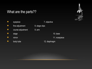
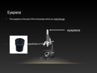
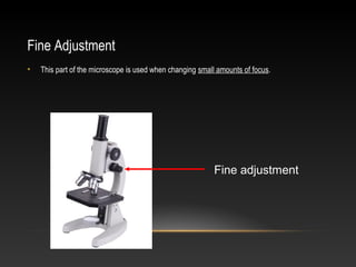
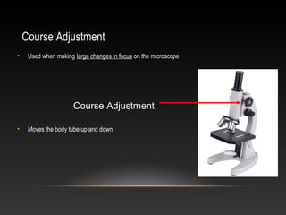
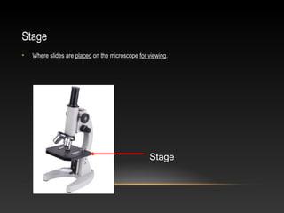
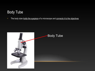
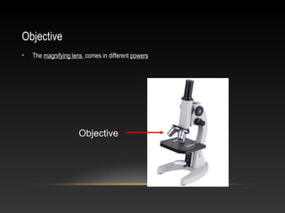
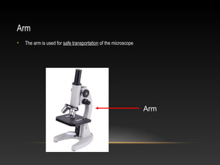
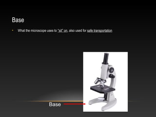
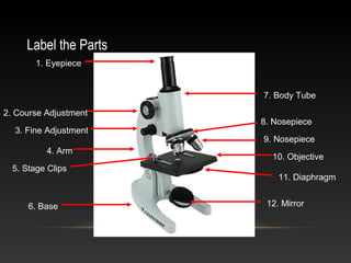
The document describes the main parts of a microscope and their functions. The eyepiece is used to view the slide and usually has a magnification of 10x. The fine and course adjustments control focus, with the fine adjustment used for small changes and the course adjustment for larger movements. The stage holds the slide and the stage clips secure it in place. The objective lenses provide different magnifications and the nosepiece allows rotation between them. The mirror, body tube, arm, base, and diaphragm also have specific microscope functions.
Compound microscopes are what most people visualize when they think about microscopes. They are available in monocular, binocular and trinocular formats. They have a number of objectives (the lens closest to the object being viewed) of varying magnifications mounted in a rotating nosepiece.
as a partial requirement for one of my subject for this semester
I would like you to view my presentation and comment as well
I will be very glad if you find my presentation interesting, or comment on how I can improve my craft, THANK YOU :)
A microscope (from the Ancient Greek: μικρός, mikrós, "small" and σκοπεῖν, skopeîn, "to look" or "see") is an instrument used to see objects that are too small to be seen by the naked eye. Microscopy is the science of investigating small objects and structures using such an instrument. Microscopic means invisible to the eye unless aided by a microscope to know more visit our website www.jaincoexport.com
The present is on Instrumentation of various microscopes such as compound microscope, stereo microscope, polarized microscope, comparison microscope, fluorescent microscope, dark field microscope, electron microscope and it also discusses about the forensic applications of each microscope briefly.
Araling panlipunan
Artikulo
Usapan
Basahin
Baguhin
Baguhin ang source
Tingnan ang nakaraan
Mula sa Wikipedia, ang malayang ensiklopedya
Ang araling panlipunan (Ingles: social studies) ay isang katagang naglalarawan sa isang malawak na mga pag-aaral sa iba't ibang larangan na kinakasangkutan ng nakaraan at kasalukuyang pakikipag-ugnayan at kaugalian ng mga tao. Sa halip na nakatuon sa lalim ng alin mang mga paksa, nagbibigay ang araling panlipunan ng isang malawak na buod ng kaugalian ng sangkatauhan. Kinikilala ang araling panlipunan bilang pangalan ng kurso na tinuturo sa paaralang elementarya at mataas na paaralan, ngunit maaaring tumukoy din ito sa pag-aaral ng partikular na aspeto ng lipunan ng tao sa ilang kolehiyo sa buong mundo.
Introduction:
RNA interference (RNAi) or Post-Transcriptional Gene Silencing (PTGS) is an important biological process for modulating eukaryotic gene expression.
It is highly conserved process of posttranscriptional gene silencing by which double stranded RNA (dsRNA) causes sequence-specific degradation of mRNA sequences.
dsRNA-induced gene silencing (RNAi) is reported in a wide range of eukaryotes ranging from worms, insects, mammals and plants.
This process mediates resistance to both endogenous parasitic and exogenous pathogenic nucleic acids, and regulates the expression of protein-coding genes.
What are small ncRNAs?
micro RNA (miRNA)
short interfering RNA (siRNA)
Properties of small non-coding RNA:
Involved in silencing mRNA transcripts.
Called “small” because they are usually only about 21-24 nucleotides long.
Synthesized by first cutting up longer precursor sequences (like the 61nt one that Lee discovered).
Silence an mRNA by base pairing with some sequence on the mRNA.
Discovery of siRNA?
The first small RNA:
In 1993 Rosalind Lee (Victor Ambros lab) was studying a non- coding gene in C. elegans, lin-4, that was involved in silencing of another gene, lin-14, at the appropriate time in the
development of the worm C. elegans.
Two small transcripts of lin-4 (22nt and 61nt) were found to be complementary to a sequence in the 3' UTR of lin-14.
Because lin-4 encoded no protein, she deduced that it must be these transcripts that are causing the silencing by RNA-RNA interactions.
Types of RNAi ( non coding RNA)
MiRNA
Length (23-25 nt)
Trans acting
Binds with target MRNA in mismatch
Translation inhibition
Si RNA
Length 21 nt.
Cis acting
Bind with target Mrna in perfect complementary sequence
Piwi-RNA
Length ; 25 to 36 nt.
Expressed in Germ Cells
Regulates trnasposomes activity
MECHANISM OF RNAI:
First the double-stranded RNA teams up with a protein complex named Dicer, which cuts the long RNA into short pieces.
Then another protein complex called RISC (RNA-induced silencing complex) discards one of the two RNA strands.
The RISC-docked, single-stranded RNA then pairs with the homologous mRNA and destroys it.
THE RISC COMPLEX:
RISC is large(>500kD) RNA multi- protein Binding complex which triggers MRNA degradation in response to MRNA
Unwinding of double stranded Si RNA by ATP independent Helicase
Active component of RISC is Ago proteins( ENDONUCLEASE) which cleave target MRNA.
DICER: endonuclease (RNase Family III)
Argonaute: Central Component of the RNA-Induced Silencing Complex (RISC)
One strand of the dsRNA produced by Dicer is retained in the RISC complex in association with Argonaute
ARGONAUTE PROTEIN :
1.PAZ(PIWI/Argonaute/ Zwille)- Recognition of target MRNA
2.PIWI (p-element induced wimpy Testis)- breaks Phosphodiester bond of mRNA.)RNAse H activity.
MiRNA:
The Double-stranded RNAs are naturally produced in eukaryotic cells during development, and they have a key role in regulating gene expression .
(May 29th, 2024) Advancements in Intravital Microscopy- Insights for Preclini...Scintica Instrumentation
Intravital microscopy (IVM) is a powerful tool utilized to study cellular behavior over time and space in vivo. Much of our understanding of cell biology has been accomplished using various in vitro and ex vivo methods; however, these studies do not necessarily reflect the natural dynamics of biological processes. Unlike traditional cell culture or fixed tissue imaging, IVM allows for the ultra-fast high-resolution imaging of cellular processes over time and space and were studied in its natural environment. Real-time visualization of biological processes in the context of an intact organism helps maintain physiological relevance and provide insights into the progression of disease, response to treatments or developmental processes.
In this webinar we give an overview of advanced applications of the IVM system in preclinical research. IVIM technology is a provider of all-in-one intravital microscopy systems and solutions optimized for in vivo imaging of live animal models at sub-micron resolution. The system’s unique features and user-friendly software enables researchers to probe fast dynamic biological processes such as immune cell tracking, cell-cell interaction as well as vascularization and tumor metastasis with exceptional detail. This webinar will also give an overview of IVM being utilized in drug development, offering a view into the intricate interaction between drugs/nanoparticles and tissues in vivo and allows for the evaluation of therapeutic intervention in a variety of tissues and organs. This interdisciplinary collaboration continues to drive the advancements of novel therapeutic strategies.
Earliest Galaxies in the JADES Origins Field: Luminosity Function and Cosmic ...Sérgio Sacani
We characterize the earliest galaxy population in the JADES Origins Field (JOF), the deepest
imaging field observed with JWST. We make use of the ancillary Hubble optical images (5 filters
spanning 0.4−0.9µm) and novel JWST images with 14 filters spanning 0.8−5µm, including 7 mediumband filters, and reaching total exposure times of up to 46 hours per filter. We combine all our data
at > 2.3µm to construct an ultradeep image, reaching as deep as ≈ 31.4 AB mag in the stack and
30.3-31.0 AB mag (5σ, r = 0.1” circular aperture) in individual filters. We measure photometric
redshifts and use robust selection criteria to identify a sample of eight galaxy candidates at redshifts
z = 11.5 − 15. These objects show compact half-light radii of R1/2 ∼ 50 − 200pc, stellar masses of
M⋆ ∼ 107−108M⊙, and star-formation rates of SFR ∼ 0.1−1 M⊙ yr−1
. Our search finds no candidates
at 15 < z < 20, placing upper limits at these redshifts. We develop a forward modeling approach to
infer the properties of the evolving luminosity function without binning in redshift or luminosity that
marginalizes over the photometric redshift uncertainty of our candidate galaxies and incorporates the
impact of non-detections. We find a z = 12 luminosity function in good agreement with prior results,
and that the luminosity function normalization and UV luminosity density decline by a factor of ∼ 2.5
from z = 12 to z = 14. We discuss the possible implications of our results in the context of theoretical
models for evolution of the dark matter halo mass function.
This pdf is about the Schizophrenia.
For more details visit on YouTube; @SELF-EXPLANATORY;
https://www.youtube.com/channel/UCAiarMZDNhe1A3Rnpr_WkzA/videos
Thanks...!
Observation of Io’s Resurfacing via Plume Deposition Using Ground-based Adapt...Sérgio Sacani
Since volcanic activity was first discovered on Io from Voyager images in 1979, changes
on Io’s surface have been monitored from both spacecraft and ground-based telescopes.
Here, we present the highest spatial resolution images of Io ever obtained from a groundbased telescope. These images, acquired by the SHARK-VIS instrument on the Large
Binocular Telescope, show evidence of a major resurfacing event on Io’s trailing hemisphere. When compared to the most recent spacecraft images, the SHARK-VIS images
show that a plume deposit from a powerful eruption at Pillan Patera has covered part
of the long-lived Pele plume deposit. Although this type of resurfacing event may be common on Io, few have been detected due to the rarity of spacecraft visits and the previously low spatial resolution available from Earth-based telescopes. The SHARK-VIS instrument ushers in a new era of high resolution imaging of Io’s surface using adaptive
optics at visible wavelengths.
Seminar of U.V. Spectroscopy by SAMIR PANDASAMIR PANDA
Spectroscopy is a branch of science dealing the study of interaction of electromagnetic radiation with matter.
Ultraviolet-visible spectroscopy refers to absorption spectroscopy or reflect spectroscopy in the UV-VIS spectral region.
Ultraviolet-visible spectroscopy is an analytical method that can measure the amount of light received by the analyte.
2. What are the parts??
eyepiece 7. objective
fine adjustment 8. stage clips
course adjustment 9. arm
stage 10. base
mirror 11. nosepiece
body tube 12. diaphragm
3. Eyepiece
• The eyepiece is the part of the microscope which you look through.
• It usually has its own magnification of 10x.
eyepiece
4. Fine Adjustment
• This part of the microscope is used when changing small amounts of focus.
Fine adjustment
5. Course Adjustment
• Used when making large changes in focus on the microscope
Course Adjustment
• Moves the body tube up and down
6. Adjustment
• Some microscopes only have ONE adjustment knob
• If so, it is a course adjustment!
7. Stage
• Where slides are placed on the microscope for viewing.
Stage
8. Mirror
• The mirror is used to reflect light from a light source up through the diaphragm
Mirror
9. Body Tube
• The body tube holds the eyepiece of a microscope and connects it to the objectives
Body Tube
10. Objective
• The magnifying lens, comes in different powers
Objective
11. Stage Clips
• The stage clips hold the slide in place
Stage Clips
12. Arm
• The arm is used for safe transportation of the microscope
Arm
13. Base
• What the microscope uses to “sit” on, also used for safe transportation
Base
14. Nosepiece
• The nosepiece holds two or more objective lenses and can be rotated to easily change
powers
Nosepiece
15. Diaphragm
• Used to vary the amount of light which passes through the slide
• Usually better in lower amounts
Diaphragm
16. Label the Parts
1. Eyepiece
2. Course Adjustment
3. Fine Adjustment
4. Arm
5. Stage Clips
6. Base
7. Body Tube
8. Nosepiece
9. Nosepiece
10. Objective
11. Diaphragm
12. Mirror