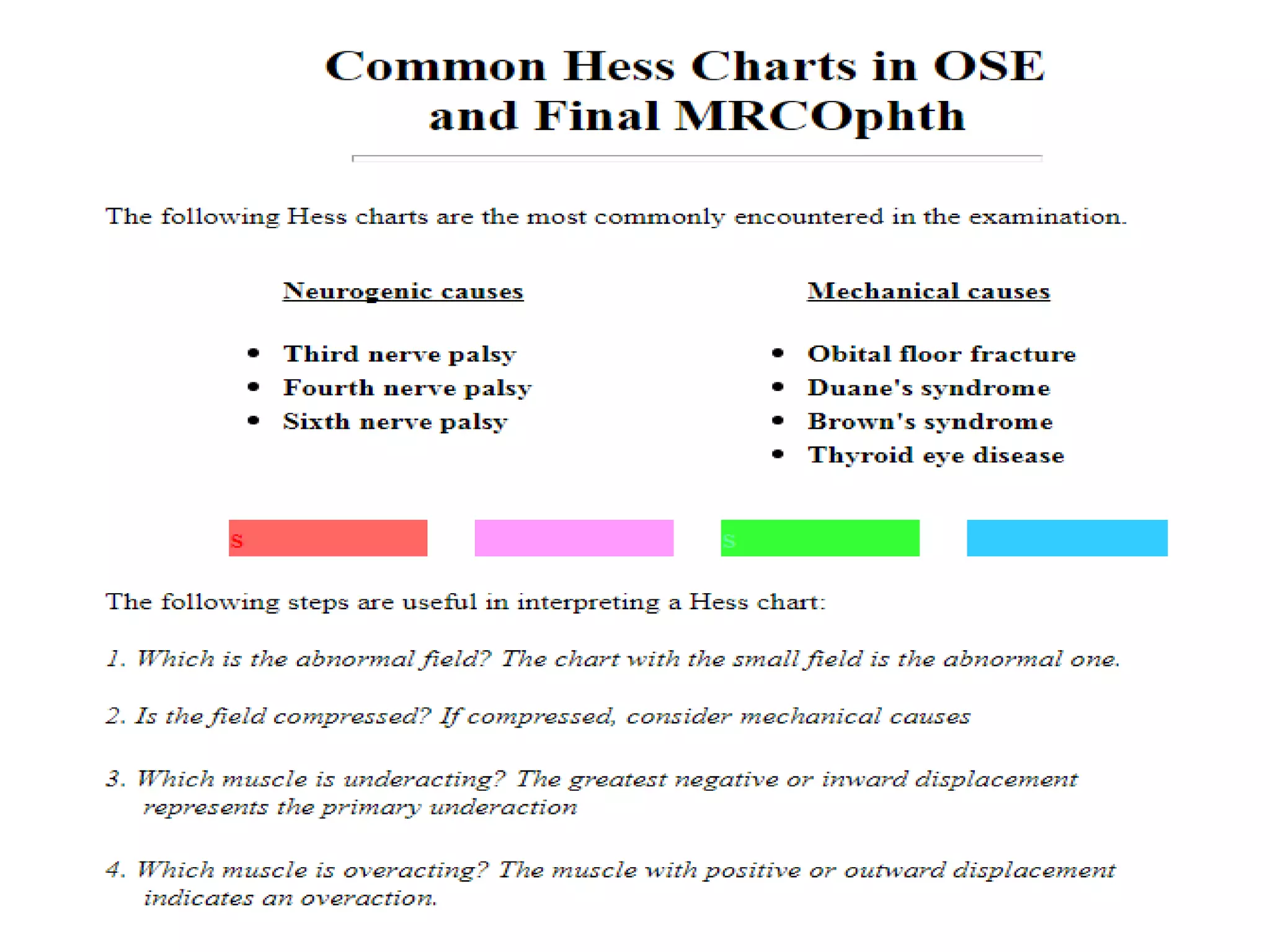
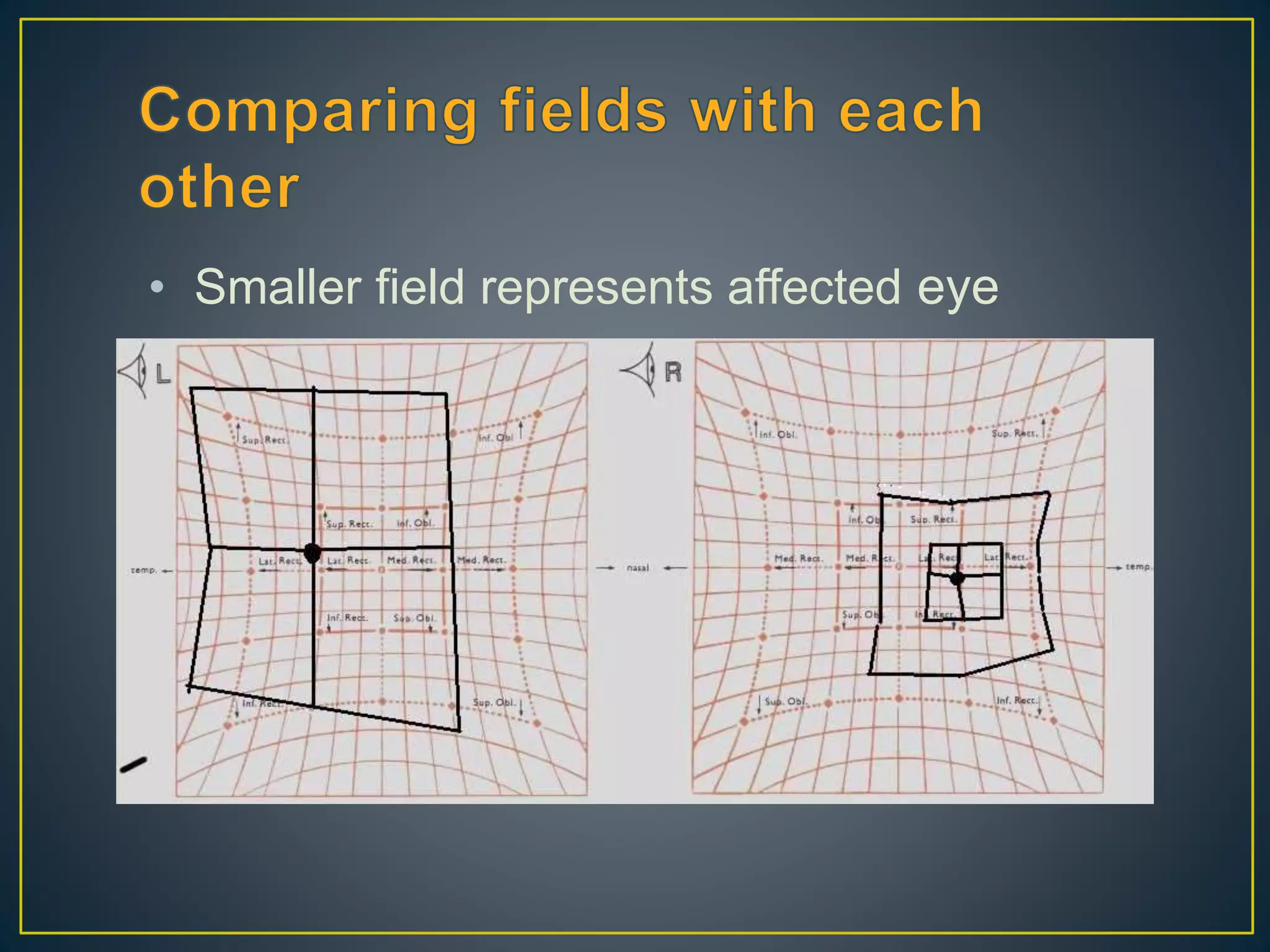
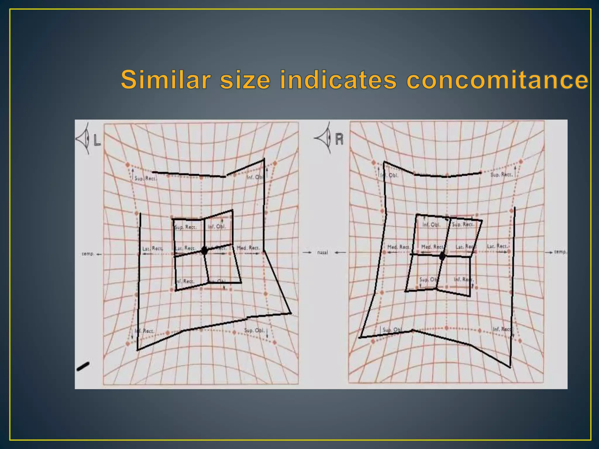
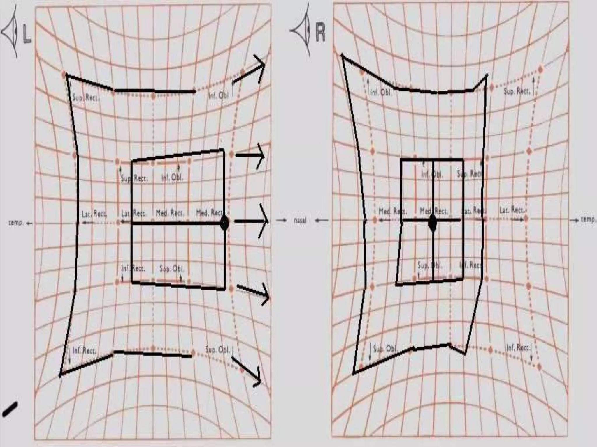
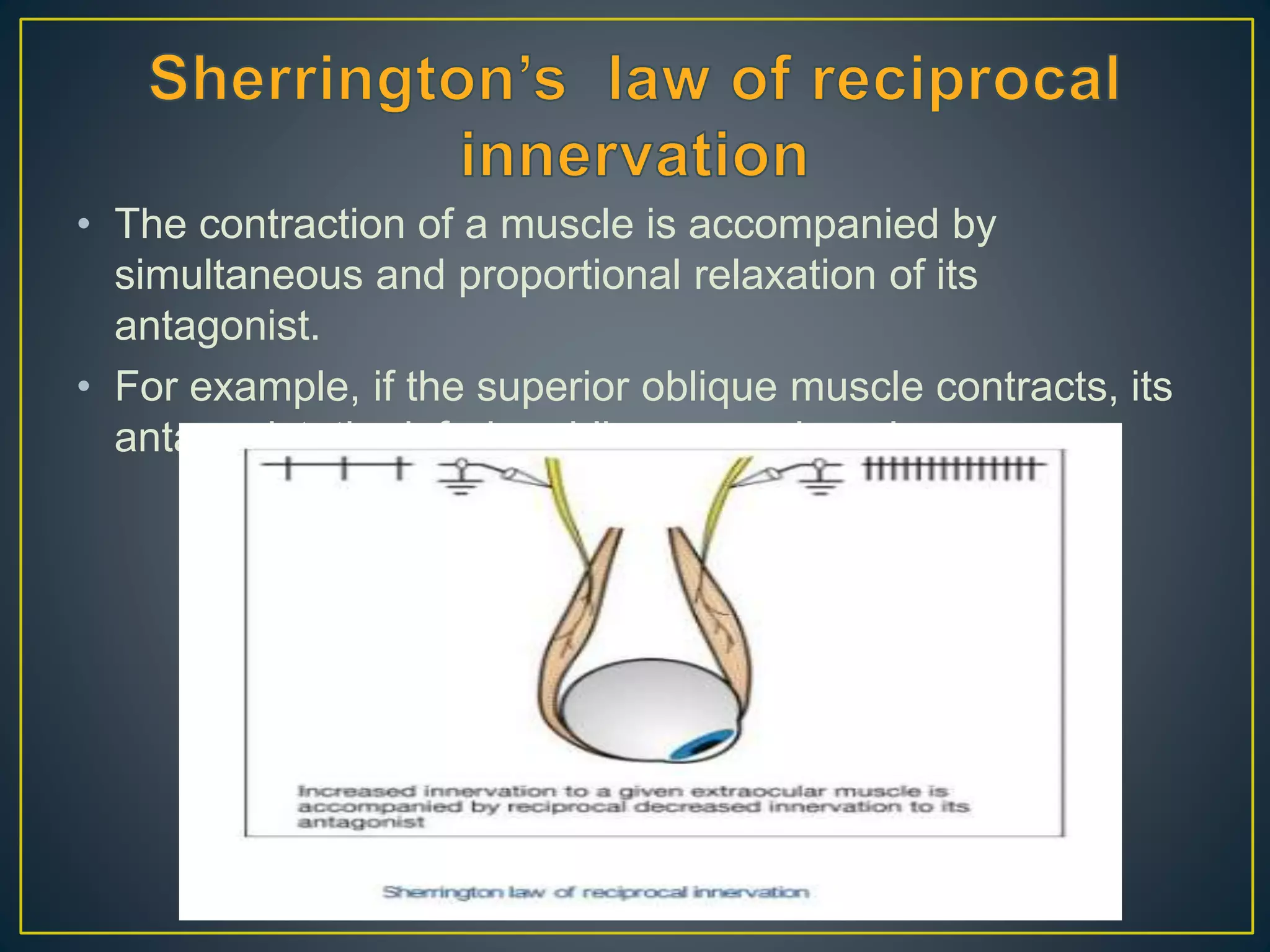
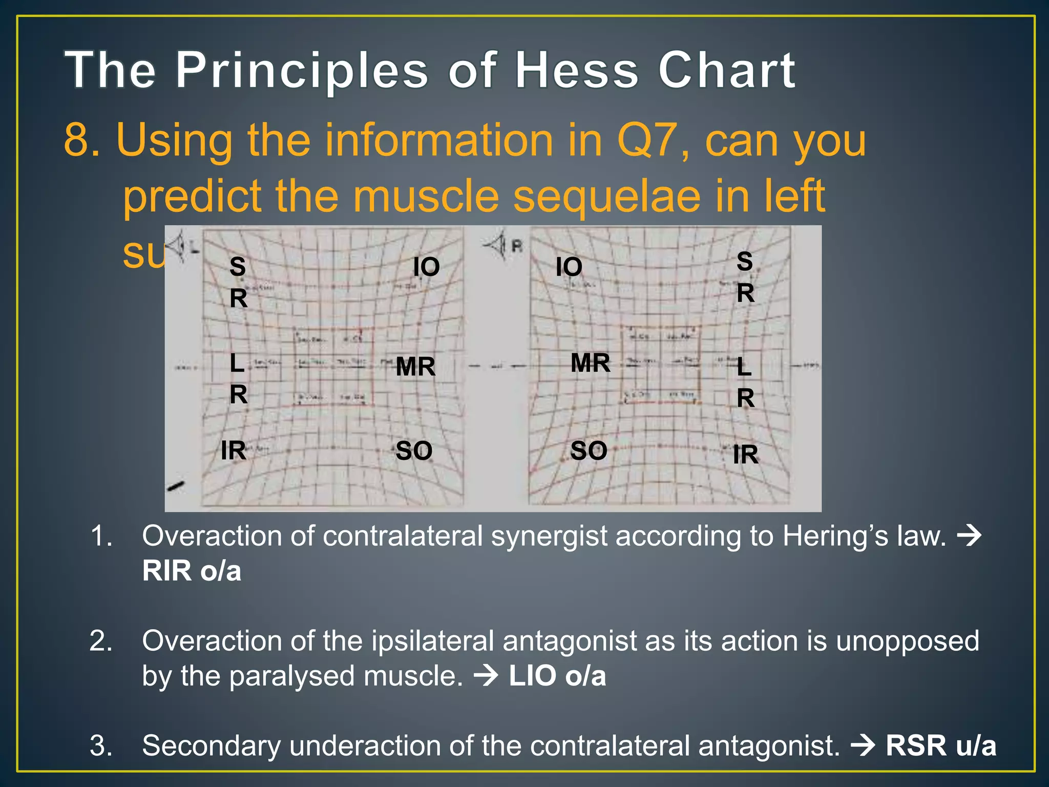
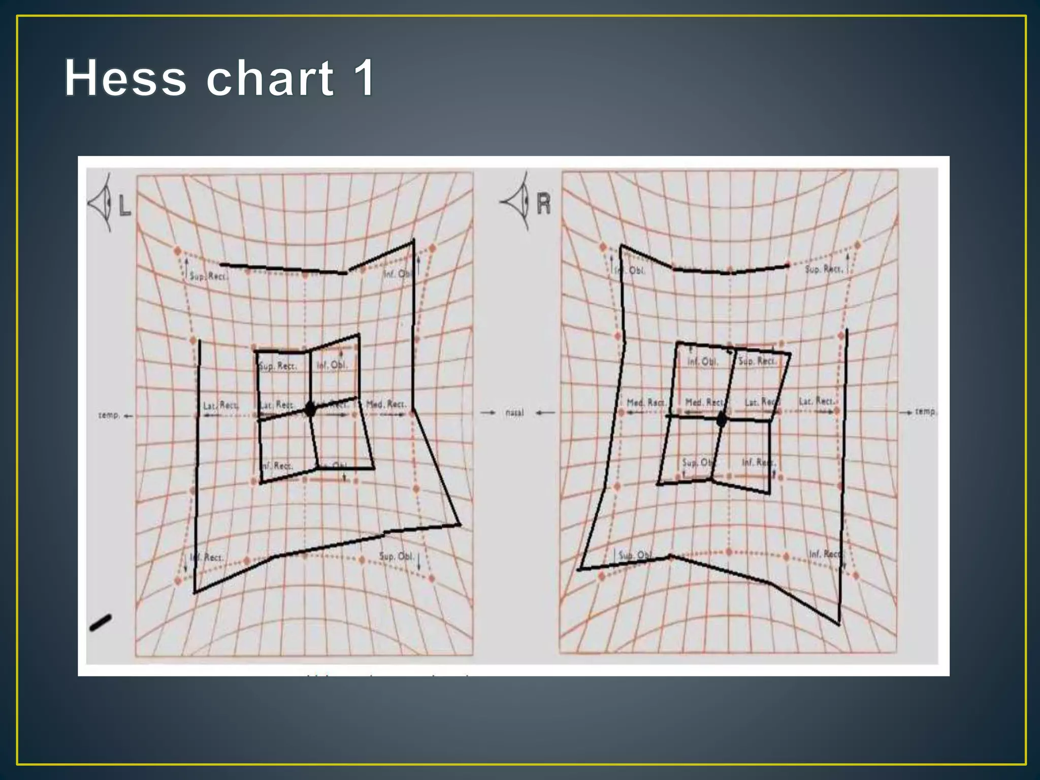
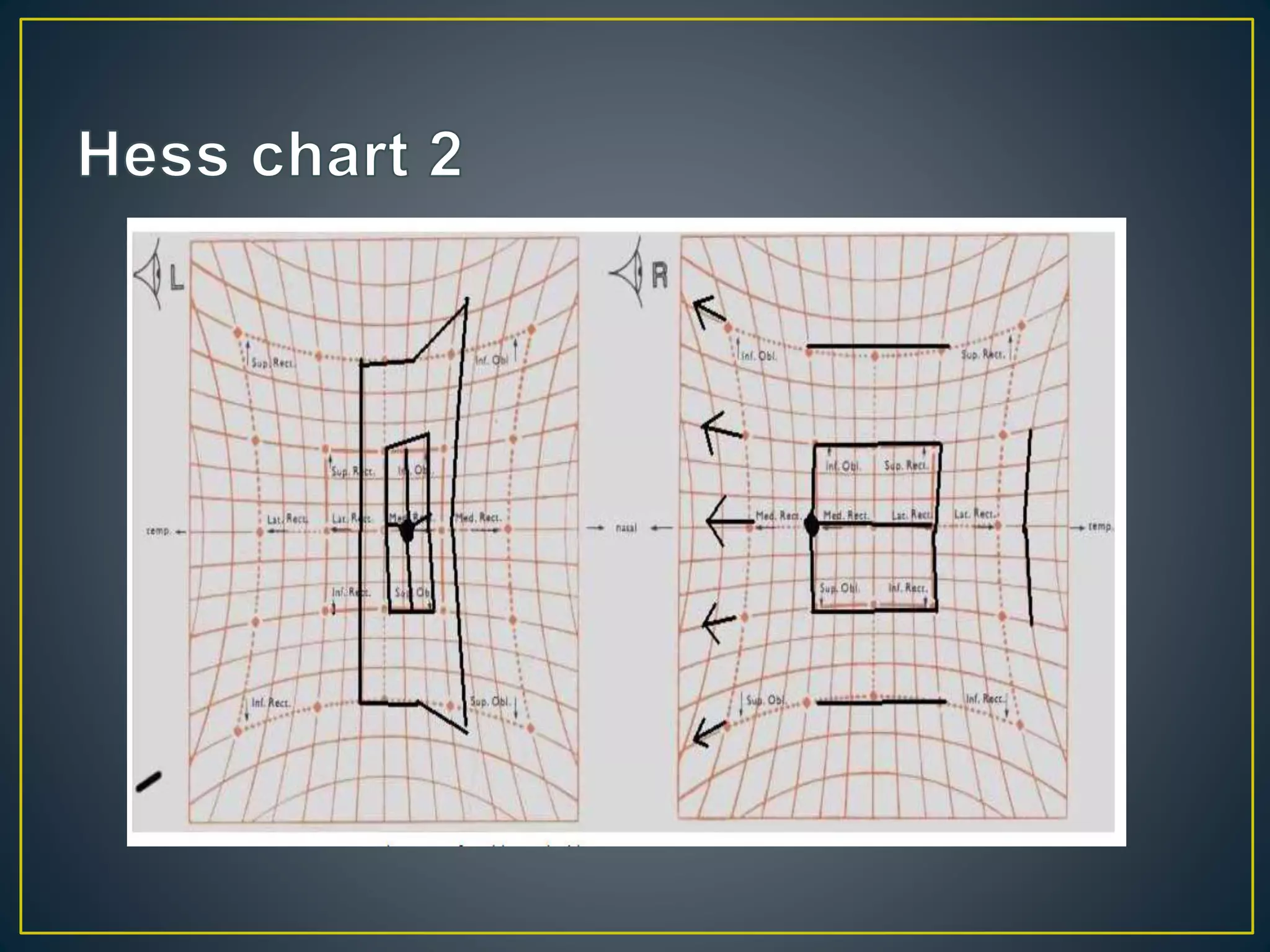
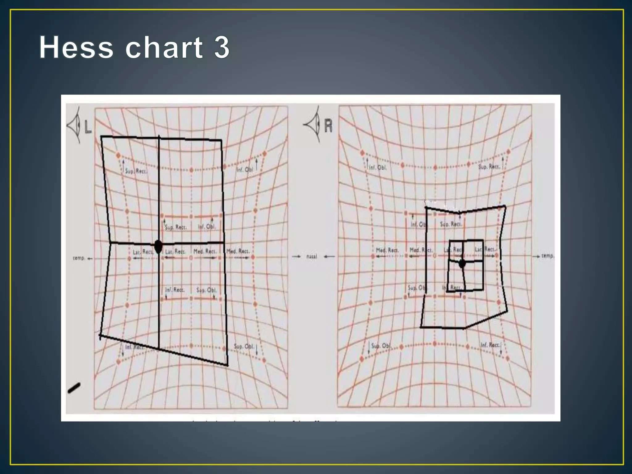
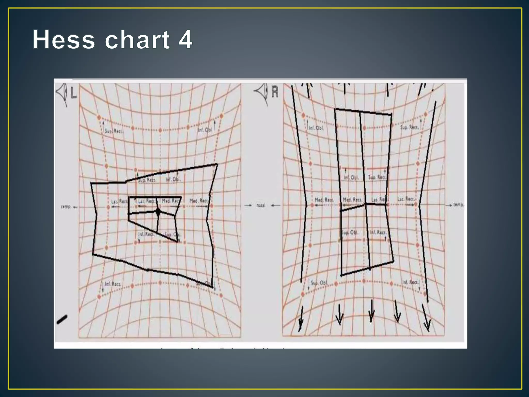
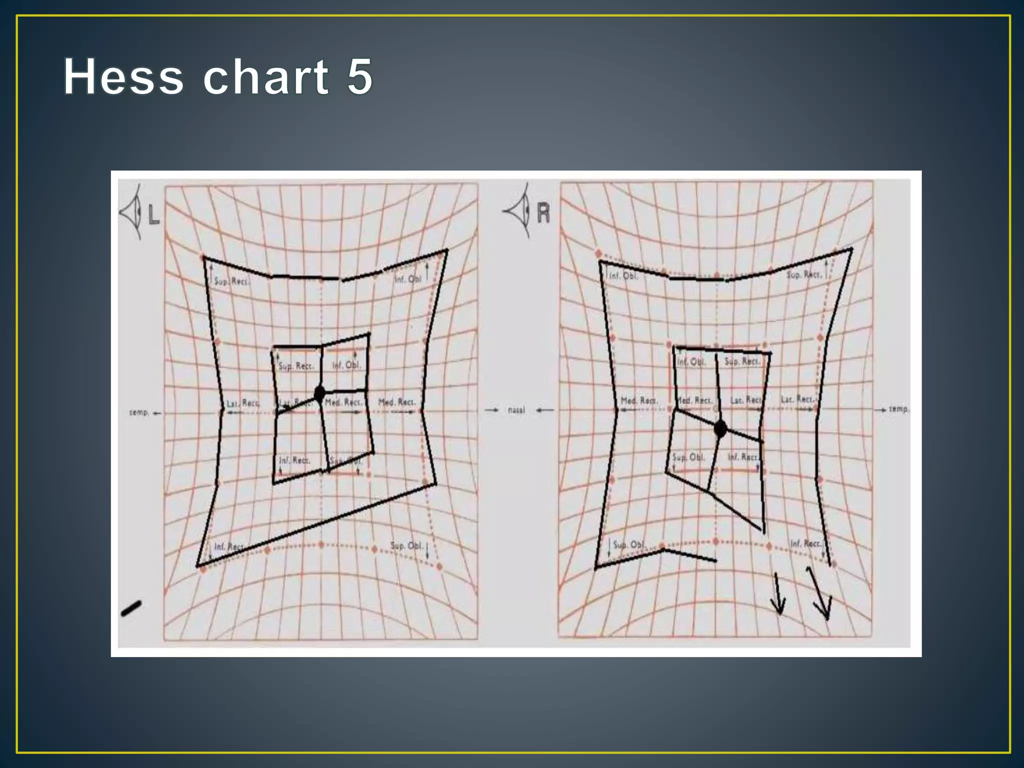
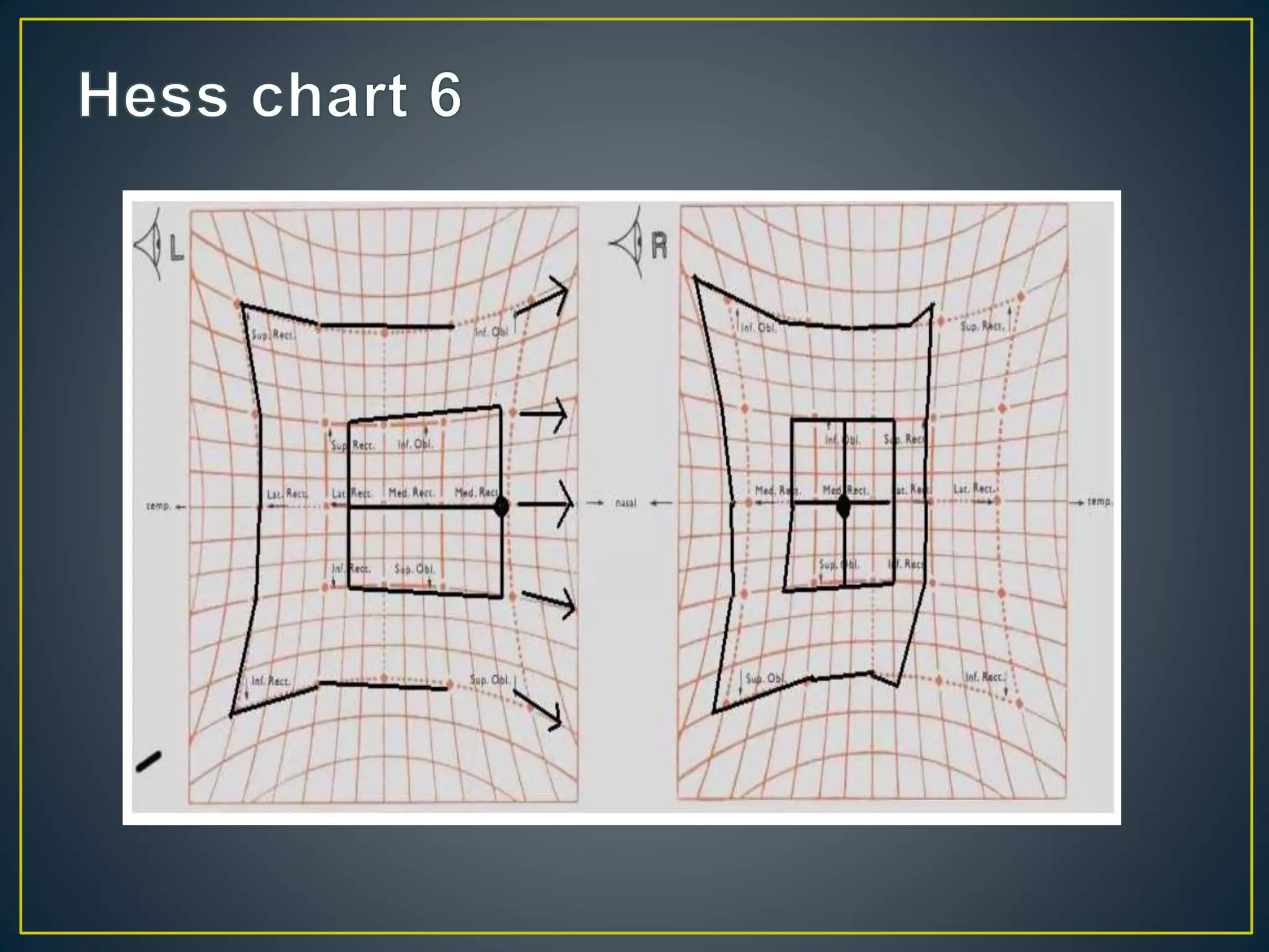
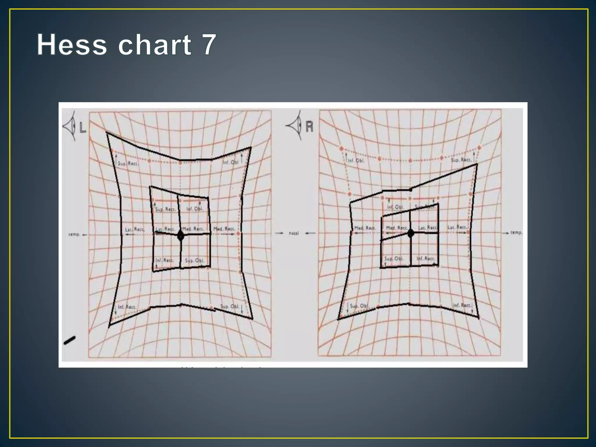
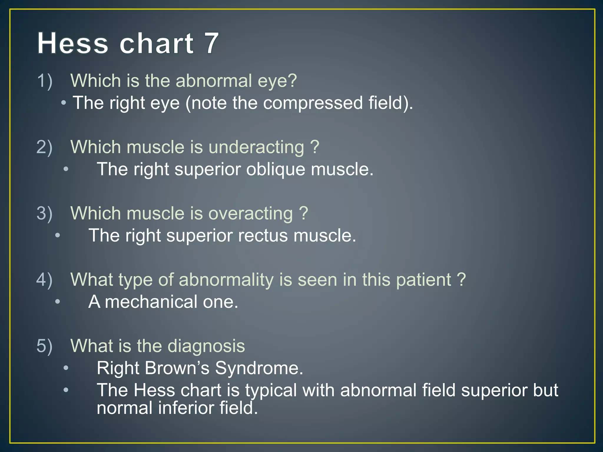
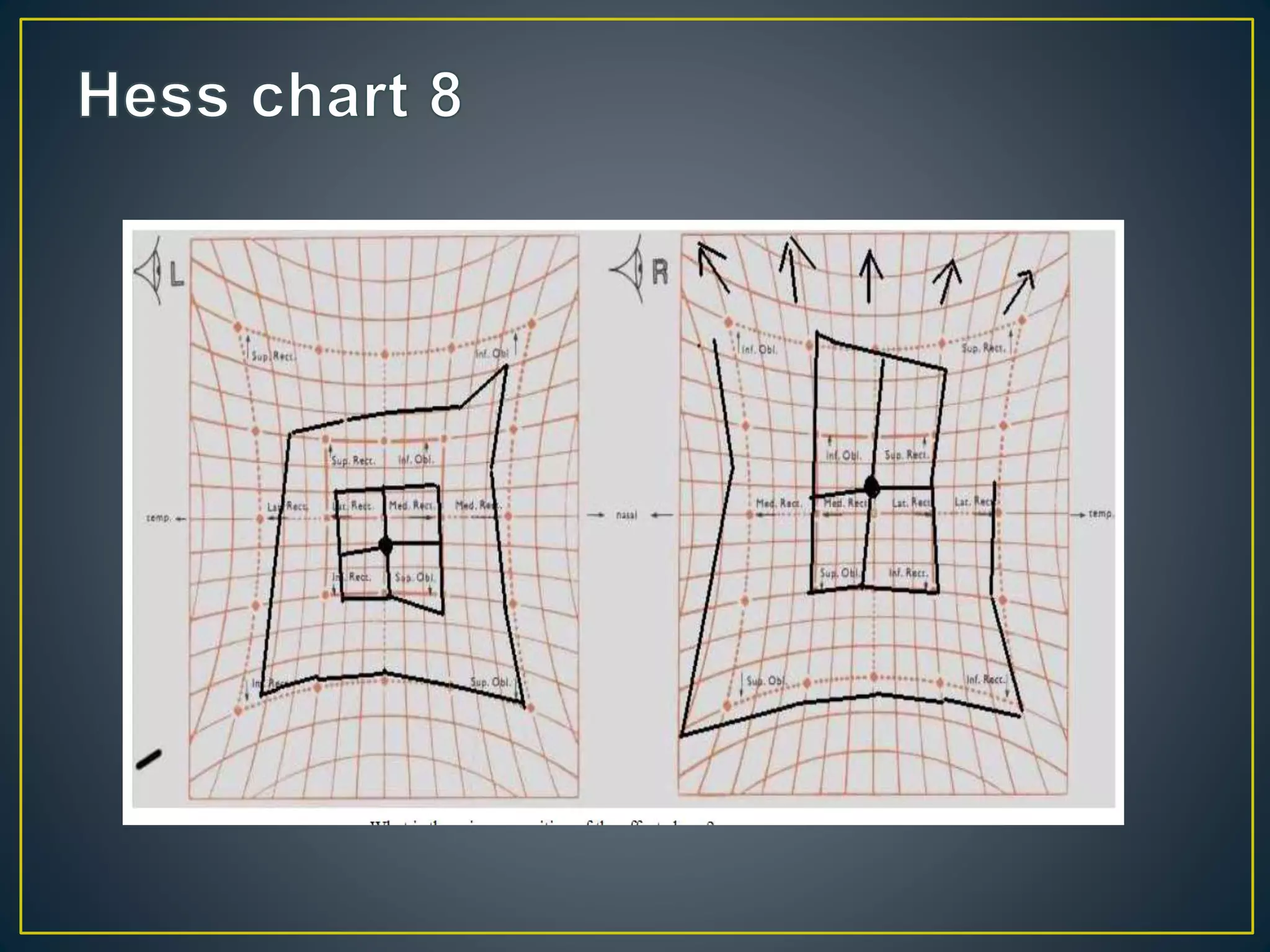
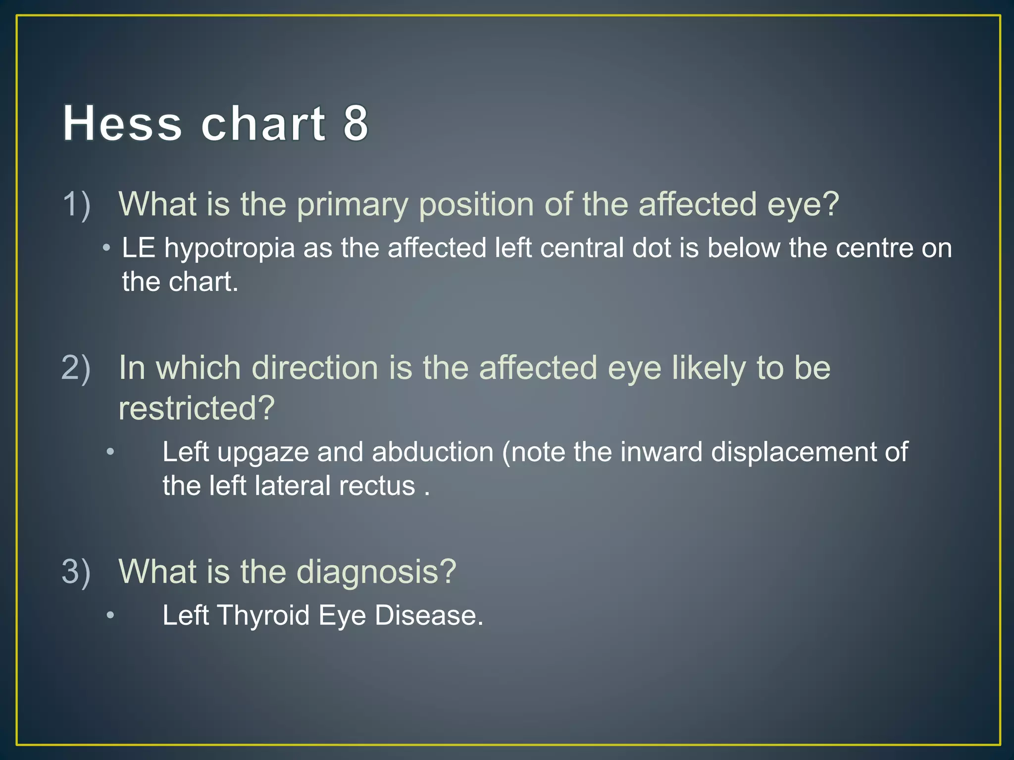
Prepared by :
Anis Suzanna Binti Mohamad
Optometrist
Ophthalmology Department,
Hospital Sultanah Bahiyah
• Smaller field represents affected eye
• Incomitance suggests recent onset
• Look at the smaller field
• The greatest inward displacement represents the
primary underaction
• The greatest outward displacement indicates an
overaction
• Look at the larger field
• The greatest outward displacement is the main
overaction and is usually of the contralateral
synergist
1. Is Hess chart useful in all cases of
strabismus?
No. It is useful only for incomitant strabismus.
• The patient must possess NRC and central
fixation
• Should always be considered together with
assessment of ocular movements
• May be performed on a Hess screen or a
Lees Screen
2. What features of the Hess chart are used
for interpretation?
The position, size, shape are compared between the two
eyes. The deviation between the two eyes can also be
calculated.
3. What does the position of fields in Hess
chart indicate?
 POSITION = DEVIATION
 The central dot in each field indicates the deviation in
the primary position.
 Each small square subtends an angle of 5 degrees at
the 50 cm working distance.
 The displacement of the pointer from the dot is a
measure of the size of the deviation.
4. What information can be obtained from
the size of the Hess chart?
 The eye with the small field is the effected eye.
 Presence of a compressed field indicates a mechanical
cause.
• Mechanical
• Compressed fields
• Limited muscle
sequelae
• Binocular single vision
is often present in the
primary position
• Neurogenic
• Less compressed
• Muscle sequelae not
limited and eventually
become concomitant
• Deviation in primary
position reflects
extent of palsy
5. What can be obtained from the shape of
Hess chart?
 Charts with sloping fields indicates the presence of A
and V patterns.
 Look at the separation of the inner filed in up and down
gaze.
6. In neurogenic strabismus, the muscle
sequelae make the Hess charts between
the two eyes become more similar in
size. What laws govern the muscle
sequelae?
 Hering’s law of equal innervation
 Sherrington’s law of reciprocal innervation
• Innervation to the extraocular muscles is equal to both
eyes.
• Thus, when a nervous impulse is sent to an ocular
muscle to contract, an equal impulse is sent to its
contralateral synergist to contract.
• The contraction of a muscle is accompanied by
simultaneous and proportional relaxation of its
antagonist.
• For example, if the superior oblique muscle contracts, its
antagonist, the inferior oblique muscle, relaxes.
7. What are the three stages in the
development of muscle sequelae?
1. Overaction of contralateral synergist according to
Hering’s law.
2. Overaction of the ipsilateral antagonist as its action is
unopposed by the paralysed muscle.
3. Secondary underaction of the contralateral antagonist.
8. Using the information in Q7, can you
predict the muscle sequelae in left
superior oblique?
1. Overaction of contralateral synergist according to Hering’s law. 
RIR o/a
2. Overaction of the ipsilateral antagonist as its action is unopposed
by the paralysed muscle.  LIO o/a
3. Secondary underaction of the contralateral antagonist.  RSR u/a
S
R
L
R
IR
IO
MR
SO
IO S
R
MR
SO IR
L
R
1) Which muscle are underacting?
• The right and the left superior oblique muscles
• The left SO is effected more than right
2) Which muscles are overacting?
• The right and the left inferior rectus
• The right and the left superior rectus
3) What is the diagnosis?
• Bilateral asymmetrical fourth nerve palsy
1) What type of strabismus is this?
• A mechanical one. The field are compressed vertically.
2) Is the eye straight in the primary position?
• The field indicates that the patient is likely to have convergent
squint as the dot on the central field is move nasally.
3) What is the diagnosis?
• Left Duane’s Syndrome of the type A (according to Brown’s
classification): with limited abduction> limited adduction.
1) What type of strabismus is this?
• A mechanical one. The right field is compressed.
2) Is the eye straight in the primary position?
• The right eye is hypotropia and exotropia (down and out).
3) What is the diagnosis?
• Right third nerve palsy
1) What type of abnormality is seen in this patient?
• A mechanical one. The left field is compressed.
2) In which direction is the affected eye likely to be
restricted?
• Left upgaze and downgaze.
3) What is the diagnosis?
• Left orbital floor fracture causing restriction of the inferior rectus
movement.
1) Which is the abnormal eye?
• The left eye (note the smaller field).
2) Which muscle is underacting ?
• The left superior oblique muscle.
3) Which muscle is overacting ?
• The right inferior rectus muscle.
4) Is the long-standing palsy ?
• No, the muscle sequelae have not fully developed.
5) What is the diagnosis
• Left superior oblique palsy.
1) Which is the abnormal eye?
• The right eye (note the smaller field).
2) Which muscle is underacting ?
• The right lateral rectus muscle.
3) Which muscle is overacting ?
• The left medial rectus muscle.
1) What is the diagnosis
• Right sixth nerve palsy.
1) Which is the abnormal eye?
• The right eye (note the compressed field).
2) Which muscle is underacting ?
• The right superior oblique muscle.
3) Which muscle is overacting ?
• The right superior rectus muscle.
4) What type of abnormality is seen in this patient ?
• A mechanical one.
5) What is the diagnosis
• Right Brown’s Syndrome.
• The Hess chart is typical with abnormal field superior but
normal inferior field.
1) What is the primary position of the affected eye?
• LE hypotropia as the affected left central dot is below the centre on
the chart.
2) In which direction is the affected eye likely to be
restricted?
• Left upgaze and abduction (note the inward displacement of
the left lateral rectus .
3) What is the diagnosis?
• Left Thyroid Eye Disease.
• Underactions and overactions
• Affected eye
• A or V Patterns
• Type of deviation in primary position
• Size of deviation – each box = 5°
• Muscle sequelae
• Differential diagnosis between recent and
longstanding
• Differential diagnosis between neurogenic and
mechanical defects
• Comparison between clinic visits
• Comparison pre- and post-operatively

Revision about Hess Chart

  • 1.
    Prepared by : AnisSuzanna Binti Mohamad Optometrist Ophthalmology Department, Hospital Sultanah Bahiyah
  • 5.
    • Smaller fieldrepresents affected eye • Incomitance suggests recent onset
  • 7.
    • Look atthe smaller field • The greatest inward displacement represents the primary underaction • The greatest outward displacement indicates an overaction • Look at the larger field • The greatest outward displacement is the main overaction and is usually of the contralateral synergist
  • 8.
    1. Is Hesschart useful in all cases of strabismus? No. It is useful only for incomitant strabismus.
  • 9.
    • The patientmust possess NRC and central fixation • Should always be considered together with assessment of ocular movements • May be performed on a Hess screen or a Lees Screen
  • 10.
    2. What featuresof the Hess chart are used for interpretation? The position, size, shape are compared between the two eyes. The deviation between the two eyes can also be calculated.
  • 11.
    3. What doesthe position of fields in Hess chart indicate?  POSITION = DEVIATION  The central dot in each field indicates the deviation in the primary position.  Each small square subtends an angle of 5 degrees at the 50 cm working distance.  The displacement of the pointer from the dot is a measure of the size of the deviation.
  • 13.
    4. What informationcan be obtained from the size of the Hess chart?  The eye with the small field is the effected eye.  Presence of a compressed field indicates a mechanical cause.
  • 14.
    • Mechanical • Compressedfields • Limited muscle sequelae • Binocular single vision is often present in the primary position • Neurogenic • Less compressed • Muscle sequelae not limited and eventually become concomitant • Deviation in primary position reflects extent of palsy
  • 15.
    5. What canbe obtained from the shape of Hess chart?  Charts with sloping fields indicates the presence of A and V patterns.  Look at the separation of the inner filed in up and down gaze.
  • 16.
    6. In neurogenicstrabismus, the muscle sequelae make the Hess charts between the two eyes become more similar in size. What laws govern the muscle sequelae?  Hering’s law of equal innervation  Sherrington’s law of reciprocal innervation
  • 17.
    • Innervation tothe extraocular muscles is equal to both eyes. • Thus, when a nervous impulse is sent to an ocular muscle to contract, an equal impulse is sent to its contralateral synergist to contract.
  • 18.
    • The contractionof a muscle is accompanied by simultaneous and proportional relaxation of its antagonist. • For example, if the superior oblique muscle contracts, its antagonist, the inferior oblique muscle, relaxes.
  • 19.
    7. What arethe three stages in the development of muscle sequelae? 1. Overaction of contralateral synergist according to Hering’s law. 2. Overaction of the ipsilateral antagonist as its action is unopposed by the paralysed muscle. 3. Secondary underaction of the contralateral antagonist.
  • 20.
    8. Using theinformation in Q7, can you predict the muscle sequelae in left superior oblique? 1. Overaction of contralateral synergist according to Hering’s law.  RIR o/a 2. Overaction of the ipsilateral antagonist as its action is unopposed by the paralysed muscle.  LIO o/a 3. Secondary underaction of the contralateral antagonist.  RSR u/a S R L R IR IO MR SO IO S R MR SO IR L R
  • 23.
    1) Which muscleare underacting? • The right and the left superior oblique muscles • The left SO is effected more than right 2) Which muscles are overacting? • The right and the left inferior rectus • The right and the left superior rectus 3) What is the diagnosis? • Bilateral asymmetrical fourth nerve palsy
  • 25.
    1) What typeof strabismus is this? • A mechanical one. The field are compressed vertically. 2) Is the eye straight in the primary position? • The field indicates that the patient is likely to have convergent squint as the dot on the central field is move nasally. 3) What is the diagnosis? • Left Duane’s Syndrome of the type A (according to Brown’s classification): with limited abduction> limited adduction.
  • 27.
    1) What typeof strabismus is this? • A mechanical one. The right field is compressed. 2) Is the eye straight in the primary position? • The right eye is hypotropia and exotropia (down and out). 3) What is the diagnosis? • Right third nerve palsy
  • 29.
    1) What typeof abnormality is seen in this patient? • A mechanical one. The left field is compressed. 2) In which direction is the affected eye likely to be restricted? • Left upgaze and downgaze. 3) What is the diagnosis? • Left orbital floor fracture causing restriction of the inferior rectus movement.
  • 31.
    1) Which isthe abnormal eye? • The left eye (note the smaller field). 2) Which muscle is underacting ? • The left superior oblique muscle. 3) Which muscle is overacting ? • The right inferior rectus muscle. 4) Is the long-standing palsy ? • No, the muscle sequelae have not fully developed. 5) What is the diagnosis • Left superior oblique palsy.
  • 33.
    1) Which isthe abnormal eye? • The right eye (note the smaller field). 2) Which muscle is underacting ? • The right lateral rectus muscle. 3) Which muscle is overacting ? • The left medial rectus muscle. 1) What is the diagnosis • Right sixth nerve palsy.
  • 35.
    1) Which isthe abnormal eye? • The right eye (note the compressed field). 2) Which muscle is underacting ? • The right superior oblique muscle. 3) Which muscle is overacting ? • The right superior rectus muscle. 4) What type of abnormality is seen in this patient ? • A mechanical one. 5) What is the diagnosis • Right Brown’s Syndrome. • The Hess chart is typical with abnormal field superior but normal inferior field.
  • 37.
    1) What isthe primary position of the affected eye? • LE hypotropia as the affected left central dot is below the centre on the chart. 2) In which direction is the affected eye likely to be restricted? • Left upgaze and abduction (note the inward displacement of the left lateral rectus . 3) What is the diagnosis? • Left Thyroid Eye Disease.
  • 39.
    • Underactions andoveractions • Affected eye • A or V Patterns • Type of deviation in primary position • Size of deviation – each box = 5° • Muscle sequelae • Differential diagnosis between recent and longstanding • Differential diagnosis between neurogenic and mechanical defects
  • 40.
    • Comparison betweenclinic visits • Comparison pre- and post-operatively

Editor's Notes

  • #4 Hess charts are common questions in the OSE and final MRCOphth. A short tutorials on its principles and some exercises should help every candidate to answer the most commonly encountered cases
  • #20 Secondary underaction of the contralateral antagonist. -This occurs for two reasons:- 1) As the ipsilateral antagonist action is unopposed, less impulse is needed to move it into its desired position and subsequently according to Hering’s law, its contralateral synergist receives less impulse and therefore underact. 2) Overaction of the contralateral synergist means that its ipsilateral antagonist will receive equal impulse to relax according to the Sherrington’s law.
  • #21 Secondary underaction of the contralateral antagonist. -This occurs for two reasons:- 1) As the ipsilateral antagonist action is unopposed, less impulse is needed to move it into its desired position and subsequently according to Hering’s law, its contralateral synergist receives less impulse and therefore underact. 2) Overaction of the contralateral synergist means that its ipsilateral antagonist will receive equal impulse to relax according to the Sherrington’s law.