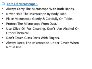
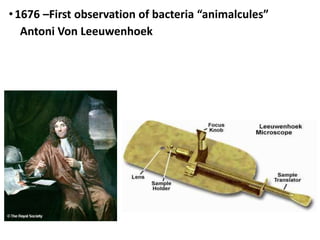
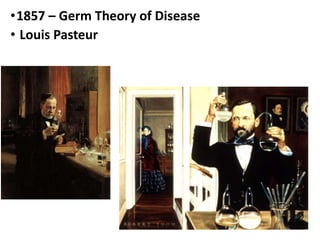
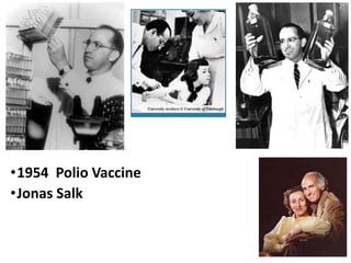
 UNIT-1
INTRODUCTION OF
MICROBIOLOGY
Ahmed Sodha
M.Sc.(N).– M.S.N.
 MICROBIOLOGY
 INTRODUCTION:-
• Microbiology term was introduced by French
Chemist Louis Pasteur .
• Microbe means the organisms too small to be
seen by naked eye.
• The term microbe is now replaced by the term
Micro-organisms.
 DEFINITION:-
• “Microbiology is the science which deals with the
study of microorganisms.”
 BRANCHES OF MICROBIOLOGY:-
 Medical Microbiology
 Industrial Microbiology
 Food Microbiology
 Soil Microbiology
 Plant Microbiology
 Medical Microbiology:- It Is The Study Of
Causative Agents Of Infectious Diseases Of Human
Beings & Their Reactions To Such Infections.
 Branches Of Medical Microbiology:-
• Bacteriology:- It Deals With Study Of Bacteria.
• Parasitology:- It Deals With Study Of Parasites.
• Mycology:- It Deals With Study Of Fungi.
• Virology:- It Deals With Study Of Viruses.
• Immunology:- It Deals With Study Of Mechanism
Of Immunity.
 Classification of Micro-organisms:-
• Micro-organisms are classified into ten groups:
1. Bacteria
2. Viruses
3. Fungi
4. Protozoa
5. Rickettsiales
6. Algae
7. Moulds
8. Yeast
9. Chlamydia
10. Mycoplasma
Bacteria
Bacteria
Viruses
Fungi
Protozoa
Rickettsiales
Algae
Algae
Moulds
Yeast
Chlamydia
Mycoplasma
 Importance Of Microbiology In Nursing:- By
Knowledge Of Microbiology Nurse Can:
• Learn The Process Of Spreading Of Infection.
• Know About The Surfaces Which Are Susceptible
To Infectious Agent.
• Keep Instruments Aseptic.
• Recognize Symptoms Of An Infection.
• Take Care Of Open Wound.
• Understand The Importance Of Immunity &
Vaccination.
 Microscope
 Microscope:- A Microscope Is A Laboratory
Instrument Used To Examine Objects That Are Too
Small To Be Seen By The Naked Eye.
 Microscopy:- Microscopy Is The Science Of
Investigating Small Objects And Structures Using
A Microscope Device.
 Antoni Van Leeuwenhoek is a ‘Father of
Microbiology’ & ‘Father of Microscopy.’
 Robert Koch (1834-1910) is also known as the
'Father of Bacteriology‘.
 Louis Pasteur is a father of ‘Pasteurization’.
 Types of Microscope:-
1. Light Microscope
2. Phase Contrast Microscope
3. Dark Ground Microscope
4. Fluorescence Microscope
5. Electron Microscope
 Parts of Microscope:- There are 3 main parts of
the microscope are:
1. Stand
2. Body
3. Optical system
 Microscope
1. Stand:- It is made of solid iron part provides
balance & stability to instrument.
2. Body:- it consist of:
i. Stage:- It is platform that supports specimen
under observation. It has central aperture for the
passage of light rays.
ii. Condenser:- It is used to concentrate the light.
iii. Iris Diaphragm:- It is present in condenser. It
regulates the amount of light falling on the
specimen.
iv. Mirror:- It reflects the light on the specimen.
3. Optical System:- It Consists Of :
i. Coarse Adjustment:- It Is Used To Raise & Lower
The Objective Lens.
ii. Fine Adjustment:- It Is Used For Delicate
Focusing.
iii. Nose Piece:- It Provided With Sockets Of
Different Magnifying Power.
iv. Eye Piece:- It Magnifies Further The Image.
 Care Of Microscope:-
• Always Carry The Microscope With Both Hands.
• Never Hold The Microscope By Body Tube.
• Place Microscope Gently & Carefully On Table.
• Protect The Microscope From Dust.
• Use Olive Oil For Cleaning, Don’t Use Alcohol Or
Other Chemical.
• Don’t Touch Glass Parts With Fingers.
• Always Keep The Microscope Under Cover When
Not In Use.
History of Microbiology
•1590 – First compound light microscope
• Zacharias Janssen
•1676 –First observation of bacteria “animalcules”
Antoni Von Leeuwenhoek
•1796 – First vaccine (smallpox)
Edward Jenner
•1857 – Germ Theory of Disease
• Louis Pasteur
•1867 Antiseptic Surgery
• Joseph Lister
•1884 Koch’s Postulates of Disease Transmission
•Robert Koch
•1885 - Vaccine against Rabies
•Louis Pasteur
•1929 Discovery of Penicillin (first antibiotic)
• Alexander Fleming
•1938 – First Electron Microscope
•The electron microscope is
capable of magnifying biological
specimens up to one million
times. These computer enhanced
images of 1. smallpox, 2. herpes
simplex, and 3. mumps are
magnified, respectively, 150,000,
150,000 and 90,000 times.
•1953-1954 Structure of DNA Revealed
•Watson & Crick
•1954 Polio Vaccine
•Jonas Salk
Introduction - Microbiology.pptx

Introduction - Microbiology.pptx

  • 1.
  • 2.
  • 3.
     INTRODUCTION:- • Microbiologyterm was introduced by French Chemist Louis Pasteur . • Microbe means the organisms too small to be seen by naked eye. • The term microbe is now replaced by the term Micro-organisms.  DEFINITION:- • “Microbiology is the science which deals with the study of microorganisms.”
  • 4.
     BRANCHES OFMICROBIOLOGY:-  Medical Microbiology  Industrial Microbiology  Food Microbiology  Soil Microbiology  Plant Microbiology
  • 5.
     Medical Microbiology:-It Is The Study Of Causative Agents Of Infectious Diseases Of Human Beings & Their Reactions To Such Infections.  Branches Of Medical Microbiology:- • Bacteriology:- It Deals With Study Of Bacteria. • Parasitology:- It Deals With Study Of Parasites. • Mycology:- It Deals With Study Of Fungi. • Virology:- It Deals With Study Of Viruses. • Immunology:- It Deals With Study Of Mechanism Of Immunity.
  • 6.
     Classification ofMicro-organisms:- • Micro-organisms are classified into ten groups: 1. Bacteria 2. Viruses 3. Fungi 4. Protozoa 5. Rickettsiales 6. Algae 7. Moulds 8. Yeast 9. Chlamydia 10. Mycoplasma
  • 7.
  • 8.
  • 9.
  • 10.
  • 11.
  • 12.
  • 13.
  • 14.
  • 15.
  • 16.
  • 17.
  • 18.
  • 19.
     Importance OfMicrobiology In Nursing:- By Knowledge Of Microbiology Nurse Can: • Learn The Process Of Spreading Of Infection. • Know About The Surfaces Which Are Susceptible To Infectious Agent. • Keep Instruments Aseptic. • Recognize Symptoms Of An Infection. • Take Care Of Open Wound. • Understand The Importance Of Immunity & Vaccination.
  • 20.
  • 21.
     Microscope:- AMicroscope Is A Laboratory Instrument Used To Examine Objects That Are Too Small To Be Seen By The Naked Eye.  Microscopy:- Microscopy Is The Science Of Investigating Small Objects And Structures Using A Microscope Device.  Antoni Van Leeuwenhoek is a ‘Father of Microbiology’ & ‘Father of Microscopy.’  Robert Koch (1834-1910) is also known as the 'Father of Bacteriology‘.  Louis Pasteur is a father of ‘Pasteurization’.
  • 22.
     Types ofMicroscope:- 1. Light Microscope 2. Phase Contrast Microscope 3. Dark Ground Microscope 4. Fluorescence Microscope 5. Electron Microscope  Parts of Microscope:- There are 3 main parts of the microscope are: 1. Stand 2. Body 3. Optical system
  • 23.
  • 24.
    1. Stand:- Itis made of solid iron part provides balance & stability to instrument. 2. Body:- it consist of: i. Stage:- It is platform that supports specimen under observation. It has central aperture for the passage of light rays. ii. Condenser:- It is used to concentrate the light. iii. Iris Diaphragm:- It is present in condenser. It regulates the amount of light falling on the specimen. iv. Mirror:- It reflects the light on the specimen.
  • 25.
    3. Optical System:-It Consists Of : i. Coarse Adjustment:- It Is Used To Raise & Lower The Objective Lens. ii. Fine Adjustment:- It Is Used For Delicate Focusing. iii. Nose Piece:- It Provided With Sockets Of Different Magnifying Power. iv. Eye Piece:- It Magnifies Further The Image.
  • 26.
     Care OfMicroscope:- • Always Carry The Microscope With Both Hands. • Never Hold The Microscope By Body Tube. • Place Microscope Gently & Carefully On Table. • Protect The Microscope From Dust. • Use Olive Oil For Cleaning, Don’t Use Alcohol Or Other Chemical. • Don’t Touch Glass Parts With Fingers. • Always Keep The Microscope Under Cover When Not In Use.
  • 27.
    History of Microbiology •1590– First compound light microscope • Zacharias Janssen
  • 28.
    •1676 –First observationof bacteria “animalcules” Antoni Von Leeuwenhoek
  • 29.
    •1796 – Firstvaccine (smallpox) Edward Jenner
  • 30.
    •1857 – GermTheory of Disease • Louis Pasteur
  • 31.
  • 32.
    •1884 Koch’s Postulatesof Disease Transmission •Robert Koch
  • 33.
    •1885 - Vaccineagainst Rabies •Louis Pasteur
  • 34.
    •1929 Discovery ofPenicillin (first antibiotic) • Alexander Fleming
  • 35.
    •1938 – FirstElectron Microscope •The electron microscope is capable of magnifying biological specimens up to one million times. These computer enhanced images of 1. smallpox, 2. herpes simplex, and 3. mumps are magnified, respectively, 150,000, 150,000 and 90,000 times.
  • 36.
    •1953-1954 Structure ofDNA Revealed •Watson & Crick
  • 37.