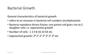
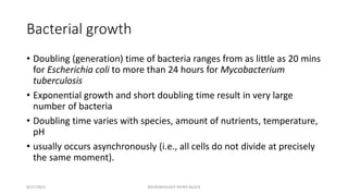
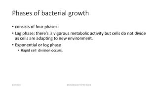
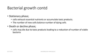
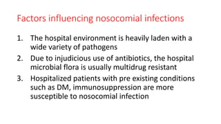
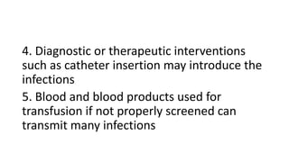
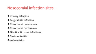
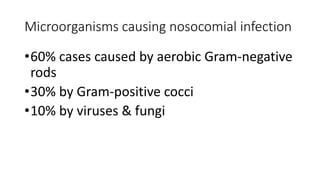
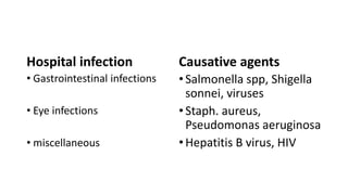
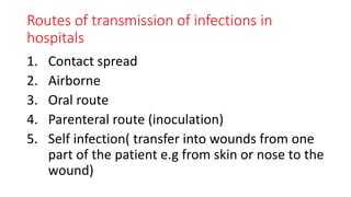
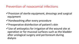
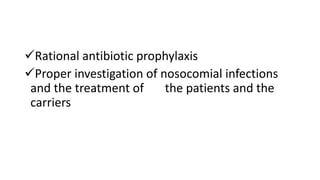
MICROBIOLOGY &
IMMUNOLOGY
DORRINE TITANY
• The first simple forms of life appeared on earth more then 3 billion
years ago.
• Their descendants have changed and developed into the several
million type of animals , plants and microorganisms are recognized.
• Microscopic forms of life are present in vast numbers in nearly
every environment like soil, water, food, air , etc.
• Microbiology is the science that dealing with the study of
microorganisms.
• Microorganisms: An organism that can be seen only with the aid of a
microscope and that typically consist of only a single cell.
• microorganisms include: • bacteria • fungi • protozoa • microscopic
algae • viruses • various parasitic worms
•Development of microscopy
• (348-322) Aristotle and other believed that living organisms could develop from Non- living materials.
• (1590) Zacharias Janssen- developed first compound microscope
•6. (1660) Robert Hooke – Published “micrographia” drawing and detailed observation of biological materials
• made with the best compound microscope
• (1676) Anton Van Leeuwenhoek- 1st person to observe microorganisms.
•7. From his teeth, he observed
• (A)& (B)- rod forms
•(C) & (D)- motion pathway
• (E)- Spherical form
•(F)- Longer type of spherical form
•(H)- Cluster -Royal Society letter (Sept 17th, 1683)
•The microscope used Simple microscope (one lens)
• Spontaneous generation controversy
• 1688: Francesco Redi (1626-1678) was an Italian physician who
refuted the idea of spontaneous generation by showing that rotting
meat carefully kept from flies will not spontaneously produce
maggots.
• 1836: Theodor Schwann (1810-1882) helped develop the cell theory
of living organisms, namely that that all living organisms are
composed of one or more cells and that the cell is the basic functional
unit of living organisms.
INTRODUCTION
•Microbiology is the study of living organisms of
microscopic size ( Louis Pasteur)
•Microbes were probably the first living things to
appear on the earth and the study of fossil
remains indicate that microbial infections and
epidemic diseases existed thousand of years ago
• Antonie van Leeuwenhoek placed the science of
microbiology on the firm basis of direct observation
using compound microscope
• He first accurately described different shapes of
bacteria ( coccal, bacillary & spiral) and pictured their
arrangement in infected material in 1683
• The scientific community acknowledge him as “ father
of protozoology and bacteriology”
ORIGIN OF MICROBIAL LIFE
• In 1856, Louis Pasteur (1822-1895) investigated the beet juice from which
alcohol was derived which was contaminated with grey material which
interfered with alcohol production. proposed the
• “Germ theory of disease”.- Ancients believed that disease was the result of a
divine punishment. Pasteur fought to convince surgeons that germs existed
and carried diseases, and dirty instruments and hands spread germs and
therefore disease. Pasteur's pasteurization process killed germs and prevented
the spread of disease
• During the course of investigation his attention was abruptly focused on the
role of microorganisms in alcohol fermentation and spoilage
• Pasteur established that different types of fermentations were due to the
activity of different kinds of microbesHis studies on fermentation of wine led him to
take interest in microbiology. finally he proved that microorganisms do not arise by
spontaneous generation. He is known as the founder or father microbiolog
• Joseph Lister (1827-1912) introduced antiseptics in surgery. By
spraying carbolic acid on surgical instruments, wounds and dressings,
he reduced surgical mortality due to bacterial infection considerably.
He is known as father of antiseptic surgery.
• Robert Koch(1843-1910) (father of bacteriology) German
bacteriologist was the first to cultivate anthrax bacteria outside the
body using blood serum at body temperature. introduced methods
for isolation of pure strains of bacteria. He introduced staining
techniques. He is known as father of bacteriology
• Beginning with Pasteur’s work, discoveries included relationship between
microbes and disease, immunity, and antimicrobial drugs Robert Koch
Identified a bacterium as cause of anthrax Introduced “Koch’s Postulates”
and the concept that a disease is caused by a single organism.
• Beginning with Pasteur’s work, discoveries included relationship between
microbes and disease, immunity, and antimicrobial drugs
• Robert Koch Identified a bacterium as cause of anthrax Introduced
“Koch’s Postulates” and the concept that a disease is caused by a single
organism.
• Joseph Lister Introduced the “antiseptic technique”. Use of phenol
(carbolic acid) as disinfectant. Martinus Beijerinck Discovered “viruses”
(toxins, poisons). Infectious agents in tobacco plant fluids.
• John Tyndall (1820-1893). Explained the need for
prolonged heating to eliminate microbial life from
infusions.
• He concluded, by exposing infusions to heat for
varying times, that bacteria existed in two forms- a
heat stable form and a heat sensitive form.
Intermittent heating now called tyndallization, killed
both forms
•Ferdinand Cohn (1828-1898) described heat
stable forms as spores. Spores as well as
vegetative forms were responsible for the
appearance of microbial life in inadequately
heated infusions
•Joseph Lister (1827-1912) introduced antiseptics
in surgery
•For this work he is known as “ father of
antiseptic surgery”
THE DEVELOPING SCIENCE OF
MICROBIOLOGY
• Pasteur introduced the techniques of sterilization and
developed steam sterilizer, hot air oven and autoclave
• Robert Koch (1843-1910) perfected the bacteriological
techniques, staining procedures and methods of
obtaining bacteria in pure culture using solid media
during his studies on the culture and characteristics of
anthrax bacillus
KOCH’S POSTULATES
• A microorganism can be accepted as the causative agent of
an infectious disease only if following postulates are
satisfied:
The microorganism must be present in the lesions in every
case of the infectious disease
It should be possible to isolate the microorganism in pure
culture from the lesions
Inoculation of the pure culture by a suitable route into a
suitable laboratory animal should produce a similar disease
It should again be possible to re-isolate the
microorganism in pure culture from the lesions
produced in the experimental animals
Specific antibodies to the organism should be
demonstrable in the serum of the patient
suffering from the disease
CONTRIBUTIONS OF VARIOUS SCIENTISTS IN
THE FIELD OF MICROBIOLOGY
•A large number of scientists has contributed in
the field of microbiology. Principal contributions
of some of them are described in the following
slides
Louis Pasteur
•Coined the term microbiology
•Coined the term vaccine
•He used various forms of nutrient fluid to grow
microorganisms and showed that a medium
suitable for one might be unsuitable for another
•Proved that all forms of life arose from their alike
and not de novo
• In 1860-64, he gave experimental evidence that
fermentation and putrefaction are effects of microbial
growth
• In 1863-65,he devised the process of destroying
bacteria, known as pasteurization. This process is
employed throughout the civilized world today to
preserve milk and certain other perishable foods.
•In 1877, Koch and Pasteur demonstrated that
anthrax is caused by bacteria
•In 1881, he developed live attenuated anthrax
vaccine
•In 1881, pneumococci were first noticed by
Pasteur and Sternberg independently
Robert Koch
• In 1876, he reported the isolation of anthrax bacillus
in pure culture, formation and germination of its
spores & the proof of its infectiousness
• He introduced the method of making smears of
bacteria on glass slides, and of staining them with
aniline dyes
• In 1881, he described means of cultivating bacteria on
solid media
• The hanging drop method of studying bacteria as used today
is a product of his genius
• In 1882, he discovered tubercle bacillus
• In 1883, he discovered causative agents of cholera ( Vibrio
cholerae)
• In 1884, he expounded Koch’s postulates or laws by which an
organism may be proved to be the cause of a particular
disease
•In invented the hot air oven and steam
sterilizer. He developed methods for testing
antiseptics and to distinguish between
bacteriostatic and bactericidal
concentrations
Antonie van Leeuwenhoek (1632-1723)
• He became expert in the grinding of magnifying glasses
• His microscopes were superior to any of that time
• He observed, drew and measured a large number of the
living organisms including bacteria and protozoa in materials
such as rain water, saliva
• He was the first to accurately describe different shapes of
the bacteria (coccal, bacillary and spiral) and pictured their
arrangement in infected material.
Edward Jenner (1749-1823)
•He introduced the modern method of
vaccination to prevent smallpox
•He observed that milkmaids who contracted
cowpox or vaccinia while milking were
subsequently immune to smallpox
• He performed a vaccination against smallpox by
transferring material from a cowpox pustule on the
hand of the milkmaid, Sarah Nelmes, to the arm of a
small boy named James Phipps, his gardener’s son. Six
weeks later the boy was inoculated with smallpox,
• He failed to develop the disease
Paul Ehrlich (1854-1915)
•In 1879, he applied stains to cells and tissues for
the purpose of revealing their function
•In 1882, he reported the acid fastness of
tubercle bacillus
•In 1898, he proposed side chain theory of
antibody production
IMPORTANCE & RELEVANCE TO NURSING
•Nurses are involved in controlling infection in hospital, so nurses must know about microbiology.
•• To know about harmful and harmless microorganisms to human being.
•3. • Nurses apply the microbiology knowledge in health care for drug production, diagnosis
•and sterilization methods and cleanliness.
•• Nurses use hot water or anti-septic as a measure to sterilize the surgical knives,
• needles, scissors and other metals instruments
•• Microbiology also gives knowledge to nurses on how to handle a patient and
•his samples infected with communicable diseases.
• to free from microbes.
•4. • It also helps detect diseases like Tuberculosis by simple skin test namely the Mantoux test.
• • Also diagnostic tests like Elisa, electrophoreis and radioimmuno assay
•also use principles of microbiology for identification of disease.
MICROBIOLOGY
8/27/2023 MICROBIOLOGY INTRO BLOCK
Main objective
• To enable the learner acquire knowledge on microorganisms.
• Develop skills and attitudes to be able to promote health.
• Prevent illness, diagnose, manage and rehabilitate patients suffering
from infections.
8/27/2023 MICROBIOLOGY INTRO BLOCK
Specific Objectives
1. Describe the historical development of microbiology
2. State the different classifications of micro-organisms.
3. Describe the structure and other characteristics of microorganisms.
4. Explain the identification of microorganisms in the laboratory
5. Describe immunological process
6. Describe infection prevention
7. Describe medical parasitology
8/27/2023 MICROBIOLOGY INTRO BLOCK
Introduction
• Inside your intestines, in the mouth and your skin there reside > 1014
microbial cells(100 times) the number of cells that make up the
human body
• We are not conscious of these colleagues as we pass them round
every time we shake hands, speak or touch a surface
• Inoculation with just one microbe of the wrong type in the wrong way
may kill you, yet we tolerate and indeed thrive on constant
appropriate exposure to these unforeseen world
8/27/2023 MICROBIOLOGY INTRO BLOCK
• Because microbes are generally hidden from our senses, an
appreciation of microbiology demands imagination
• Advances in microbiology have transformed diagnosis, prevention,
and cure of infection and has improved human health.
• In developed countries, infections may be considered insignificant. In
developing countries about 10 million children die each year from
infections of diarrhea, measles, tetanus, diphtheria and whooping
cough alone. Others are TB, cholera, typhoid
8/27/2023 MICROBIOLOGY INTRO BLOCK
Definition
• Microbiology is derived from the Greek;
• Micro – small
• Bios – life
• Logia – study
• It is the study of microscopic organisms either unicellular (single cell),
multicellular or acellular
• It encompasses numerous subdisciplines;
• Virology
• Mycology
• Parasitology
• Bacteriology
8/27/2023 MICROBIOLOGY INTRO BLOCK
Branches of microbiology
• Pure
• Applied
• Pure
• Deals with study of microorganisms on their own right. Bacteriology,
mycology, protozoology, parasitology, immunology, virology
8/27/2023 MICROBIOLOGY INTRO BLOCK
Applied
• Medical microbiology
• Study of pathogenic microbes and role of microbes in human illness
• Others
• Pharmaceutical microbiology- production of antibiotics, enzymes,
vitamins, vaccines and other pharmaceutical products
8/27/2023 MICROBIOLOGY INTRO BLOCK
Applied others;
• Industrial microbiology
• Microbial biotechnology
• Food microbiology
• Agricultural microbiology
• Veterinary microbiology
• Environmental microbiology
8/27/2023 MICROBIOLOGY INTRO BLOCK
History of microbiology
• Infection and microbiology followed different development for
centuries
• We tend to map hthis story against the recorded efforts of prominent
individuals, though others contributed
• 3c BC – Hippocrates; epedemics
• 16c AD – Fracastoro: proposed seeds of contagion not spirits
8/27/2023 MICROBIOLOGY INTRO BLOCK
• Microbiology
• 17c – Leeuwenhoek: microorganisms seen by microscopy by using
homemade microscope.
• Discipline of microbiology was formally established in 2nd half of 19th
century
• Before that, the following events demonstrate how infection was
transmitted;
8/27/2023 MICROBIOLOGY INTRO BLOCK
• 18c- John Hunter (1728-1793): demonstrated transmission of
syphilis and gonorrhea
• Edward Jenner (1749 -1823); used cowpox to prevent smallpox. This
established concept of immunization
• John Snow- (1813 -1858) showed that preventing access to a water
source linked to cholera outbreak terminated new infections
8/27/2023 MICROBIOLOGY INTRO BLOCK
• This was confirmed by Semmelweis (1818-1865): who showed that
physical measures can prevent the transmission of infection e.g.
applying simple hygiene measures.
• 19c – Pasteur(1822 -1895) and Robert Koch (1843-1910):played a
central role in establishing microbial causation of infectious disease
• Pasteur: microbes are necessary for fermentation; also developed
live attenuated anthrax vaccine.
8/27/2023 MICROBIOLOGY INTRO BLOCK
• Joseph Lister (1827 -1912) a British surgeon developed asepsis aimed
at destroying microorganisms responsible for infection during surgery
• Robert Koch
• Established techniques required to isolate and propagate pure
cultures of specific bacteria: TB, cholera, Anthrax
8/27/2023 MICROBIOLOGY INTRO BLOCK
• Formulated proposals on how specific microbes might be recognized
as the cause of specific disease
• Austin Bradford Hill (1897 -1991) developed an algorithm which was
an approach to determining the role of specific molecules in
pathogenesis
8/27/2023 MICROBIOLOGY INTRO BLOCK
• Organisms that are capable of causing disease are termed as
pathogens to distinguish them from non pathogenic organisms
• In the 20th century, the list of specific human pathogens has extended
to include several hundred microorganisms
8/27/2023 MICROBIOLOGY INTRO BLOCK
• Through technology e.g. tissue culture and development of electron
microscope viruses can be recognized and led to more precise
understanding of nature and function of microbes
• Revolution in molecular biology following DNA structure identification
by James Watson, Francis Crick, Maurice Wilkins and Rosalind
Franklin in 1953 has enabled analytical capability
8/27/2023 MICROBIOLOGY INTRO BLOCK
Hygiene, treatment and prevention of
infection
• Hygiene was appreciated as a means of preventing infection e.g.
establishment of aseptic practices
• Discovery of phagocytes and antibodies led to the use of antibodies
for protection of another host e.g. use of antitoxin in diphtheria and
tetanus
8/27/2023 MICROBIOLOGY INTRO BLOCK
• In 20th century antibiotics were discovered e.g.
• 1943;streptomycin
• After 2nd world war; chloramphenicol, tetracycline and many other
natural, synthetic and semisynthetic antimicrobial compounds
• Progress in development of antiviral, antifungal and antiparasitic
compounds have been slower
8/27/2023 MICROBIOLOGY INTRO BLOCK
Sources of microorganisms
8/27/2023 MICROBIOLOGY INTRO BLOCK
Source/reservoir
• It is the natural habitat of the organism
• These may/may not be the same as the immediate host
• It can be an infected person or animal or the soil.
• We consider reservoirs in control of infection
• Elimination of the organism in the reservoir will lead to eradiation of
infection
• Elimination from the reservoir provides control of infection
Sources of infection
• Most sources of infection are exogenous other are endogenous
• Endogenous; result from organisms that are normally resident in the
patient
• Endogenous organisms constitute normal flora or microbiota of the
host
• Disturbance of the normal flora may allow opportunistic pathogens
• The reservoir and the infection are the same and transmission is
unnecessary
8/27/2023 MICROBIOLOGY INTRO BLOCK
• Exogenous; in this case the infection comes from external source
• Zoonosis; the natural habitat of the organisms is another animal
• the study of the ecology and transmission of disease is termed as
epidemiology
8/27/2023 MICROBIOLOGY INTRO BLOCK
Mode/Route of Transmission
• The way in which an organism leaves the
source/reservoir (the infected host) and passes
to a new susceptible host.
• The main routes of transmission are;
• Direct contact with an infected person
• And indirect transmission;
• Vector aided e.g. insects such as mosquitoes
• Fecal Contamination of water and food
• Contact with animals or their products
Indirect transmission
• Transplacental
• Blood contact
• Airborne
• Contamination of inanimate objects e.g. medical equipment or bed
linen
8/27/2023 MICROBIOLOGY INTRO BLOCK
Purpose of microorganisms
8/27/2023 MICROBIOLOGY INTRO BLOCK
• Microorganisms can be both beneficial and harmful to humans.
• They play an important role in our daily lives and food supply.
8/27/2023
Benefits of microorganisms
• Production of medicines e.g. penicillin (antibiotic)
• Some act as normal flora in various body and act as to prevent the
body against harmful microorganisms e.g. gut
• Food and beverages industry;
• yeast for baking bread, brewing beer, making wine, alcohol production;
• Bacteria ;yoghurt; Lactobacillus bulgaricus
• Algae – used in production of icecream and toothpaste
8/27/2023 MICROBIOLOGY INTRO BLOCK
Benefits cont’d
• Biotechnology
• Decomposition and nitrifying bacteria
• They decompose organic waste and dead bodies into ammonia
• Nitrifying bacteria change ammonia into nitrates for plants to make protein
• (find out more benefits of microorganisms)
• Harmful effects of microorganisms
8/27/2023 MICROBIOLOGY INTRO BLOCK
• Cause disease
• Bacterial cholera, sore throat, Tb
• Viral AIDS
• Fungal: athletes feet, ringworm
• Protozoa: malaria, sleeping sickness
• Lead to food decay – may lead to food poisoning
• Infection of wounds
8/27/2023 MICROBIOLOGY INTRO BLOCK
Structure of microorganisms
8/27/2023 MICROBIOLOGY INTRO BLOCK
General properties of microorganisms
• belong to the Protista biologic kingdom.
• include some eukaryotes and prokaryotes, viruses, viroids, and
prions.
8/27/2023 MICROBIOLOGY INTRO BLOCK
Eukaryotic cells
• contain organelles and a nucleus bounded by a
nuclear membrane.
• contain complex phospholipids, sphingolipids,
histones, and sterols.
• lack a cell wall (plant cells and fungi have a cell
wall).
• have multiple diploid chromosomes and
nucleosomes.
• have relatively long-lived mRNA
• have 80S ribosomes
• Include fungi, protozoa
8/27/2023 MICROBIOLOGY INTRO BLOCK
Prokaryotic cells
• have no organelles, no membrane-enclosed
nucleus, and no histones; in rare cases, they
contain complex phospholipids, sphingolipids, and
sterols.
• have 70S ribosomes.
• have a cell wall composed of peptidoglycan-
containing muramic acid.
• are haploid with a single chromosome.
• have short-lived, unprocessed mRNA.
• Include typical bacteria, mycoplasmas, rickettsia,
chlamydiae
8/27/2023 MICROBIOLOGY INTRO BLOCK
Prokaryotic cells cont’d
• Typical bacteria
• have normal peptidoglycan.
• may be normal flora or may be pathogenic in humans.
• do not have a sexual growth cycle; however, some can produce asexual
spores.
• Mycoplasmas
• are the smallest and simplest of the bacteria that are self-replicating.
• lack a cell wall.
• are the only prokaryotes that contain sterols.
8/27/2023 MICROBIOLOGY INTRO BLOCK
Prokaryotic cells cont’d
• Rickettsia organisms
• are obligate intracellular bacteria that are incapable of self-replication.
• depend on the host cell for ATP production.
• Chlamydiae
• are bacteria-like obligate intracellular pathogens with a complex growth cycle
involving intracellular and extracellular forms.
• depend on the host cell for ATP production.
8/27/2023 MICROBIOLOGY INTRO BLOCK
Viruses
• are not cells and are not visible with the light microscope.
• are obligate intracellular parasites.
• contain no organelles or biosynthetic machinery, except for a few
enzymes.
• contain either RNA or DNA as genetic material.
• are called bacteriophages (or phages)
8/27/2023 MICROBIOLOGY INTRO BLOCK
Bacteria
8/27/2023 MICROBIOLOGY INTRO BLOCK
Bacterial Structure
• A. Bacterial shape
• can usually be determined with appropriate staining
and a light microscope.
• There are 3 basic groups
• Coccus (cocci); round
• Cocci that grow in clusters are called staphylococci
• Cocci that grow in chains are called streptococci
• Bacillus; rod-like
• spirochetes; spiral
• may be pleomorphic (variable in shape) with some
species, such as Bacteroides.
8/27/2023 MICROBIOLOGY INTRO BLOCK
Shape cont’d
• is used, along with other properties, to identify bacteria.
• is determined by the mechanism of cell wall assembly.
• may be altered by antibiotics that affect cell wall
biosynthesis (e.g., penicillin).
8/27/2023 MICROBIOLOGY INTRO BLOCK
Cell (cytoplasmic) membrane
• is a typical phospholipid bilayer.
• contains the cytochromes and enzymes involved in
electron transport and oxidative phosphorylation.
• contains carrier lipids and enzymes involved in cell
wall biosynthesis.
• contains enzymes involved in phospholipid
synthesis and DNA replication.
• contains chemoreceptors.
8/27/2023 MICROBIOLOGY INTRO BLOCK
Functions of cell membrane
1. is responsible for selective permeability and
active transport, which are facilitated by
membrane-bound permeases, binding proteins,
and various transport systems.
2. is the site of action of certain antibiotics, such as
polymyxin.
3. Energy generation by oxidative phosphorylation
4. Synthesis of precursors of cell wall
5. Secretion of enzymes and toxins
8/27/2023 MICROBIOLOGY INTRO BLOCK
Mesosomes
• are convoluted invaginations of the plasma membrane.
• function in DNA replication and cell division as well as in secretion.
• are termed septal mesosomes if they occur at the septum (cross-wall)
or lateral mesosomes if they are nonseptal.
8/27/2023 MICROBIOLOGY INTRO BLOCK
Bacterial cytoplasm
• Contains two distinct areas when seen in the
electron microscope:
• An amorphous matrix that contains ribosomes and
various types of nutritional storage granules,
plasmids
• an inner nucleoid region composed of DNA
• contains no organelles.
8/27/2023 MICROBIOLOGY INTRO BLOCK
Bacterial nucleus(nucleoid)
• is not surrounded by a nuclear membrane, nor does it contain a
mitotic apparatus.
• Double stranded DNA
8/27/2023 MICROBIOLOGY INTRO BLOCK
Bacterial ribosomes
• Sizes of 70S, composed of 30S and 50S subunits
• are the sites of action of many antibiotics that inhibit protein
synthesis.
• Are sites of protein synthesis
8/27/2023 MICROBIOLOGY INTRO BLOCK
Plasmids
• are small, circular, non chromosomal, double-stranded DNA
molecules.
• are capable of self-replication.
• are most frequently extra chromosomal, but may become integrated
into bacterial DNA.
• contain genes for the following functions and structures of medical
importance;
8/27/2023 MICROBIOLOGY INTRO BLOCK
Plasmids contd..
• antibiotic resistance
• Resistance to heavy metals e.g. mercury
• Resistance to UV light
• Pili (fimbriae) which mediate the adherence of bacteria to
epithelial cells
• Exotoxins including several enterotoxins
8/27/2023 MICROBIOLOGY INTRO BLOCK
Granules
•Cytoplasm contains several different types
of granules that serve as storage areas for
nutrients and stain characteristically with
certain dyes
8/27/2023 MICROBIOLOGY INTRO BLOCK
Transposons
• are small pieces of DNA that move between the DNA of
bacteriophages, bacteria
• are bacteria or plasmids not capable of self-replication, they replicate
as part of the recipient DNA
• code for antibiotic resistance enzymes, metabolic enzymes, or toxins.
• may alter expression of neighboring genes or cause mutations to
genes into which they are inserted.
8/27/2023 MICROBIOLOGY INTRO BLOCK
Cell envelope
• always includes a cell membrane and a
peptidoglycan layer.
• includes an outer membrane layer in gram-negative
bacteria.
• may include a capsule
• contains antigens that frequently induce a specific
antibody response.
8/27/2023 MICROBIOLOGY INTRO BLOCK
Cell wall
• Outermost component common to all bacteria
• It is relatively rigid
• Supports the weak cytoplasmic membrane and confers gram- staining
xtics
• is composed of peptidoglycan, teichoic and teichuronic acids, and
polysaccharides in gram-positive bacteria.
8/27/2023 MICROBIOLOGY INTRO BLOCK
Cell wall
• is composed of peptidoglycan, lipoprotein, and an outer phospholipid
membrane that contains lipopolysaccharide in gram-negative
bacteria.
• contains penicillin-binding proteins
• Plays an important role in cell division
8/27/2023 MICROBIOLOGY INTRO BLOCK
Peptidoglycan
• is found in all bacterial cell walls, except Mycoplasma.
• Derived from peptides, sugars
• Give support to the cell
• Composed of alternate N-acetlyglucosamine and N-acetlymuramic
acid
• It s a good target for antibacterial drugs
• comprises up to 50% of the cell wall of gram-positive bacteria but
only 2% -10% of the cell wall of gram-negative
8/27/2023 MICROBIOLOGY INTRO BLOCK
Teichoic and teichuronic acids
• are found in gram-positive cell walls or membranes.
• are chemically bonded to peptidoglycan (wall teichoic acid) or
• membrane glycolipid (lipoteichoic acid)
• Teichoic acid are responsible for pathogenesis of gram + bacteria
• lipoteichoic acid helps to anchor the cell wall to the cytoplasmic
membrane.
8/27/2023 MICROBIOLOGY INTRO BLOCK
• Lipoprotein
• cross-links the peptidoglycan and outer membrane in gram-
negative bacteria.
8/27/2023 MICROBIOLOGY INTRO BLOCK
• Periplasmic space
• is found in gram-negative cells.
• refers to the area between the cell membrane and the outer
membrane.
• contains hydrated peptidoglycan, penicillin-binding proteins,
hydrolytic enzymes (including beta-lactamases)
8/27/2023 MICROBIOLOGY INTRO BLOCK
• Outer membrane
• is found in gram-negative cells.
• is a phospholipid bilayer
• protects cells from many things, including harmful enzymes and
some antibiotics, and prevents leakage of periplasmic proteins.
8/27/2023 MICROBIOLOGY INTRO BLOCK
• Lipopolysaccharide
• is found in the outer leaflet of the outer membrane of gram-
negative cells.
• is also called endotoxin; the toxicity is associated with the lipid A.
• The endotoxin is responsible for pathogenesis in gram negative
bacteria
• contains major surface antigenic determinants, including O
antigen found in the polysaccharide component.
8/27/2023 MICROBIOLOGY INTRO BLOCK
• External layers
• Capsule
• is a well-defined structure of polysaccharide surrounding a bacterial
cell and is external to the cell wall.
• protects the bacteria from phagocytosis and plays a role in bacterial
adherence.
8/27/2023 MICROBIOLOGY INTRO BLOCK
External layers
• Glycocalyx
• refers to a loose network of polysaccharide fibrils that surrounds
some bacterial cell walls.
• is sometimes called a slime layer.
• is associated with adhesive properties of the bacterial cell.
• is synthesized by surface enzymes.
• contains prominent antigenic sites.
8/27/2023 MICROBIOLOGY INTRO BLOCK
Appendages
• Flagella
• are protein appendages for locomotion.
• may be located in only one area of a cell (polar) or over the entire
bacterial cell surface (peritrichous).
• contain prominent antigenic determinants.
8/27/2023 MICROBIOLOGY INTRO BLOCK
Pili (fimbriae)
• are rigid surface appendages composed mainly of a protein called
pilin.
• exist in two classes: ordinary pili (adhesins) and sex pili, involved in
attachment of donor and recipient bacteria in conjugation.
8/27/2023 MICROBIOLOGY INTRO BLOCK
Pili (fimbriae)
• Are the colonization antigens or virulence factors associated with
some bacterial species, such as Streptococcus pyogenes and Neisseria
gonorrhoeae.
• May confer antiphagocytic properties, like the M protein of S
pyogenes.
8/27/2023 MICROBIOLOGY INTRO BLOCK
Endospores
•are formed as a survival response to certain
adverse nutritional conditions, such as depletion of
a certain resource e.g carbon
•are metabolically inactive bacterial cells that are
highly resistant to desiccation, radiation, heat and
various chemicals.
8/27/2023 MICROBIOLOGY INTRO BLOCK
Endospores
• possess a core that contains bacterial DNA, a small
amount of cytoplasm, cell membrane, very little
water, peptidoglycan, a thick keratin like coat
responsible for the resistance
8/27/2023 MICROBIOLOGY INTRO BLOCK
Endospores
• germinate under favorable nutritional conditions after an activation
process that involves damage to the spore coat.
• Produced by members of only 2 genera of bacteria of medical
importance; Bacillus and Clostridium, both Gram-positive
• are not reproductive structures
8/27/2023 MICROBIOLOGY INTRO BLOCK
Cell walls of gram positive and gram
negative bacteria
Gram positive bacteria
• Thicker; multilayer
peptidoglycan layer
• Have teichoic acids
• Do not have
lipopolysaccharide
(endotoxin)
Gram negative bacteria
• Thinner; single layer
peptidoglycan layer
• Have no teichoic acids
• Have lipopolysaccharide
(endotoxin)
8/27/2023 MICROBIOLOGY INTRO BLOCK
Cell walls of acid-fast bacteria
• Mycobacteria e.g. Mycobacterium tuberculosis, have an unusual cell
wall, resulting in their inability to be Gram –stained.
• These bacteria are said to be acid-fast, because they resist
decolorization with acid-alcohol after being stained with
carbolfuschin.
• This property is related to the high concentration of lipids called
mycolic acids
8/27/2023 MICROBIOLOGY INTRO BLOCK
Bacterial growth
8/27/2023 MICROBIOLOGY INTRO BLOCK
Bacterial Growth
General characteristics of bacterial growth
• refers to an increase in bacterial cell numbers (multiplication)
• Bacteria reproduce binary fission; one parent cell gives rise to 2
daughter cells i.e. exponential growth
• Number of cells: 1 2 4 8 16 32 64 etc
• Exponential growth: 20 21 22 23 24 25 26 etc
8/27/2023 MICROBIOLOGY INTRO BLOCK
Bacterial growth
• Doubling (generation) time of bacteria ranges from as little as 20 mins
for Escherichia coli to more than 24 hours for Mycobacterium
tuberculosis
• Exponential growth and short doubling time result in very large
number of bacteria
• Doubling time varies with species, amount of nutrients, temperature,
pH
• usually occurs asynchronously (i.e., all cells do not divide at precisely
the same moment).
8/27/2023 MICROBIOLOGY INTRO BLOCK
Phases of bacterial growth
• consists of four phases:
• Lag phase; there’s is vigorous metabolic activity but cells do not divide
as cells are adapting to new environment.
• Exponential or log phase
• Rapid cell division occurs.
8/27/2023 MICROBIOLOGY INTRO BLOCK
Bacterial growth contd
• Stationary phase;
• cells exhaust essential nutrients or accumulate toxic products.
• The number of new cells balance number of dying cells
• Death or decline phase;
• cells may die due to toxic products leading to a reduction of number of viable
bacteria
8/27/2023 MICROBIOLOGY INTRO BLOCK
Bacterial growth curve
8/27/2023 MICROBIOLOGY INTRO BLOCK
Oxygen requirements
• Obligate aerobes
• refer to bacteria that require oxygen for growth.
• contain the enzyme superoxide dismutase, which protects them
from the toxic O2
-.
8/27/2023 MICROBIOLOGY INTRO BLOCK
• Obligate anaerobes
• Cannot grow in presence of oxygen;
• lack superoxide dismutase, catalase, or both
• require a substance other than oxygen as a hydrogen acceptor
during the generation of metabolic energy.
8/27/2023 MICROBIOLOGY INTRO BLOCK
Obligate anaerobes
• use fermentation pathways with distinctive metabolic products.
• outnumber aerobes 1000:1 in the gut and 100:1 in the mouth.
• comprise 99% of the total fecal flora
• usually cause polymicrobial infections (i.e., infections involving more
than one genus or species).
8/27/2023 MICROBIOLOGY INTRO BLOCK
Facultative anaerobes
• grow in the presence or absence of oxygen.
• shift from a fermentative to a respiratory metabolism in the presence
of air.
• display the Pasteur effect, in which the energy needs of the cell are
met by consuming less glucose under a respiratory metabolism than
under a fermentative metabolism.
• include most pathogenic bacteria.
8/27/2023 MICROBIOLOGY INTRO BLOCK
Classification of bacteria
8/27/2023 MICROBIOLOGY INTRO BLOCK
• Current classification of bacteria is based primarily on morphologic
and biochemical xtics
• These are;
• Nature of cell wall,
• Staining xtics
• Ability to grow in presence of absence of oxygen and
• Ability to form spores
8/27/2023 MICROBIOLOGY INTRO BLOCK
Cell wall
• i.e. rigid, flexible or absent
• Bacteria with rigid thick walls can be subdivided into free-living
bacteria which are capable of growing on lab medium in absence of
human or other animal cells
• Non free living bacteria which are obligate intracellular parasites
8/27/2023 MICROBIOLOGY INTRO BLOCK
Staining and shape
• Gram –positive and Gram negative cocci and rods with different
oxygen requirements and spore forming abilities
• Others;
• Bacteria with flexible thin walls
• Bacteria without cell walls
8/27/2023 MICROBIOLOGY INTRO BLOCK
Normal flora
8/27/2023 MICROBIOLOGY INTRO BLOCK
Definition
• It is a term used to describe various bacteria and fungi that are
permanent residents of certain body sites especially skin, oropharynx,
colon and vagina
• Viruses and parasites are not considered members
• They inhabit surfaces exposed to the environment: skin, oropharynx,
intestinal tract, vagina
• They differ in the number and type at various anatomic sites.
8/27/2023 MICROBIOLOGY INTRO BLOCK
Carrier state and normal flora
• Carrier state refers to a situation in which individuals in whom
pathogenic organisms are present in significant numbers and
therefore are a source of infection for others
8/27/2023 MICROBIOLOGY INTRO BLOCK
Medically important members of the normal
flora
• Skin
• Staph. Epidermidis
• Staph aureus
• Yeasts e.g candida albicans
• Nose
• Staph aureus
• Mouth
• Strep viridans
• Dental plaque
• Strep mutans
• Gingival crevices
• Various anaerobes e.g. bacterodes, fusobacterium
8/27/2023 MICROBIOLOGY INTRO BLOCK
• Throat
• Viridans streptococcus
• Colon
• Bacteroides fragilis, Escherichia coli
• Vagina
• Lactobacillus
• Candida ablicans
• Urethra
• Staph epidermidis
• Various gram negative rods e.g. Esherichia coli
• Various streptococci
• Corynebacteria (dipheroids)
8/27/2023 MICROBIOLOGY INTRO BLOCK
Importance of normal flora
• note They can cause disease especially in immune compromised
individuals
• They constitute protective host defense mechanism.
• Serve a nutritional function. Intestinal bacteria produce several B
vitamins and Vit K
8/27/2023 MICROBIOLOGY INTRO BLOCK
Pathogenesis
8/27/2023 MICROBIOLOGY INTRO BLOCK
Pathogenesis
• Pathogen; microbe that can cause disease
• Opportunistic pathogen; can cause disease only in
immunocompromised people
• Virulence; a measure of microbes ability to cause disease i.e. a highly
virulent organisms need fewer organisms to cause disease (i.e.
infectious dose is smaller in highly virulent organisms)
• Infection; the organism has entered the body of a host. It may or may
not lead to s/s
8/27/2023 MICROBIOLOGY INTRO BLOCK
Why do people get infectious diseases?
• Occurs when microbes overpower host defense mechanism; which
depend on;
• Number of organisms a host is exposed to
• Virulence of the organism (dependent on virulence factors)
• Reduction in the host defense mechanism (innate and acquired)
• AIDS
• Drug induced immunosuppression
• Autoimmune diseases
• Cancer pts on chemotherapy
• Some infections are asymptomatic, a person acquires an organism but
no s/s.
8/27/2023 MICROBIOLOGY INTRO BLOCK
Pathogenesis
• Bacteria cause symptoms of disease by 2 main mechanisms;
• Production of toxins (exotoxins and endotoxins)
• Induction of inflammation
• Exotoxins are polypeptides released by the cell
• Endotoxins are lipopolysaccharides which form an integral part of the
cell.
• Both toxins can produce s/s presence
8/27/2023 MICROBIOLOGY INTRO BLOCK
Determinants of bacterial pathogenesis
• Transmission
• Direct contact
• Indirect contact
• Main portal of entry into the body are;
• Respiratory tract
• The GIT
• The skin
• The genital tract
8/27/2023 MICROBIOLOGY INTRO BLOCK
• Adherence to cell surfaces;
• Pili – main mechanism by which bacteria adhere to human cells
• Glycocalyx ; a polysaccharide secreted by some strains of bacteria. it
mediates strong attacchement to specific structures e.g. heart valves,
catheters
8/27/2023 MICROBIOLOGY INTRO BLOCK
• Invasion, inflammation and intracellular survival
• Invasion of tissue is enhanced by enzymes secreted by bacteria e.g.
• hyaluronidase produced by Streptococcus pyogenes degrades the hyaluronic
acid in the subcutaneous tissue
• IGA protease destroy secretory IgA
• Capsule; antiphagocytic
• Inflammation; host defense against induced by presence of bacteria
in the body
8/27/2023 MICROBIOLOGY INTRO BLOCK
Two types of inflammation
Granulomatous
Pyogenic
• Pyogenic inflammation is against pus producing bacteria e.g. strep
pyogenes
• Granulomatous inflammation; done against intracellular granuloma
producing bacteria e.g. M.tb
8/27/2023 MICROBIOLOGY INTRO BLOCK
• Evasion
• Bacteria can survive host defenses by a process called intracellular
survival using different mechanisms e.g. inhibition of fusion of the
phagosome with the lysosome which allows organisms avoid
degraditive enzymes in the lysosome
8/27/2023 MICROBIOLOGY INTRO BLOCK
Stages of bacterial infection
A generalized sequence of stages is;
1. Transmission from external source into portal of entry
2. Evasion of primary host defenses e.g. skin or stomach acid
3. Adherence to mucus membranes e.g. by use of pili
4. Colonization by growth of bacteria at the site of adherence
5. Disease symptoms caused by toxin production or invasion
accompanied by inflammation
6. Host responses
7. Progression or resolution of the disease
8/27/2023 MICROBIOLOGY INTRO BLOCK
NOSOCOMIAL INFECTIONS
•Is defined as infection developing in a patient
after admission to the hospital, which was
neither present nor in its incubation period
when the subject entered the hospital
•For most bacterial infections, an onset of
symptoms more than 48 hours after admission is
evidence of nosocomial acquisition
Factors influencing nosocomial infections
1. The hospital environment is heavily laden with a
wide variety of pathogens
2. Due to injudicious use of antibiotics, the hospital
microbial flora is usually multidrug resistant
3. Hospitalized patients with pre existing conditions
such as DM, immunosuppression are more
susceptible to nosocomial infection
4. Diagnostic or therapeutic interventions
such as catheter insertion may introduce the
infections
5. Blood and blood products used for
transfusion if not properly screened can
transmit many infections
Nosocomial infection sites
Urinary infection
Surgical site infection
Nosocomial pneumonia
Nosocomial bacteremia
Skin & soft tissue infections
Gastroenteritis
endometritis
Microorganisms causing nosocomial infection
•60% cases caused by aerobic Gram-negative
rods
•30% by Gram-positive cocci
•10% by viruses & fungi
Hospital infection
•Urinary tract infections
Causative agent
•Escherichia coli,
Klebsiella, Proteus,
Pseudomonas,
coagulase-negative
Staphylococci, Candida
albicans
Hospital infections
•Respiratory infections
Causative agents
•Haemophilus
influenzae, Strep.
Pneumoniae, Staph.
aureus, Klebsiella,
Enterobacter, Serratia,
Proteus, Escherichia
Hospital infection
•Wound and skin sepsis
Causative agents
•Staph. aureus, E. coli,
Proteus, Bacteroides,
Enterococci,
Coagulase-negative
staphylococci
Hospital infection
•Burn infections
Causative agent
•Staph. aureus, P.
aeruginosa,
Acinetobacter, Strep.
pyogenes
Hospital infection
• Gastrointestinal infections
• Eye infections
• miscellaneous
Causative agents
•Salmonella spp, Shigella
sonnei, viruses
•Staph. aureus,
Pseudomonas aeruginosa
•Hepatitis B virus, HIV
Routes of transmission of infections in
hospitals
1. Contact spread
2. Airborne
3. Oral route
4. Parenteral route (inoculation)
5. Self infection( transfer into wounds from one
part of the patient e.g from skin or nose to the
wound)
Prevention of nosocomial infections
Provision of sterile equipment, dressings and surgical
equipment
Handwashing after every procedure
Preoperative disinfection of patient’s skin
Use of antiseptics for irrigation of the wound site at
operation or for mucosal surfaces such as the bladder
after urological surgery and peritoneum during
dialysis
Rational antibiotic prophylaxis
Proper investigation of nosocomial infections
and the treatment of the patients and the
carriers
IMMUNOLOGY
Objectives
Immunological processes and clinical importance;
Body defense mechanisms
innate immunity
cellular immunity
Humoral immunity
Complement systems
Cell mediated immunity
Hypersensitivity states
Anaphylactic shock
Cytotoxic immune complex
Delayed hypersensitivity
Immunological tolerance:
Definition and types
 Autoimmune diseases:
Definition and classification
Transplantation-immunity:
Definition and classification
Introduction
•The main function of the immune system is to
prevent or limit infections by microorganisms e.g
bacteria, fungi, viruses
•Without our body's innate defense and
specific resistance we would not be able to
ward off damage or disease or to defend
ourselves against pathogens
Definitions
•An antigen is anything that stimulates an
immune response
•Epitope ; it is the part of an antigen that is
recognized by specific antibodies
•Antibody; these are protein complexes
(immunologlobulins) produced against specific
antigens
Non specific immune system
• Present at birth
• Provide immediate but general protection against invading microbes
• These are the first lines of general defense.
• There are five main non-specific defense mechanisms:
defense at body surfaces
phagocytosis
natural antimicrobial substances
the inflammatory response
immunological surveillance.
Defense at body surfaces
• Include;
• intact and healthy skin and mucous membranes
• Mucus secreted by mucous membranes
• Sebum and sweat contain antibacterial and antifungal
substances.
• Hairs in the nose, cilia in the respiratory tract
• The one-way flow of urine
Phagocytosis
•Process of cell eating
•Effected by phagocytic defense cells such as
macrophages and neutrophils
•These cells are non-selective in their targets
•Macrophages also link non-specific and specific
defense mechanisms by acting as antigen
presenting cells
•They display their antigen on their own cell
surface to stimulate T -lymphocytes
Natural antimicrobial substances
•Include;
•Hydrochloric acid in stomach
•Lysozyme
•small protein with antibacterial properties
present in granulocytes, tears, and other body
secretions
•Antibodies; These are present in nasal secretions
and saliva, and inactivate bacteria
•Saliva;
•washes away food debri
•Its slightly acid medium is antibacterial.
•Interferons;
•Are chemicals produced by T-lymphocytes
and by cells that have been invaded by viruses.
•They prevent viral replication within infected
cells, and the spread of viruses to healthy
cells.
Complement
•It is a system of about 20 proteins found in the
blood and tissues.
•It is activated by the presence of immune
complexes and by foreign sugars on bacterial cell
walls.
Functions
•binds to, and damages, bacterial cell walls,
destroying the microbe
•binds to bacterial cell walls, stimulating
phagocytosis by neutrophils and macrophages
•attracts phagocytic cells such as neutrophils into an
area of infection, i.e. stimulates chemotaxis.
The inflammatory response
•This is the physiological response to tissue damage
•It most commonly takes place when microbes have
overcome other non-specific defense mechanisms.
•Its purpose is protective:
•to isolate, inactivate and remove both the causative
agent and damaged tissue so that healing can take
place.
Causes of inflammation
•The numerous causes;
•Pathogens
•physical agents, e.g. heat, cold, mechanical injury,
ultraviolet and ionizing radiation
•chemical agents
•organic, e.g. microbial toxins and organic poisons,
such as weed killers
•inorganic, e.g. strong acids, alkalis.
Acute inflammation
•is typically of short duration, e.g. days to a few weeks,
and may range from mild to very severe, depending on
the extent of the tissue damage.
•Events in acute inflammation:
•increased blood flow
•accumulation of tissue fluid
•migration of leukocytes
•Increased core temperature
•pain and suppuration.
Inflammatory response
Chemicals released during inflammation
• Prostaglandins
• Histamine
• Bradykinin
• Serotonin
• Heparin
• NOTE: refer to Ross and Wilson Anatomy and Physiology textbook for
the functions of each of the above chemicals.
Immunological surveillance
• Done by lymphocytes, called natural killer (NK) cells,
• NK constantly patrol the body searching for abnormal
cells e.g. virus infected cell or cancer cell
• Detected abnormal cells are killed by the NK cell.
• NK cell are less selective than T- and B- lymphocytes
Specific immunity
• Develops in adult body upon encounter with invaded
microbe or specific invader
• Activated when non specific immunity is
overwhelmed
• Has two key attributes not seen with non-specific
defenses:
•Specificity ;
•memory
•Specificity
•immune response is directed against one antigen and
no others
•Immunological memory
•This means that the immune response on subsequent
exposures to the same antigen is generally faster and
more powerful
Cell types in specific immunity
• The cell type involved in immunity is the lymphocyte
• This cell is manufactured in the bone marrow
• They are further processed into two functionally
distinct types:
•T-lymphocyte
•B lymphocyte.
T-lymphocytes
•These are processed by the thymus gland
•The hormone thymosin promotes process
•a mature T-lymphocyte has been programmed
to recognize only one type of antigen
•T-lymphocytes provide cell-mediated immunity
B-lymphocytes
•These are both produced and processed in the
bone marrow.
•They produce antibodies (immunoglobulins),
•each B-lymphocyte targets one specific antigen
• B-lymphocytes provide antibody-mediated
immunity
Cell-mediated immunity
•Immunity in which T lymphocytes deal with the
antigen
•When they encounter their antigen for the first
time, T-lymphocytes become sensitized to it.
•If the antigen has come from outside the body, it
needs to be ‘presented’ to the T-lymphocyte by
APCs or cancer cell markers
• When an antigen is presented to the T-lymphocyte, it
stimulates the division and proliferation (clonal
expansion) of the T-lymphocyte
• Four main types of specialized T-lymphocyte are
produced, each of which is still directed against the
original antigen, but which will tackle it in different
ways.
Major histocompatibility complex
• group of genes that code for proteins found on the surfaces of cells
that help the immune system recognize foreign substances. MHC
proteins are found in all higher vertebrates. In human beings the
complex is also called the human leukocyte antigen (HLA) system.
Two major types of MHC
• Class I MHC molecules span the membrane of almost every cell in an
organism, while class II molecules are restricted to cells of the
immune system called macrophages and lymphocytes.
• In humans these molecules are encoded by several genes all clustered
in the same region on chromosome 6.
• Each gene has an unusually large number of alleles (alternate forms
of a gene that produce alternate forms of the protein). As a result, it
is very rare for two individuals to have the same set of MHC
molecules, which are collectively called a tissue type.
FUNCTION
• MHC molecules are important components of the immune system
because they allow T lymphocytes to detect cells, such as
macrophages, that have ingested infectious microorganisms.
• When a macrophage engulfs a microorganism, it partially digests it
and displays peptide fragments of the microbe on its surface, bound
to MHC molecules. The T lymphocyte recognizes the foreign fragment
attached to the MHC molecule and binds to it, stimulating an immune
response.
• In uninfected healthy cells, the MHC molecule presents peptides
from its own cell (self peptides), to which T cells do not normally
react.
•Memory T-cells
•They provide cell mediated immunity by
responding rapidly to another encounter with the
same antigen.
•Cytotoxic T-cells; The main role of cytotoxic T-
lymphocytes is in destruction of abnormal body
cells, e.g. infected cells and cancer cells.
Helper T-cells
•Are the commonest of the T-lymphocytes
•their main functions include:
•production of special chemicals called
cytokines, e.g. interleukins and interferons,
which support and promote cytotoxic T-
lymphocytes and macrophages
•cooperating with B-lymphocytes to produce
antibodies;
•Suppressor T-cells/Regulatory T-cells
•These cells act as ‘brakes’, turning off activated T-
and B-lymphocytes.
•This limits the powerful and potentially damaging
effects of the immune response.
Antibody-mediated (humoral)
immunity
INTRO BLOCK IMMUNOLOGY
• Carried out by antibodies produced by plasma B cells.
• B-lymphocytes recognize and bind antigen particles
without having to be presented with them by an
antigen-presenting cell.
• Once its antigen has been detected and bound, and
with the help of an activated helper T lymphocyte, the
B-lymphocyte enlarges and begins to divide (clonal
expansion).
•It produces two functionally distinct types
of cells;
•plasma cells and
•memory B-cells.
Plasma cells
•These secrete massive quantities of antibodies
(immunoglobulins) into the blood.
•Antibodies are carried throughout the tissues.
•Plasma produce millions of molecules of only
one type of antibody, which targets the specific
antigen that originally bound to the B-
lymphocyte.
Functions of antibodies
1. bind to antigens, labelling them as targets for
other defense cells such as cytotoxic T-cells
macrophages
2. bind to bacterial toxins, neutralizing them
3. activate complement system.
Classes of immunoglobulins
• There are five classes each with distinct chemical structures and
biological functions within the human body
• Immunoglubulin G
• Immunoglubulin A
• Immunoglubulin M
• Immunoglubulin D
• Immunoglubulin E
• (refer to Ross and Wilson Anat. textbook and state function of each)
IgG
• Most Abs to infection belong to this class.
• It is widely distributed in the tissue fluids and are equally
available in the intra and extravascular spaces.
• It can cross the placenta, and so it provides passive
immunity to the newborn.
IgM
•This is the first type produced by the maturing
fetus, and it is the main type responsible for the
primary immune response.
•It is mainly intravascular but it does not cross the
placenta.
IgA
Found in high concentration in the external
secretions: Colostrum, Saliva, tears and
intestinal and bronchial secretions. Because
of this, IgA is part of the first line of defense
against infectious agents.
IgE
Very low in serum and tissue fluids. It has a
particular affinity to fix to tissues and so it is
able to sensitize mast cells so that upon contact
with Ags, the biologically active material present
in mast cells is released. Because of this it is called
a "reagin".
IgD
•Ab-activity has rarely been
demonstrated, and the biologic
function is uncertain
Memory B-cells
•They rapidly respond to another
encounter with the same
antigen by stimulating the
production of antibody-
secreting plasma cells.
Acquired immunity
• Immunity may be acquired naturally or artificially and
both forms may be active or passive
• The immune response to an antigen following the first
exposure (primary immunization) is called the primary
response.
• Second and subsequent exposures give rise to a
secondary response
INTRO BLOCK IMMUNOLOGY
The primary response
•There is a slow and delayed rise in antibody
levels, peaking 1–2 weeks after infection.
•This delayed response reflects the time required
to activate the T-lymphocyte system, which then
stimulates B lymphocyte division.
The secondary response
•On subsequent exposures to the same antigen,
the immune response is much faster and
significantly more powerful, because of memory
B-lymphocytes
Active immunity
•It means that the individual has responded
to an antigen and produced his own
antibodies, lymphocytes are activated and
the memory cells formed provide long-
lasting resistance.
Passive immunity
•In passive immunity the individual is given
antibodies produced by someone else. The
antibodies eventually break down, so passive
immunity is relatively brief.
Active naturally acquired
immunity
•The body may be stimulated to produce its
own antibodies by:
•Having the disease.
•Having a subclinical infection.
Active artificially acquired immunity
• This type of immunity develops in response to the
administration of dead or live artificially weakened
pathogens (vaccines) or deactivated toxins (toxoids).
• The vaccines and toxoids stimulate the development
of immunity but they cannot cause the disease.
• Many infectious diseases can be prevented by artificial
immunization- Vaccine administration
Active artificially acquired immunity
•Active immunization against some infectious disorders
gives lifelong immunity, e.g.
• diphtheria, whooping cough or mumps.
•In other infections revaccination is necessary.
•In older or poorly nourished individuals, lymphocyte
production, especially B-lymphocytes, is reduced and
the primary and secondary response may be
inadequate.
Passive artificially acquired immunity
•In this type, ready-made antibodies, in human or
animal serum, are injected into the recipient.
•The source of the antibodies may be an
individual who has recovered from the infection,
or animals, commonly horses, that have been
artificially actively immunized.
•Specific immunoglobulins (antiserum) may be
administered prophylactically to prevent the
development of disease in people who have
been exposed to the infection, or therapeutically
after the disease has developed.
Abnormal immune function
Hypersensitivity (allergy)
• Allergy is powerful immune response to an antigen
(allergen).
• The allergen; is a substance (antigen) that can cause
allergic response
• It is usually harmless (e.g. house dust, animal fur,
grass pollen).
• It is therefore usually the immune response that
causes the damage to the body, not the allergen itself.
• Allergic responses are exaggerated versions of normal
immune function
• Symptoms can be mild or overwhelming
INTRO BLOCK IMMUNOLOGY
Hypersensitivity (allergy) types
Type I hypersensitivity (immediate hypersensitivity )
• This occurs in individuals who have inherited very high levels
of immunoglobulin E (IgE)
• When exposed to an allergen, e.g. house dust, these high
levels of antibody activate mast cells and basophils which
release their granular contents especially histamine
INTRO BLOCK IMMUNOLOGY
•Example; anaphylaxis-
•There is profound bronchoconstriction and
shock due to extensive vasodilation.
•The condition can lead to death.
INTRO BLOCK IMMUNOLOGY
Type II, cytotoxic hypersensitivity
•When an antibody reacts with an
antigen on a cell surface, that cell is
marked for destruction by e.g.
phagocytosis, or destruction by lytic
enzymes.
•This is the usual procedure in the
elimination of foreign agents
INTRO BLOCK IMMUNOLOGY
• If the antibodies are directed against self-antigens the
result is destruction of the body's own tissues
(autoimmune disease).
• Type II mechanisms cause other conditions, e.g.
hemolytic disease of the newborn and transfusion
reactions
INTRO BLOCK IMMUNOLOGY
Type III, immune-complex-mediated
hypersensitivity
• It occurs due to presence of immune complexes
• Immune complexes are usually cleared efficiently from the
blood by phagocytosis.
• If they are not they can be deposited in tissues, e.g. kidneys,
skin, joints and the eye, where they set up an inflammatory
reaction in these sites
INTRO BLOCK IMMUNOLOGY
• Examples
• glomerulonephritis; complexes lodge in and block the
glomeruli.
• Sensitivity to penicillin; antibodies bind to penicillin and
the symptoms are the result of deposition of immune
complexes in tissues – rashes, joint pains and sometimes
haematuria.
INTRO BLOCK IMMUNOLOGY
Type IV, delayed type hypersensitivity
• It is an overreaction of T-lymphocytes to an antigen.
• It does not involve antibodies as in types I- III
• It occurs when the actively aggressive cytotoxic T cells
are uncontrolled, they damage normal tissues.
INTRO BLOCK IMMUNOLOGY
• Examples
•Contact dermatitis --- tuberculin test
•Graft rejection
•Tuberculosis infection
• The response is delayed i.e. starts hours or days after
contact with an antigen and often lasts for days.
INTRO BLOCK IMMUNOLOGY
Autoimmune diseases
• These are diseases that occur when immune reactions to host
antigens develop
• Most of autoimmune diseases are antibody mediated
• Diseases that are autoimmune are examples of type II hypersensitivity
• They include;
• Rheumatoid arthritis
• Myasthenia gravis
• Hashimotos’ disease
• Graves disease
INTRO BLOCK IMMUNOLOGY
Rheumatoid arthritis
•The body produces antibodies to the synovial
membranes
•The antibody called rheumatoid factor can be
detected in the blood.
•It leads to chronically inflamed joints that are
stiff, painful and swollen.
INTRO BLOCK IMMUNOLOGY
• Hashimoto's disease
• The body makes antibodies to thyroglobulin,
leading to destruction of thyroid hormone,
and hypo secretion of the thyroid.
• Graves' disease
• The body makes antibodies to thyroid cells.
• Unlike Hashimoto's disease, however, the
effect of the antibodies is to stimulate the
gland, with a resultant hyperthyroidism
INTRO BLOCK IMMUNOLOGY
•Autoimmune hemolytic anaemia
•In this, individuals make antibodies
to their own red blood cells, leading
to hemolytic anaemia.
INTRO BLOCK IMMUNOLOGY
Myasthenia gravis
• antibodies are produced that bind to and block the
acetylcholine receptors of neuromuscular junctions.
• The transmission of nerve impulses to muscle fibers is
therefore blocked.
• This causes progressive and extensive muscle weakness
• E.g. ptosis if eyelid muscles are affected
INTRO BLOCK IMMUNOLOGY
Immunodeficiency
• Means compromise of the immune system
• It can be;
• primary i.e. genetically mediated and occurs in infancy
• Secondary i.e. acquired later in life as a result of;
Malnutrition , Acute infections, HIV
• There is a tendency for recurrent infections
INTRO BLOCK IMMUNOLOGY
Acquired immune deficiency syndrome (AIDS)
• HIV has an affinity for cells that have a protein receptor called CD4 in
their membrane e.g.
• T-lymphocytes, monocytes, macrophages, some B-lymphocytes and, possibly,
cells in the GIT and neuroglial cells in the brain.
• Helper T-cells are the main cells involved.
• When infected their number is reduced, causing suppression of both
antibody-mediated and cell-mediated immunity
• This leads to widespread opportunistic infections
INTRO BLOCK IMMUNOLOGY
Immunological tolerance
INTRO BLOCK IMMUNOLOGY
defined
•It is a state of unresponsiveness for a particular
antigen
INTRO BLOCK IMMUNOLOGY
Tolerance importance
• Active tolerance mechanisms are required to prevent inflammatory
responses to many antigens e.g. food and airborne antigens
• The most important aspect is self tolerance; prevents autoimmune
attacks
• self tolerance occurs during development of immune system by self-
non-self discrimination
• Tolerance has negative effects; it allows pathogens to successfully
infect a host and avoid elimination
INTRO BLOCK IMMUNOLOGY
Mechanism of tolerance
• Central tolerance –
• takes place in thymus (T cells) and Bone marrow (B cells)
• It is the main way immune system learns to discriminate self from non self
• Peripheral tolerance
• It occurs after lymphocytes leave their primary lymphoid organs
• Its key in preventing over-reactivity of immune system to various env. agents
e.g. allergens
INTRO BLOCK IMMUNOLOGY
Transplantation
INTRO BLOCK IMMUNOLOGY
Definition
• Process of taking cells, tissues, and organs (grafts)
from one individual and placing them to another
different individual
• Major limitation in the process and success of
transplantation is the immune response of the
recipient to the donor tissue
INTRO BLOCK IMMUNOLOGY
Types of grafts
•Autologous
•Tissues form one site within an individual to
another site of the same individual e.g. skin graft
•Always permanently accepted i.e. always
“taken’’
INTRO BLOCK IMMUNOLOGY
•Allogenic
•Grafts transplanted between two genetically
different individuals of the same species
•Usually rejected unless recipient given
immunosuppressive drugs
INTRO BLOCK IMMUNOLOGY
Xenografts
• Transplantation of the tissues between 2 individuals of
the different species
• Always rejected
Syngeneic ; transfer of tissue between genetically
identical individuals
• Always “taken”
INTRO BLOCK IMMUNOLOGY
Transplant rejection
• Unless immunosuppression measures are taken, allografts are
rejected by a process called allograft reaction
• Types
• Acute
• Chronic
• Hyperacute
INTRO BLOCK IMMUNOLOGY
Acute rejection
• Vascularization of graft is normally initially, but in 10 -14 days, there is
reduction in circulation
• Process is mediated by T cells and antibodies
• T cells play a central role in acute rejection by responding to
alloantigen
• They direct lysis of graft cells and activate inflammation  injure the
graft
• Antibodies contribute to rejection of certain transplants e.g. bone
marrow
INTRO BLOCK IMMUNOLOGY
Hyperacute rejection
• Typically occurs within minutes of engraftment
• Its due to preformed anti ABO antibodies in the
recipient with ABO antigens on the graft
• There's spasms and occlusion of blood vessels; hence
loss of blood supply to the graft
INTRO BLOCK IMMUNOLOGY
Chronic rejection
• Occurs months to years after graft implantation
• There is atherosclerosis of vascular endothelium
• It is less well defined than in either acute or hyper
acute
• Caused by multiple causes; antibodies and
lymphocytes
INTRO BLOCK IMMUNOLOGY
End thank you
INTRO BLOCK IMMUNOLOGY

MICROBIOLOGY & IMMUNOLOGY.pptx

  • 1.
  • 2.
    • The firstsimple forms of life appeared on earth more then 3 billion years ago. • Their descendants have changed and developed into the several million type of animals , plants and microorganisms are recognized. • Microscopic forms of life are present in vast numbers in nearly every environment like soil, water, food, air , etc.
  • 3.
    • Microbiology isthe science that dealing with the study of microorganisms. • Microorganisms: An organism that can be seen only with the aid of a microscope and that typically consist of only a single cell. • microorganisms include: • bacteria • fungi • protozoa • microscopic algae • viruses • various parasitic worms
  • 4.
    •Development of microscopy •(348-322) Aristotle and other believed that living organisms could develop from Non- living materials. • (1590) Zacharias Janssen- developed first compound microscope •6. (1660) Robert Hooke – Published “micrographia” drawing and detailed observation of biological materials • made with the best compound microscope • (1676) Anton Van Leeuwenhoek- 1st person to observe microorganisms. •7. From his teeth, he observed • (A)& (B)- rod forms •(C) & (D)- motion pathway • (E)- Spherical form •(F)- Longer type of spherical form •(H)- Cluster -Royal Society letter (Sept 17th, 1683) •The microscope used Simple microscope (one lens)
  • 5.
    • Spontaneous generationcontroversy • 1688: Francesco Redi (1626-1678) was an Italian physician who refuted the idea of spontaneous generation by showing that rotting meat carefully kept from flies will not spontaneously produce maggots. • 1836: Theodor Schwann (1810-1882) helped develop the cell theory of living organisms, namely that that all living organisms are composed of one or more cells and that the cell is the basic functional unit of living organisms.
  • 6.
    INTRODUCTION •Microbiology is thestudy of living organisms of microscopic size ( Louis Pasteur) •Microbes were probably the first living things to appear on the earth and the study of fossil remains indicate that microbial infections and epidemic diseases existed thousand of years ago
  • 7.
    • Antonie vanLeeuwenhoek placed the science of microbiology on the firm basis of direct observation using compound microscope • He first accurately described different shapes of bacteria ( coccal, bacillary & spiral) and pictured their arrangement in infected material in 1683 • The scientific community acknowledge him as “ father of protozoology and bacteriology”
  • 8.
    ORIGIN OF MICROBIALLIFE • In 1856, Louis Pasteur (1822-1895) investigated the beet juice from which alcohol was derived which was contaminated with grey material which interfered with alcohol production. proposed the • “Germ theory of disease”.- Ancients believed that disease was the result of a divine punishment. Pasteur fought to convince surgeons that germs existed and carried diseases, and dirty instruments and hands spread germs and therefore disease. Pasteur's pasteurization process killed germs and prevented the spread of disease • During the course of investigation his attention was abruptly focused on the role of microorganisms in alcohol fermentation and spoilage • Pasteur established that different types of fermentations were due to the activity of different kinds of microbesHis studies on fermentation of wine led him to take interest in microbiology. finally he proved that microorganisms do not arise by spontaneous generation. He is known as the founder or father microbiolog
  • 9.
    • Joseph Lister(1827-1912) introduced antiseptics in surgery. By spraying carbolic acid on surgical instruments, wounds and dressings, he reduced surgical mortality due to bacterial infection considerably. He is known as father of antiseptic surgery. • Robert Koch(1843-1910) (father of bacteriology) German bacteriologist was the first to cultivate anthrax bacteria outside the body using blood serum at body temperature. introduced methods for isolation of pure strains of bacteria. He introduced staining techniques. He is known as father of bacteriology
  • 10.
    • Beginning withPasteur’s work, discoveries included relationship between microbes and disease, immunity, and antimicrobial drugs Robert Koch Identified a bacterium as cause of anthrax Introduced “Koch’s Postulates” and the concept that a disease is caused by a single organism. • Beginning with Pasteur’s work, discoveries included relationship between microbes and disease, immunity, and antimicrobial drugs • Robert Koch Identified a bacterium as cause of anthrax Introduced “Koch’s Postulates” and the concept that a disease is caused by a single organism. • Joseph Lister Introduced the “antiseptic technique”. Use of phenol (carbolic acid) as disinfectant. Martinus Beijerinck Discovered “viruses” (toxins, poisons). Infectious agents in tobacco plant fluids.
  • 12.
    • John Tyndall(1820-1893). Explained the need for prolonged heating to eliminate microbial life from infusions. • He concluded, by exposing infusions to heat for varying times, that bacteria existed in two forms- a heat stable form and a heat sensitive form. Intermittent heating now called tyndallization, killed both forms
  • 13.
    •Ferdinand Cohn (1828-1898)described heat stable forms as spores. Spores as well as vegetative forms were responsible for the appearance of microbial life in inadequately heated infusions
  • 14.
    •Joseph Lister (1827-1912)introduced antiseptics in surgery •For this work he is known as “ father of antiseptic surgery”
  • 15.
    THE DEVELOPING SCIENCEOF MICROBIOLOGY • Pasteur introduced the techniques of sterilization and developed steam sterilizer, hot air oven and autoclave • Robert Koch (1843-1910) perfected the bacteriological techniques, staining procedures and methods of obtaining bacteria in pure culture using solid media during his studies on the culture and characteristics of anthrax bacillus
  • 16.
    KOCH’S POSTULATES • Amicroorganism can be accepted as the causative agent of an infectious disease only if following postulates are satisfied: The microorganism must be present in the lesions in every case of the infectious disease It should be possible to isolate the microorganism in pure culture from the lesions Inoculation of the pure culture by a suitable route into a suitable laboratory animal should produce a similar disease
  • 17.
    It should againbe possible to re-isolate the microorganism in pure culture from the lesions produced in the experimental animals Specific antibodies to the organism should be demonstrable in the serum of the patient suffering from the disease
  • 18.
    CONTRIBUTIONS OF VARIOUSSCIENTISTS IN THE FIELD OF MICROBIOLOGY •A large number of scientists has contributed in the field of microbiology. Principal contributions of some of them are described in the following slides
  • 19.
    Louis Pasteur •Coined theterm microbiology •Coined the term vaccine •He used various forms of nutrient fluid to grow microorganisms and showed that a medium suitable for one might be unsuitable for another •Proved that all forms of life arose from their alike and not de novo
  • 20.
    • In 1860-64,he gave experimental evidence that fermentation and putrefaction are effects of microbial growth • In 1863-65,he devised the process of destroying bacteria, known as pasteurization. This process is employed throughout the civilized world today to preserve milk and certain other perishable foods.
  • 21.
    •In 1877, Kochand Pasteur demonstrated that anthrax is caused by bacteria •In 1881, he developed live attenuated anthrax vaccine •In 1881, pneumococci were first noticed by Pasteur and Sternberg independently
  • 22.
    Robert Koch • In1876, he reported the isolation of anthrax bacillus in pure culture, formation and germination of its spores & the proof of its infectiousness • He introduced the method of making smears of bacteria on glass slides, and of staining them with aniline dyes • In 1881, he described means of cultivating bacteria on solid media
  • 23.
    • The hangingdrop method of studying bacteria as used today is a product of his genius • In 1882, he discovered tubercle bacillus • In 1883, he discovered causative agents of cholera ( Vibrio cholerae) • In 1884, he expounded Koch’s postulates or laws by which an organism may be proved to be the cause of a particular disease
  • 24.
    •In invented thehot air oven and steam sterilizer. He developed methods for testing antiseptics and to distinguish between bacteriostatic and bactericidal concentrations
  • 25.
    Antonie van Leeuwenhoek(1632-1723) • He became expert in the grinding of magnifying glasses • His microscopes were superior to any of that time • He observed, drew and measured a large number of the living organisms including bacteria and protozoa in materials such as rain water, saliva • He was the first to accurately describe different shapes of the bacteria (coccal, bacillary and spiral) and pictured their arrangement in infected material.
  • 26.
    Edward Jenner (1749-1823) •Heintroduced the modern method of vaccination to prevent smallpox •He observed that milkmaids who contracted cowpox or vaccinia while milking were subsequently immune to smallpox
  • 27.
    • He performeda vaccination against smallpox by transferring material from a cowpox pustule on the hand of the milkmaid, Sarah Nelmes, to the arm of a small boy named James Phipps, his gardener’s son. Six weeks later the boy was inoculated with smallpox, • He failed to develop the disease
  • 28.
    Paul Ehrlich (1854-1915) •In1879, he applied stains to cells and tissues for the purpose of revealing their function •In 1882, he reported the acid fastness of tubercle bacillus •In 1898, he proposed side chain theory of antibody production
  • 29.
    IMPORTANCE & RELEVANCETO NURSING •Nurses are involved in controlling infection in hospital, so nurses must know about microbiology. •• To know about harmful and harmless microorganisms to human being. •3. • Nurses apply the microbiology knowledge in health care for drug production, diagnosis •and sterilization methods and cleanliness. •• Nurses use hot water or anti-septic as a measure to sterilize the surgical knives, • needles, scissors and other metals instruments •• Microbiology also gives knowledge to nurses on how to handle a patient and •his samples infected with communicable diseases. • to free from microbes. •4. • It also helps detect diseases like Tuberculosis by simple skin test namely the Mantoux test. • • Also diagnostic tests like Elisa, electrophoreis and radioimmuno assay •also use principles of microbiology for identification of disease.
  • 30.
  • 31.
    Main objective • Toenable the learner acquire knowledge on microorganisms. • Develop skills and attitudes to be able to promote health. • Prevent illness, diagnose, manage and rehabilitate patients suffering from infections. 8/27/2023 MICROBIOLOGY INTRO BLOCK
  • 32.
    Specific Objectives 1. Describethe historical development of microbiology 2. State the different classifications of micro-organisms. 3. Describe the structure and other characteristics of microorganisms. 4. Explain the identification of microorganisms in the laboratory 5. Describe immunological process 6. Describe infection prevention 7. Describe medical parasitology 8/27/2023 MICROBIOLOGY INTRO BLOCK
  • 33.
    Introduction • Inside yourintestines, in the mouth and your skin there reside > 1014 microbial cells(100 times) the number of cells that make up the human body • We are not conscious of these colleagues as we pass them round every time we shake hands, speak or touch a surface • Inoculation with just one microbe of the wrong type in the wrong way may kill you, yet we tolerate and indeed thrive on constant appropriate exposure to these unforeseen world 8/27/2023 MICROBIOLOGY INTRO BLOCK
  • 34.
    • Because microbesare generally hidden from our senses, an appreciation of microbiology demands imagination • Advances in microbiology have transformed diagnosis, prevention, and cure of infection and has improved human health. • In developed countries, infections may be considered insignificant. In developing countries about 10 million children die each year from infections of diarrhea, measles, tetanus, diphtheria and whooping cough alone. Others are TB, cholera, typhoid 8/27/2023 MICROBIOLOGY INTRO BLOCK
  • 35.
    Definition • Microbiology isderived from the Greek; • Micro – small • Bios – life • Logia – study • It is the study of microscopic organisms either unicellular (single cell), multicellular or acellular • It encompasses numerous subdisciplines; • Virology • Mycology • Parasitology • Bacteriology 8/27/2023 MICROBIOLOGY INTRO BLOCK
  • 36.
    Branches of microbiology •Pure • Applied • Pure • Deals with study of microorganisms on their own right. Bacteriology, mycology, protozoology, parasitology, immunology, virology 8/27/2023 MICROBIOLOGY INTRO BLOCK
  • 37.
    Applied • Medical microbiology •Study of pathogenic microbes and role of microbes in human illness • Others • Pharmaceutical microbiology- production of antibiotics, enzymes, vitamins, vaccines and other pharmaceutical products 8/27/2023 MICROBIOLOGY INTRO BLOCK
  • 38.
    Applied others; • Industrialmicrobiology • Microbial biotechnology • Food microbiology • Agricultural microbiology • Veterinary microbiology • Environmental microbiology 8/27/2023 MICROBIOLOGY INTRO BLOCK
  • 39.
    History of microbiology •Infection and microbiology followed different development for centuries • We tend to map hthis story against the recorded efforts of prominent individuals, though others contributed • 3c BC – Hippocrates; epedemics • 16c AD – Fracastoro: proposed seeds of contagion not spirits 8/27/2023 MICROBIOLOGY INTRO BLOCK
  • 40.
    • Microbiology • 17c– Leeuwenhoek: microorganisms seen by microscopy by using homemade microscope. • Discipline of microbiology was formally established in 2nd half of 19th century • Before that, the following events demonstrate how infection was transmitted; 8/27/2023 MICROBIOLOGY INTRO BLOCK
  • 41.
    • 18c- JohnHunter (1728-1793): demonstrated transmission of syphilis and gonorrhea • Edward Jenner (1749 -1823); used cowpox to prevent smallpox. This established concept of immunization • John Snow- (1813 -1858) showed that preventing access to a water source linked to cholera outbreak terminated new infections 8/27/2023 MICROBIOLOGY INTRO BLOCK
  • 42.
    • This wasconfirmed by Semmelweis (1818-1865): who showed that physical measures can prevent the transmission of infection e.g. applying simple hygiene measures. • 19c – Pasteur(1822 -1895) and Robert Koch (1843-1910):played a central role in establishing microbial causation of infectious disease • Pasteur: microbes are necessary for fermentation; also developed live attenuated anthrax vaccine. 8/27/2023 MICROBIOLOGY INTRO BLOCK
  • 43.
    • Joseph Lister(1827 -1912) a British surgeon developed asepsis aimed at destroying microorganisms responsible for infection during surgery • Robert Koch • Established techniques required to isolate and propagate pure cultures of specific bacteria: TB, cholera, Anthrax 8/27/2023 MICROBIOLOGY INTRO BLOCK
  • 44.
    • Formulated proposalson how specific microbes might be recognized as the cause of specific disease • Austin Bradford Hill (1897 -1991) developed an algorithm which was an approach to determining the role of specific molecules in pathogenesis 8/27/2023 MICROBIOLOGY INTRO BLOCK
  • 45.
    • Organisms thatare capable of causing disease are termed as pathogens to distinguish them from non pathogenic organisms • In the 20th century, the list of specific human pathogens has extended to include several hundred microorganisms 8/27/2023 MICROBIOLOGY INTRO BLOCK
  • 46.
    • Through technologye.g. tissue culture and development of electron microscope viruses can be recognized and led to more precise understanding of nature and function of microbes • Revolution in molecular biology following DNA structure identification by James Watson, Francis Crick, Maurice Wilkins and Rosalind Franklin in 1953 has enabled analytical capability 8/27/2023 MICROBIOLOGY INTRO BLOCK
  • 47.
    Hygiene, treatment andprevention of infection • Hygiene was appreciated as a means of preventing infection e.g. establishment of aseptic practices • Discovery of phagocytes and antibodies led to the use of antibodies for protection of another host e.g. use of antitoxin in diphtheria and tetanus 8/27/2023 MICROBIOLOGY INTRO BLOCK
  • 48.
    • In 20thcentury antibiotics were discovered e.g. • 1943;streptomycin • After 2nd world war; chloramphenicol, tetracycline and many other natural, synthetic and semisynthetic antimicrobial compounds • Progress in development of antiviral, antifungal and antiparasitic compounds have been slower 8/27/2023 MICROBIOLOGY INTRO BLOCK
  • 49.
    Sources of microorganisms 8/27/2023MICROBIOLOGY INTRO BLOCK
  • 50.
    Source/reservoir • It isthe natural habitat of the organism • These may/may not be the same as the immediate host • It can be an infected person or animal or the soil. • We consider reservoirs in control of infection • Elimination of the organism in the reservoir will lead to eradiation of infection • Elimination from the reservoir provides control of infection
  • 51.
    Sources of infection •Most sources of infection are exogenous other are endogenous • Endogenous; result from organisms that are normally resident in the patient • Endogenous organisms constitute normal flora or microbiota of the host • Disturbance of the normal flora may allow opportunistic pathogens • The reservoir and the infection are the same and transmission is unnecessary 8/27/2023 MICROBIOLOGY INTRO BLOCK
  • 52.
    • Exogenous; inthis case the infection comes from external source • Zoonosis; the natural habitat of the organisms is another animal • the study of the ecology and transmission of disease is termed as epidemiology 8/27/2023 MICROBIOLOGY INTRO BLOCK
  • 53.
    Mode/Route of Transmission •The way in which an organism leaves the source/reservoir (the infected host) and passes to a new susceptible host.
  • 54.
    • The mainroutes of transmission are; • Direct contact with an infected person • And indirect transmission; • Vector aided e.g. insects such as mosquitoes • Fecal Contamination of water and food • Contact with animals or their products
  • 55.
    Indirect transmission • Transplacental •Blood contact • Airborne • Contamination of inanimate objects e.g. medical equipment or bed linen 8/27/2023 MICROBIOLOGY INTRO BLOCK
  • 56.
    Purpose of microorganisms 8/27/2023MICROBIOLOGY INTRO BLOCK
  • 57.
    • Microorganisms canbe both beneficial and harmful to humans. • They play an important role in our daily lives and food supply. 8/27/2023
  • 58.
    Benefits of microorganisms •Production of medicines e.g. penicillin (antibiotic) • Some act as normal flora in various body and act as to prevent the body against harmful microorganisms e.g. gut • Food and beverages industry; • yeast for baking bread, brewing beer, making wine, alcohol production; • Bacteria ;yoghurt; Lactobacillus bulgaricus • Algae – used in production of icecream and toothpaste 8/27/2023 MICROBIOLOGY INTRO BLOCK
  • 59.
    Benefits cont’d • Biotechnology •Decomposition and nitrifying bacteria • They decompose organic waste and dead bodies into ammonia • Nitrifying bacteria change ammonia into nitrates for plants to make protein • (find out more benefits of microorganisms) • Harmful effects of microorganisms 8/27/2023 MICROBIOLOGY INTRO BLOCK
  • 60.
    • Cause disease •Bacterial cholera, sore throat, Tb • Viral AIDS • Fungal: athletes feet, ringworm • Protozoa: malaria, sleeping sickness • Lead to food decay – may lead to food poisoning • Infection of wounds 8/27/2023 MICROBIOLOGY INTRO BLOCK
  • 61.
    Structure of microorganisms 8/27/2023MICROBIOLOGY INTRO BLOCK
  • 62.
    General properties ofmicroorganisms • belong to the Protista biologic kingdom. • include some eukaryotes and prokaryotes, viruses, viroids, and prions. 8/27/2023 MICROBIOLOGY INTRO BLOCK
  • 63.
    Eukaryotic cells • containorganelles and a nucleus bounded by a nuclear membrane. • contain complex phospholipids, sphingolipids, histones, and sterols. • lack a cell wall (plant cells and fungi have a cell wall). • have multiple diploid chromosomes and nucleosomes. • have relatively long-lived mRNA • have 80S ribosomes • Include fungi, protozoa 8/27/2023 MICROBIOLOGY INTRO BLOCK
  • 64.
    Prokaryotic cells • haveno organelles, no membrane-enclosed nucleus, and no histones; in rare cases, they contain complex phospholipids, sphingolipids, and sterols. • have 70S ribosomes. • have a cell wall composed of peptidoglycan- containing muramic acid. • are haploid with a single chromosome. • have short-lived, unprocessed mRNA. • Include typical bacteria, mycoplasmas, rickettsia, chlamydiae 8/27/2023 MICROBIOLOGY INTRO BLOCK
  • 65.
    Prokaryotic cells cont’d •Typical bacteria • have normal peptidoglycan. • may be normal flora or may be pathogenic in humans. • do not have a sexual growth cycle; however, some can produce asexual spores. • Mycoplasmas • are the smallest and simplest of the bacteria that are self-replicating. • lack a cell wall. • are the only prokaryotes that contain sterols. 8/27/2023 MICROBIOLOGY INTRO BLOCK
  • 66.
    Prokaryotic cells cont’d •Rickettsia organisms • are obligate intracellular bacteria that are incapable of self-replication. • depend on the host cell for ATP production. • Chlamydiae • are bacteria-like obligate intracellular pathogens with a complex growth cycle involving intracellular and extracellular forms. • depend on the host cell for ATP production. 8/27/2023 MICROBIOLOGY INTRO BLOCK
  • 67.
    Viruses • are notcells and are not visible with the light microscope. • are obligate intracellular parasites. • contain no organelles or biosynthetic machinery, except for a few enzymes. • contain either RNA or DNA as genetic material. • are called bacteriophages (or phages) 8/27/2023 MICROBIOLOGY INTRO BLOCK
  • 68.
  • 69.
  • 70.
    Bacterial Structure • A.Bacterial shape • can usually be determined with appropriate staining and a light microscope. • There are 3 basic groups • Coccus (cocci); round • Cocci that grow in clusters are called staphylococci • Cocci that grow in chains are called streptococci • Bacillus; rod-like • spirochetes; spiral • may be pleomorphic (variable in shape) with some species, such as Bacteroides. 8/27/2023 MICROBIOLOGY INTRO BLOCK
  • 71.
    Shape cont’d • isused, along with other properties, to identify bacteria. • is determined by the mechanism of cell wall assembly. • may be altered by antibiotics that affect cell wall biosynthesis (e.g., penicillin). 8/27/2023 MICROBIOLOGY INTRO BLOCK
  • 72.
    Cell (cytoplasmic) membrane •is a typical phospholipid bilayer. • contains the cytochromes and enzymes involved in electron transport and oxidative phosphorylation. • contains carrier lipids and enzymes involved in cell wall biosynthesis. • contains enzymes involved in phospholipid synthesis and DNA replication. • contains chemoreceptors. 8/27/2023 MICROBIOLOGY INTRO BLOCK
  • 73.
    Functions of cellmembrane 1. is responsible for selective permeability and active transport, which are facilitated by membrane-bound permeases, binding proteins, and various transport systems. 2. is the site of action of certain antibiotics, such as polymyxin. 3. Energy generation by oxidative phosphorylation 4. Synthesis of precursors of cell wall 5. Secretion of enzymes and toxins 8/27/2023 MICROBIOLOGY INTRO BLOCK
  • 74.
    Mesosomes • are convolutedinvaginations of the plasma membrane. • function in DNA replication and cell division as well as in secretion. • are termed septal mesosomes if they occur at the septum (cross-wall) or lateral mesosomes if they are nonseptal. 8/27/2023 MICROBIOLOGY INTRO BLOCK
  • 75.
    Bacterial cytoplasm • Containstwo distinct areas when seen in the electron microscope: • An amorphous matrix that contains ribosomes and various types of nutritional storage granules, plasmids • an inner nucleoid region composed of DNA • contains no organelles. 8/27/2023 MICROBIOLOGY INTRO BLOCK
  • 76.
    Bacterial nucleus(nucleoid) • isnot surrounded by a nuclear membrane, nor does it contain a mitotic apparatus. • Double stranded DNA 8/27/2023 MICROBIOLOGY INTRO BLOCK
  • 77.
    Bacterial ribosomes • Sizesof 70S, composed of 30S and 50S subunits • are the sites of action of many antibiotics that inhibit protein synthesis. • Are sites of protein synthesis 8/27/2023 MICROBIOLOGY INTRO BLOCK
  • 78.
    Plasmids • are small,circular, non chromosomal, double-stranded DNA molecules. • are capable of self-replication. • are most frequently extra chromosomal, but may become integrated into bacterial DNA. • contain genes for the following functions and structures of medical importance; 8/27/2023 MICROBIOLOGY INTRO BLOCK
  • 79.
    Plasmids contd.. • antibioticresistance • Resistance to heavy metals e.g. mercury • Resistance to UV light • Pili (fimbriae) which mediate the adherence of bacteria to epithelial cells • Exotoxins including several enterotoxins 8/27/2023 MICROBIOLOGY INTRO BLOCK
  • 80.
    Granules •Cytoplasm contains severaldifferent types of granules that serve as storage areas for nutrients and stain characteristically with certain dyes 8/27/2023 MICROBIOLOGY INTRO BLOCK
  • 81.
    Transposons • are smallpieces of DNA that move between the DNA of bacteriophages, bacteria • are bacteria or plasmids not capable of self-replication, they replicate as part of the recipient DNA • code for antibiotic resistance enzymes, metabolic enzymes, or toxins. • may alter expression of neighboring genes or cause mutations to genes into which they are inserted. 8/27/2023 MICROBIOLOGY INTRO BLOCK
  • 82.
    Cell envelope • alwaysincludes a cell membrane and a peptidoglycan layer. • includes an outer membrane layer in gram-negative bacteria. • may include a capsule • contains antigens that frequently induce a specific antibody response. 8/27/2023 MICROBIOLOGY INTRO BLOCK
  • 83.
    Cell wall • Outermostcomponent common to all bacteria • It is relatively rigid • Supports the weak cytoplasmic membrane and confers gram- staining xtics • is composed of peptidoglycan, teichoic and teichuronic acids, and polysaccharides in gram-positive bacteria. 8/27/2023 MICROBIOLOGY INTRO BLOCK
  • 84.
    Cell wall • iscomposed of peptidoglycan, lipoprotein, and an outer phospholipid membrane that contains lipopolysaccharide in gram-negative bacteria. • contains penicillin-binding proteins • Plays an important role in cell division 8/27/2023 MICROBIOLOGY INTRO BLOCK
  • 85.
  • 86.
  • 87.
    Peptidoglycan • is foundin all bacterial cell walls, except Mycoplasma. • Derived from peptides, sugars • Give support to the cell • Composed of alternate N-acetlyglucosamine and N-acetlymuramic acid • It s a good target for antibacterial drugs • comprises up to 50% of the cell wall of gram-positive bacteria but only 2% -10% of the cell wall of gram-negative 8/27/2023 MICROBIOLOGY INTRO BLOCK
  • 88.
    Teichoic and teichuronicacids • are found in gram-positive cell walls or membranes. • are chemically bonded to peptidoglycan (wall teichoic acid) or • membrane glycolipid (lipoteichoic acid) • Teichoic acid are responsible for pathogenesis of gram + bacteria • lipoteichoic acid helps to anchor the cell wall to the cytoplasmic membrane. 8/27/2023 MICROBIOLOGY INTRO BLOCK
  • 89.
    • Lipoprotein • cross-linksthe peptidoglycan and outer membrane in gram- negative bacteria. 8/27/2023 MICROBIOLOGY INTRO BLOCK
  • 90.
  • 91.
    • Periplasmic space •is found in gram-negative cells. • refers to the area between the cell membrane and the outer membrane. • contains hydrated peptidoglycan, penicillin-binding proteins, hydrolytic enzymes (including beta-lactamases) 8/27/2023 MICROBIOLOGY INTRO BLOCK
  • 92.
    • Outer membrane •is found in gram-negative cells. • is a phospholipid bilayer • protects cells from many things, including harmful enzymes and some antibiotics, and prevents leakage of periplasmic proteins. 8/27/2023 MICROBIOLOGY INTRO BLOCK
  • 93.
    • Lipopolysaccharide • isfound in the outer leaflet of the outer membrane of gram- negative cells. • is also called endotoxin; the toxicity is associated with the lipid A. • The endotoxin is responsible for pathogenesis in gram negative bacteria • contains major surface antigenic determinants, including O antigen found in the polysaccharide component. 8/27/2023 MICROBIOLOGY INTRO BLOCK
  • 94.
    • External layers •Capsule • is a well-defined structure of polysaccharide surrounding a bacterial cell and is external to the cell wall. • protects the bacteria from phagocytosis and plays a role in bacterial adherence. 8/27/2023 MICROBIOLOGY INTRO BLOCK
  • 95.
    External layers • Glycocalyx •refers to a loose network of polysaccharide fibrils that surrounds some bacterial cell walls. • is sometimes called a slime layer. • is associated with adhesive properties of the bacterial cell. • is synthesized by surface enzymes. • contains prominent antigenic sites. 8/27/2023 MICROBIOLOGY INTRO BLOCK
  • 96.
    Appendages • Flagella • areprotein appendages for locomotion. • may be located in only one area of a cell (polar) or over the entire bacterial cell surface (peritrichous). • contain prominent antigenic determinants. 8/27/2023 MICROBIOLOGY INTRO BLOCK
  • 97.
    Pili (fimbriae) • arerigid surface appendages composed mainly of a protein called pilin. • exist in two classes: ordinary pili (adhesins) and sex pili, involved in attachment of donor and recipient bacteria in conjugation. 8/27/2023 MICROBIOLOGY INTRO BLOCK
  • 98.
    Pili (fimbriae) • Arethe colonization antigens or virulence factors associated with some bacterial species, such as Streptococcus pyogenes and Neisseria gonorrhoeae. • May confer antiphagocytic properties, like the M protein of S pyogenes. 8/27/2023 MICROBIOLOGY INTRO BLOCK
  • 99.
    Endospores •are formed asa survival response to certain adverse nutritional conditions, such as depletion of a certain resource e.g carbon •are metabolically inactive bacterial cells that are highly resistant to desiccation, radiation, heat and various chemicals. 8/27/2023 MICROBIOLOGY INTRO BLOCK
  • 100.
    Endospores • possess acore that contains bacterial DNA, a small amount of cytoplasm, cell membrane, very little water, peptidoglycan, a thick keratin like coat responsible for the resistance 8/27/2023 MICROBIOLOGY INTRO BLOCK
  • 101.
    Endospores • germinate underfavorable nutritional conditions after an activation process that involves damage to the spore coat. • Produced by members of only 2 genera of bacteria of medical importance; Bacillus and Clostridium, both Gram-positive • are not reproductive structures 8/27/2023 MICROBIOLOGY INTRO BLOCK
  • 102.
    Cell walls ofgram positive and gram negative bacteria Gram positive bacteria • Thicker; multilayer peptidoglycan layer • Have teichoic acids • Do not have lipopolysaccharide (endotoxin) Gram negative bacteria • Thinner; single layer peptidoglycan layer • Have no teichoic acids • Have lipopolysaccharide (endotoxin) 8/27/2023 MICROBIOLOGY INTRO BLOCK
  • 103.
    Cell walls ofacid-fast bacteria • Mycobacteria e.g. Mycobacterium tuberculosis, have an unusual cell wall, resulting in their inability to be Gram –stained. • These bacteria are said to be acid-fast, because they resist decolorization with acid-alcohol after being stained with carbolfuschin. • This property is related to the high concentration of lipids called mycolic acids 8/27/2023 MICROBIOLOGY INTRO BLOCK
  • 104.
  • 105.
    Bacterial Growth General characteristicsof bacterial growth • refers to an increase in bacterial cell numbers (multiplication) • Bacteria reproduce binary fission; one parent cell gives rise to 2 daughter cells i.e. exponential growth • Number of cells: 1 2 4 8 16 32 64 etc • Exponential growth: 20 21 22 23 24 25 26 etc 8/27/2023 MICROBIOLOGY INTRO BLOCK
  • 106.
    Bacterial growth • Doubling(generation) time of bacteria ranges from as little as 20 mins for Escherichia coli to more than 24 hours for Mycobacterium tuberculosis • Exponential growth and short doubling time result in very large number of bacteria • Doubling time varies with species, amount of nutrients, temperature, pH • usually occurs asynchronously (i.e., all cells do not divide at precisely the same moment). 8/27/2023 MICROBIOLOGY INTRO BLOCK
  • 107.
    Phases of bacterialgrowth • consists of four phases: • Lag phase; there’s is vigorous metabolic activity but cells do not divide as cells are adapting to new environment. • Exponential or log phase • Rapid cell division occurs. 8/27/2023 MICROBIOLOGY INTRO BLOCK
  • 108.
    Bacterial growth contd •Stationary phase; • cells exhaust essential nutrients or accumulate toxic products. • The number of new cells balance number of dying cells • Death or decline phase; • cells may die due to toxic products leading to a reduction of number of viable bacteria 8/27/2023 MICROBIOLOGY INTRO BLOCK
  • 109.
    Bacterial growth curve 8/27/2023MICROBIOLOGY INTRO BLOCK
  • 110.
  • 111.
    Oxygen requirements • Obligateaerobes • refer to bacteria that require oxygen for growth. • contain the enzyme superoxide dismutase, which protects them from the toxic O2 -. 8/27/2023 MICROBIOLOGY INTRO BLOCK
  • 112.
    • Obligate anaerobes •Cannot grow in presence of oxygen; • lack superoxide dismutase, catalase, or both • require a substance other than oxygen as a hydrogen acceptor during the generation of metabolic energy. 8/27/2023 MICROBIOLOGY INTRO BLOCK
  • 113.
    Obligate anaerobes • usefermentation pathways with distinctive metabolic products. • outnumber aerobes 1000:1 in the gut and 100:1 in the mouth. • comprise 99% of the total fecal flora • usually cause polymicrobial infections (i.e., infections involving more than one genus or species). 8/27/2023 MICROBIOLOGY INTRO BLOCK
  • 114.
    Facultative anaerobes • growin the presence or absence of oxygen. • shift from a fermentative to a respiratory metabolism in the presence of air. • display the Pasteur effect, in which the energy needs of the cell are met by consuming less glucose under a respiratory metabolism than under a fermentative metabolism. • include most pathogenic bacteria. 8/27/2023 MICROBIOLOGY INTRO BLOCK
  • 115.
    Classification of bacteria 8/27/2023MICROBIOLOGY INTRO BLOCK
  • 116.
    • Current classificationof bacteria is based primarily on morphologic and biochemical xtics • These are; • Nature of cell wall, • Staining xtics • Ability to grow in presence of absence of oxygen and • Ability to form spores 8/27/2023 MICROBIOLOGY INTRO BLOCK
  • 117.
    Cell wall • i.e.rigid, flexible or absent • Bacteria with rigid thick walls can be subdivided into free-living bacteria which are capable of growing on lab medium in absence of human or other animal cells • Non free living bacteria which are obligate intracellular parasites 8/27/2023 MICROBIOLOGY INTRO BLOCK
  • 118.
    Staining and shape •Gram –positive and Gram negative cocci and rods with different oxygen requirements and spore forming abilities • Others; • Bacteria with flexible thin walls • Bacteria without cell walls 8/27/2023 MICROBIOLOGY INTRO BLOCK
  • 119.
  • 120.
    Definition • It isa term used to describe various bacteria and fungi that are permanent residents of certain body sites especially skin, oropharynx, colon and vagina • Viruses and parasites are not considered members • They inhabit surfaces exposed to the environment: skin, oropharynx, intestinal tract, vagina • They differ in the number and type at various anatomic sites. 8/27/2023 MICROBIOLOGY INTRO BLOCK
  • 121.
    Carrier state andnormal flora • Carrier state refers to a situation in which individuals in whom pathogenic organisms are present in significant numbers and therefore are a source of infection for others 8/27/2023 MICROBIOLOGY INTRO BLOCK
  • 122.
    Medically important membersof the normal flora • Skin • Staph. Epidermidis • Staph aureus • Yeasts e.g candida albicans • Nose • Staph aureus • Mouth • Strep viridans • Dental plaque • Strep mutans • Gingival crevices • Various anaerobes e.g. bacterodes, fusobacterium 8/27/2023 MICROBIOLOGY INTRO BLOCK
  • 123.
    • Throat • Viridansstreptococcus • Colon • Bacteroides fragilis, Escherichia coli • Vagina • Lactobacillus • Candida ablicans • Urethra • Staph epidermidis • Various gram negative rods e.g. Esherichia coli • Various streptococci • Corynebacteria (dipheroids) 8/27/2023 MICROBIOLOGY INTRO BLOCK
  • 124.
    Importance of normalflora • note They can cause disease especially in immune compromised individuals • They constitute protective host defense mechanism. • Serve a nutritional function. Intestinal bacteria produce several B vitamins and Vit K 8/27/2023 MICROBIOLOGY INTRO BLOCK
  • 125.
  • 126.
    Pathogenesis • Pathogen; microbethat can cause disease • Opportunistic pathogen; can cause disease only in immunocompromised people • Virulence; a measure of microbes ability to cause disease i.e. a highly virulent organisms need fewer organisms to cause disease (i.e. infectious dose is smaller in highly virulent organisms) • Infection; the organism has entered the body of a host. It may or may not lead to s/s 8/27/2023 MICROBIOLOGY INTRO BLOCK
  • 127.
    Why do peopleget infectious diseases? • Occurs when microbes overpower host defense mechanism; which depend on; • Number of organisms a host is exposed to • Virulence of the organism (dependent on virulence factors) • Reduction in the host defense mechanism (innate and acquired) • AIDS • Drug induced immunosuppression • Autoimmune diseases • Cancer pts on chemotherapy • Some infections are asymptomatic, a person acquires an organism but no s/s. 8/27/2023 MICROBIOLOGY INTRO BLOCK
  • 128.
    Pathogenesis • Bacteria causesymptoms of disease by 2 main mechanisms; • Production of toxins (exotoxins and endotoxins) • Induction of inflammation • Exotoxins are polypeptides released by the cell • Endotoxins are lipopolysaccharides which form an integral part of the cell. • Both toxins can produce s/s presence 8/27/2023 MICROBIOLOGY INTRO BLOCK
  • 129.
    Determinants of bacterialpathogenesis • Transmission • Direct contact • Indirect contact • Main portal of entry into the body are; • Respiratory tract • The GIT • The skin • The genital tract 8/27/2023 MICROBIOLOGY INTRO BLOCK
  • 130.
    • Adherence tocell surfaces; • Pili – main mechanism by which bacteria adhere to human cells • Glycocalyx ; a polysaccharide secreted by some strains of bacteria. it mediates strong attacchement to specific structures e.g. heart valves, catheters 8/27/2023 MICROBIOLOGY INTRO BLOCK
  • 131.
    • Invasion, inflammationand intracellular survival • Invasion of tissue is enhanced by enzymes secreted by bacteria e.g. • hyaluronidase produced by Streptococcus pyogenes degrades the hyaluronic acid in the subcutaneous tissue • IGA protease destroy secretory IgA • Capsule; antiphagocytic • Inflammation; host defense against induced by presence of bacteria in the body 8/27/2023 MICROBIOLOGY INTRO BLOCK
  • 132.
    Two types ofinflammation Granulomatous Pyogenic • Pyogenic inflammation is against pus producing bacteria e.g. strep pyogenes • Granulomatous inflammation; done against intracellular granuloma producing bacteria e.g. M.tb 8/27/2023 MICROBIOLOGY INTRO BLOCK
  • 133.
    • Evasion • Bacteriacan survive host defenses by a process called intracellular survival using different mechanisms e.g. inhibition of fusion of the phagosome with the lysosome which allows organisms avoid degraditive enzymes in the lysosome 8/27/2023 MICROBIOLOGY INTRO BLOCK
  • 134.
    Stages of bacterialinfection A generalized sequence of stages is; 1. Transmission from external source into portal of entry 2. Evasion of primary host defenses e.g. skin or stomach acid 3. Adherence to mucus membranes e.g. by use of pili 4. Colonization by growth of bacteria at the site of adherence 5. Disease symptoms caused by toxin production or invasion accompanied by inflammation 6. Host responses 7. Progression or resolution of the disease 8/27/2023 MICROBIOLOGY INTRO BLOCK
  • 135.
  • 136.
    •Is defined asinfection developing in a patient after admission to the hospital, which was neither present nor in its incubation period when the subject entered the hospital •For most bacterial infections, an onset of symptoms more than 48 hours after admission is evidence of nosocomial acquisition
  • 137.
    Factors influencing nosocomialinfections 1. The hospital environment is heavily laden with a wide variety of pathogens 2. Due to injudicious use of antibiotics, the hospital microbial flora is usually multidrug resistant 3. Hospitalized patients with pre existing conditions such as DM, immunosuppression are more susceptible to nosocomial infection
  • 138.
    4. Diagnostic ortherapeutic interventions such as catheter insertion may introduce the infections 5. Blood and blood products used for transfusion if not properly screened can transmit many infections
  • 139.
    Nosocomial infection sites Urinaryinfection Surgical site infection Nosocomial pneumonia Nosocomial bacteremia Skin & soft tissue infections Gastroenteritis endometritis
  • 140.
    Microorganisms causing nosocomialinfection •60% cases caused by aerobic Gram-negative rods •30% by Gram-positive cocci •10% by viruses & fungi
  • 141.
    Hospital infection •Urinary tractinfections Causative agent •Escherichia coli, Klebsiella, Proteus, Pseudomonas, coagulase-negative Staphylococci, Candida albicans
  • 142.
    Hospital infections •Respiratory infections Causativeagents •Haemophilus influenzae, Strep. Pneumoniae, Staph. aureus, Klebsiella, Enterobacter, Serratia, Proteus, Escherichia
  • 143.
    Hospital infection •Wound andskin sepsis Causative agents •Staph. aureus, E. coli, Proteus, Bacteroides, Enterococci, Coagulase-negative staphylococci
  • 144.
    Hospital infection •Burn infections Causativeagent •Staph. aureus, P. aeruginosa, Acinetobacter, Strep. pyogenes
  • 145.
    Hospital infection • Gastrointestinalinfections • Eye infections • miscellaneous Causative agents •Salmonella spp, Shigella sonnei, viruses •Staph. aureus, Pseudomonas aeruginosa •Hepatitis B virus, HIV
  • 146.
    Routes of transmissionof infections in hospitals 1. Contact spread 2. Airborne 3. Oral route 4. Parenteral route (inoculation) 5. Self infection( transfer into wounds from one part of the patient e.g from skin or nose to the wound)
  • 147.
    Prevention of nosocomialinfections Provision of sterile equipment, dressings and surgical equipment Handwashing after every procedure Preoperative disinfection of patient’s skin Use of antiseptics for irrigation of the wound site at operation or for mucosal surfaces such as the bladder after urological surgery and peritoneum during dialysis
  • 148.
    Rational antibiotic prophylaxis Properinvestigation of nosocomial infections and the treatment of the patients and the carriers
  • 149.
  • 150.
    Objectives Immunological processes andclinical importance; Body defense mechanisms innate immunity cellular immunity Humoral immunity Complement systems Cell mediated immunity Hypersensitivity states Anaphylactic shock Cytotoxic immune complex Delayed hypersensitivity
  • 151.
    Immunological tolerance: Definition andtypes  Autoimmune diseases: Definition and classification Transplantation-immunity: Definition and classification
  • 152.
    Introduction •The main functionof the immune system is to prevent or limit infections by microorganisms e.g bacteria, fungi, viruses
  • 153.
    •Without our body'sinnate defense and specific resistance we would not be able to ward off damage or disease or to defend ourselves against pathogens
  • 158.
    Definitions •An antigen isanything that stimulates an immune response •Epitope ; it is the part of an antigen that is recognized by specific antibodies •Antibody; these are protein complexes (immunologlobulins) produced against specific antigens
  • 159.
    Non specific immunesystem • Present at birth • Provide immediate but general protection against invading microbes • These are the first lines of general defense. • There are five main non-specific defense mechanisms: defense at body surfaces phagocytosis natural antimicrobial substances the inflammatory response immunological surveillance.
  • 160.
    Defense at bodysurfaces • Include; • intact and healthy skin and mucous membranes • Mucus secreted by mucous membranes • Sebum and sweat contain antibacterial and antifungal substances. • Hairs in the nose, cilia in the respiratory tract • The one-way flow of urine
  • 161.
    Phagocytosis •Process of celleating •Effected by phagocytic defense cells such as macrophages and neutrophils •These cells are non-selective in their targets
  • 162.
    •Macrophages also linknon-specific and specific defense mechanisms by acting as antigen presenting cells •They display their antigen on their own cell surface to stimulate T -lymphocytes
  • 164.
    Natural antimicrobial substances •Include; •Hydrochloricacid in stomach •Lysozyme •small protein with antibacterial properties present in granulocytes, tears, and other body secretions
  • 165.
    •Antibodies; These arepresent in nasal secretions and saliva, and inactivate bacteria •Saliva; •washes away food debri •Its slightly acid medium is antibacterial.
  • 166.
    •Interferons; •Are chemicals producedby T-lymphocytes and by cells that have been invaded by viruses. •They prevent viral replication within infected cells, and the spread of viruses to healthy cells.
  • 167.
    Complement •It is asystem of about 20 proteins found in the blood and tissues. •It is activated by the presence of immune complexes and by foreign sugars on bacterial cell walls.
  • 172.
    Functions •binds to, anddamages, bacterial cell walls, destroying the microbe •binds to bacterial cell walls, stimulating phagocytosis by neutrophils and macrophages •attracts phagocytic cells such as neutrophils into an area of infection, i.e. stimulates chemotaxis.
  • 173.
    The inflammatory response •Thisis the physiological response to tissue damage •It most commonly takes place when microbes have overcome other non-specific defense mechanisms. •Its purpose is protective: •to isolate, inactivate and remove both the causative agent and damaged tissue so that healing can take place.
  • 174.
    Causes of inflammation •Thenumerous causes; •Pathogens •physical agents, e.g. heat, cold, mechanical injury, ultraviolet and ionizing radiation •chemical agents •organic, e.g. microbial toxins and organic poisons, such as weed killers •inorganic, e.g. strong acids, alkalis.
  • 175.
    Acute inflammation •is typicallyof short duration, e.g. days to a few weeks, and may range from mild to very severe, depending on the extent of the tissue damage. •Events in acute inflammation: •increased blood flow •accumulation of tissue fluid •migration of leukocytes •Increased core temperature •pain and suppuration.
  • 176.
  • 177.
    Chemicals released duringinflammation • Prostaglandins • Histamine • Bradykinin • Serotonin • Heparin • NOTE: refer to Ross and Wilson Anatomy and Physiology textbook for the functions of each of the above chemicals.
  • 178.
    Immunological surveillance • Doneby lymphocytes, called natural killer (NK) cells, • NK constantly patrol the body searching for abnormal cells e.g. virus infected cell or cancer cell • Detected abnormal cells are killed by the NK cell. • NK cell are less selective than T- and B- lymphocytes
  • 179.
  • 180.
    • Develops inadult body upon encounter with invaded microbe or specific invader • Activated when non specific immunity is overwhelmed • Has two key attributes not seen with non-specific defenses: •Specificity ; •memory
  • 181.
    •Specificity •immune response isdirected against one antigen and no others •Immunological memory •This means that the immune response on subsequent exposures to the same antigen is generally faster and more powerful
  • 182.
    Cell types inspecific immunity • The cell type involved in immunity is the lymphocyte • This cell is manufactured in the bone marrow • They are further processed into two functionally distinct types: •T-lymphocyte •B lymphocyte.
  • 183.
    T-lymphocytes •These are processedby the thymus gland •The hormone thymosin promotes process •a mature T-lymphocyte has been programmed to recognize only one type of antigen •T-lymphocytes provide cell-mediated immunity
  • 187.
    B-lymphocytes •These are bothproduced and processed in the bone marrow. •They produce antibodies (immunoglobulins), •each B-lymphocyte targets one specific antigen • B-lymphocytes provide antibody-mediated immunity
  • 188.
    Cell-mediated immunity •Immunity inwhich T lymphocytes deal with the antigen •When they encounter their antigen for the first time, T-lymphocytes become sensitized to it. •If the antigen has come from outside the body, it needs to be ‘presented’ to the T-lymphocyte by APCs or cancer cell markers
  • 189.
    • When anantigen is presented to the T-lymphocyte, it stimulates the division and proliferation (clonal expansion) of the T-lymphocyte • Four main types of specialized T-lymphocyte are produced, each of which is still directed against the original antigen, but which will tackle it in different ways.
  • 192.
    Major histocompatibility complex •group of genes that code for proteins found on the surfaces of cells that help the immune system recognize foreign substances. MHC proteins are found in all higher vertebrates. In human beings the complex is also called the human leukocyte antigen (HLA) system.
  • 193.
    Two major typesof MHC • Class I MHC molecules span the membrane of almost every cell in an organism, while class II molecules are restricted to cells of the immune system called macrophages and lymphocytes. • In humans these molecules are encoded by several genes all clustered in the same region on chromosome 6. • Each gene has an unusually large number of alleles (alternate forms of a gene that produce alternate forms of the protein). As a result, it is very rare for two individuals to have the same set of MHC molecules, which are collectively called a tissue type.
  • 194.
    FUNCTION • MHC moleculesare important components of the immune system because they allow T lymphocytes to detect cells, such as macrophages, that have ingested infectious microorganisms. • When a macrophage engulfs a microorganism, it partially digests it and displays peptide fragments of the microbe on its surface, bound to MHC molecules. The T lymphocyte recognizes the foreign fragment attached to the MHC molecule and binds to it, stimulating an immune response. • In uninfected healthy cells, the MHC molecule presents peptides from its own cell (self peptides), to which T cells do not normally react.
  • 213.
    •Memory T-cells •They providecell mediated immunity by responding rapidly to another encounter with the same antigen. •Cytotoxic T-cells; The main role of cytotoxic T- lymphocytes is in destruction of abnormal body cells, e.g. infected cells and cancer cells.
  • 215.
    Helper T-cells •Are thecommonest of the T-lymphocytes •their main functions include: •production of special chemicals called cytokines, e.g. interleukins and interferons, which support and promote cytotoxic T- lymphocytes and macrophages •cooperating with B-lymphocytes to produce antibodies;
  • 216.
    •Suppressor T-cells/Regulatory T-cells •Thesecells act as ‘brakes’, turning off activated T- and B-lymphocytes. •This limits the powerful and potentially damaging effects of the immune response.
  • 217.
  • 219.
    • Carried outby antibodies produced by plasma B cells. • B-lymphocytes recognize and bind antigen particles without having to be presented with them by an antigen-presenting cell. • Once its antigen has been detected and bound, and with the help of an activated helper T lymphocyte, the B-lymphocyte enlarges and begins to divide (clonal expansion).
  • 220.
    •It produces twofunctionally distinct types of cells; •plasma cells and •memory B-cells.
  • 222.
    Plasma cells •These secretemassive quantities of antibodies (immunoglobulins) into the blood. •Antibodies are carried throughout the tissues. •Plasma produce millions of molecules of only one type of antibody, which targets the specific antigen that originally bound to the B- lymphocyte.
  • 230.
    Functions of antibodies 1.bind to antigens, labelling them as targets for other defense cells such as cytotoxic T-cells macrophages 2. bind to bacterial toxins, neutralizing them 3. activate complement system.
  • 231.
    Classes of immunoglobulins •There are five classes each with distinct chemical structures and biological functions within the human body • Immunoglubulin G • Immunoglubulin A • Immunoglubulin M • Immunoglubulin D • Immunoglubulin E • (refer to Ross and Wilson Anat. textbook and state function of each)
  • 232.
    IgG • Most Absto infection belong to this class. • It is widely distributed in the tissue fluids and are equally available in the intra and extravascular spaces. • It can cross the placenta, and so it provides passive immunity to the newborn.
  • 233.
    IgM •This is thefirst type produced by the maturing fetus, and it is the main type responsible for the primary immune response. •It is mainly intravascular but it does not cross the placenta.
  • 234.
    IgA Found in highconcentration in the external secretions: Colostrum, Saliva, tears and intestinal and bronchial secretions. Because of this, IgA is part of the first line of defense against infectious agents.
  • 235.
    IgE Very low inserum and tissue fluids. It has a particular affinity to fix to tissues and so it is able to sensitize mast cells so that upon contact with Ags, the biologically active material present in mast cells is released. Because of this it is called a "reagin".
  • 236.
    IgD •Ab-activity has rarelybeen demonstrated, and the biologic function is uncertain
  • 237.
    Memory B-cells •They rapidlyrespond to another encounter with the same antigen by stimulating the production of antibody- secreting plasma cells.
  • 238.
    Acquired immunity • Immunitymay be acquired naturally or artificially and both forms may be active or passive • The immune response to an antigen following the first exposure (primary immunization) is called the primary response. • Second and subsequent exposures give rise to a secondary response
  • 239.
  • 240.
    The primary response •Thereis a slow and delayed rise in antibody levels, peaking 1–2 weeks after infection. •This delayed response reflects the time required to activate the T-lymphocyte system, which then stimulates B lymphocyte division.
  • 241.
    The secondary response •Onsubsequent exposures to the same antigen, the immune response is much faster and significantly more powerful, because of memory B-lymphocytes
  • 242.
    Active immunity •It meansthat the individual has responded to an antigen and produced his own antibodies, lymphocytes are activated and the memory cells formed provide long- lasting resistance.
  • 243.
    Passive immunity •In passiveimmunity the individual is given antibodies produced by someone else. The antibodies eventually break down, so passive immunity is relatively brief.
  • 244.
    Active naturally acquired immunity •Thebody may be stimulated to produce its own antibodies by: •Having the disease. •Having a subclinical infection.
  • 245.
    Active artificially acquiredimmunity • This type of immunity develops in response to the administration of dead or live artificially weakened pathogens (vaccines) or deactivated toxins (toxoids). • The vaccines and toxoids stimulate the development of immunity but they cannot cause the disease. • Many infectious diseases can be prevented by artificial immunization- Vaccine administration
  • 246.
    Active artificially acquiredimmunity •Active immunization against some infectious disorders gives lifelong immunity, e.g. • diphtheria, whooping cough or mumps. •In other infections revaccination is necessary. •In older or poorly nourished individuals, lymphocyte production, especially B-lymphocytes, is reduced and the primary and secondary response may be inadequate.
  • 247.
    Passive artificially acquiredimmunity •In this type, ready-made antibodies, in human or animal serum, are injected into the recipient. •The source of the antibodies may be an individual who has recovered from the infection, or animals, commonly horses, that have been artificially actively immunized.
  • 248.
    •Specific immunoglobulins (antiserum)may be administered prophylactically to prevent the development of disease in people who have been exposed to the infection, or therapeutically after the disease has developed.
  • 249.
  • 250.
    Hypersensitivity (allergy) • Allergyis powerful immune response to an antigen (allergen). • The allergen; is a substance (antigen) that can cause allergic response • It is usually harmless (e.g. house dust, animal fur, grass pollen).
  • 251.
    • It istherefore usually the immune response that causes the damage to the body, not the allergen itself. • Allergic responses are exaggerated versions of normal immune function • Symptoms can be mild or overwhelming INTRO BLOCK IMMUNOLOGY
  • 252.
    Hypersensitivity (allergy) types TypeI hypersensitivity (immediate hypersensitivity ) • This occurs in individuals who have inherited very high levels of immunoglobulin E (IgE) • When exposed to an allergen, e.g. house dust, these high levels of antibody activate mast cells and basophils which release their granular contents especially histamine INTRO BLOCK IMMUNOLOGY
  • 253.
    •Example; anaphylaxis- •There isprofound bronchoconstriction and shock due to extensive vasodilation. •The condition can lead to death. INTRO BLOCK IMMUNOLOGY
  • 254.
    Type II, cytotoxichypersensitivity •When an antibody reacts with an antigen on a cell surface, that cell is marked for destruction by e.g. phagocytosis, or destruction by lytic enzymes. •This is the usual procedure in the elimination of foreign agents INTRO BLOCK IMMUNOLOGY
  • 255.
    • If theantibodies are directed against self-antigens the result is destruction of the body's own tissues (autoimmune disease). • Type II mechanisms cause other conditions, e.g. hemolytic disease of the newborn and transfusion reactions INTRO BLOCK IMMUNOLOGY
  • 256.
    Type III, immune-complex-mediated hypersensitivity •It occurs due to presence of immune complexes • Immune complexes are usually cleared efficiently from the blood by phagocytosis. • If they are not they can be deposited in tissues, e.g. kidneys, skin, joints and the eye, where they set up an inflammatory reaction in these sites INTRO BLOCK IMMUNOLOGY
  • 257.
    • Examples • glomerulonephritis;complexes lodge in and block the glomeruli. • Sensitivity to penicillin; antibodies bind to penicillin and the symptoms are the result of deposition of immune complexes in tissues – rashes, joint pains and sometimes haematuria. INTRO BLOCK IMMUNOLOGY
  • 258.
    Type IV, delayedtype hypersensitivity • It is an overreaction of T-lymphocytes to an antigen. • It does not involve antibodies as in types I- III • It occurs when the actively aggressive cytotoxic T cells are uncontrolled, they damage normal tissues. INTRO BLOCK IMMUNOLOGY
  • 259.
    • Examples •Contact dermatitis--- tuberculin test •Graft rejection •Tuberculosis infection • The response is delayed i.e. starts hours or days after contact with an antigen and often lasts for days. INTRO BLOCK IMMUNOLOGY
  • 260.
    Autoimmune diseases • Theseare diseases that occur when immune reactions to host antigens develop • Most of autoimmune diseases are antibody mediated • Diseases that are autoimmune are examples of type II hypersensitivity • They include; • Rheumatoid arthritis • Myasthenia gravis • Hashimotos’ disease • Graves disease INTRO BLOCK IMMUNOLOGY
  • 261.
    Rheumatoid arthritis •The bodyproduces antibodies to the synovial membranes •The antibody called rheumatoid factor can be detected in the blood. •It leads to chronically inflamed joints that are stiff, painful and swollen. INTRO BLOCK IMMUNOLOGY
  • 262.
    • Hashimoto's disease •The body makes antibodies to thyroglobulin, leading to destruction of thyroid hormone, and hypo secretion of the thyroid. • Graves' disease • The body makes antibodies to thyroid cells. • Unlike Hashimoto's disease, however, the effect of the antibodies is to stimulate the gland, with a resultant hyperthyroidism INTRO BLOCK IMMUNOLOGY
  • 263.
    •Autoimmune hemolytic anaemia •Inthis, individuals make antibodies to their own red blood cells, leading to hemolytic anaemia. INTRO BLOCK IMMUNOLOGY
  • 264.
    Myasthenia gravis • antibodiesare produced that bind to and block the acetylcholine receptors of neuromuscular junctions. • The transmission of nerve impulses to muscle fibers is therefore blocked. • This causes progressive and extensive muscle weakness • E.g. ptosis if eyelid muscles are affected INTRO BLOCK IMMUNOLOGY
  • 265.
    Immunodeficiency • Means compromiseof the immune system • It can be; • primary i.e. genetically mediated and occurs in infancy • Secondary i.e. acquired later in life as a result of; Malnutrition , Acute infections, HIV • There is a tendency for recurrent infections INTRO BLOCK IMMUNOLOGY
  • 266.
    Acquired immune deficiencysyndrome (AIDS) • HIV has an affinity for cells that have a protein receptor called CD4 in their membrane e.g. • T-lymphocytes, monocytes, macrophages, some B-lymphocytes and, possibly, cells in the GIT and neuroglial cells in the brain. • Helper T-cells are the main cells involved. • When infected their number is reduced, causing suppression of both antibody-mediated and cell-mediated immunity • This leads to widespread opportunistic infections INTRO BLOCK IMMUNOLOGY
  • 267.
  • 268.
    defined •It is astate of unresponsiveness for a particular antigen INTRO BLOCK IMMUNOLOGY
  • 269.
    Tolerance importance • Activetolerance mechanisms are required to prevent inflammatory responses to many antigens e.g. food and airborne antigens • The most important aspect is self tolerance; prevents autoimmune attacks • self tolerance occurs during development of immune system by self- non-self discrimination • Tolerance has negative effects; it allows pathogens to successfully infect a host and avoid elimination INTRO BLOCK IMMUNOLOGY
  • 270.
    Mechanism of tolerance •Central tolerance – • takes place in thymus (T cells) and Bone marrow (B cells) • It is the main way immune system learns to discriminate self from non self • Peripheral tolerance • It occurs after lymphocytes leave their primary lymphoid organs • Its key in preventing over-reactivity of immune system to various env. agents e.g. allergens INTRO BLOCK IMMUNOLOGY
  • 271.
  • 272.
    Definition • Process oftaking cells, tissues, and organs (grafts) from one individual and placing them to another different individual • Major limitation in the process and success of transplantation is the immune response of the recipient to the donor tissue INTRO BLOCK IMMUNOLOGY
  • 273.
    Types of grafts •Autologous •Tissuesform one site within an individual to another site of the same individual e.g. skin graft •Always permanently accepted i.e. always “taken’’ INTRO BLOCK IMMUNOLOGY
  • 274.
    •Allogenic •Grafts transplanted betweentwo genetically different individuals of the same species •Usually rejected unless recipient given immunosuppressive drugs INTRO BLOCK IMMUNOLOGY
  • 275.
    Xenografts • Transplantation ofthe tissues between 2 individuals of the different species • Always rejected Syngeneic ; transfer of tissue between genetically identical individuals • Always “taken” INTRO BLOCK IMMUNOLOGY
  • 276.
    Transplant rejection • Unlessimmunosuppression measures are taken, allografts are rejected by a process called allograft reaction • Types • Acute • Chronic • Hyperacute INTRO BLOCK IMMUNOLOGY
  • 277.
    Acute rejection • Vascularizationof graft is normally initially, but in 10 -14 days, there is reduction in circulation • Process is mediated by T cells and antibodies • T cells play a central role in acute rejection by responding to alloantigen • They direct lysis of graft cells and activate inflammation  injure the graft • Antibodies contribute to rejection of certain transplants e.g. bone marrow INTRO BLOCK IMMUNOLOGY
  • 278.
    Hyperacute rejection • Typicallyoccurs within minutes of engraftment • Its due to preformed anti ABO antibodies in the recipient with ABO antigens on the graft • There's spasms and occlusion of blood vessels; hence loss of blood supply to the graft INTRO BLOCK IMMUNOLOGY
  • 279.
    Chronic rejection • Occursmonths to years after graft implantation • There is atherosclerosis of vascular endothelium • It is less well defined than in either acute or hyper acute • Caused by multiple causes; antibodies and lymphocytes INTRO BLOCK IMMUNOLOGY
  • 280.
    End thank you INTROBLOCK IMMUNOLOGY

Editor's Notes

  • #63 Prion-is a self propagating misfolded conformer of a protein that is responsible for a number of diseases that affect the brain and other neural tissue
  • #78 The unit of measurement is the Svedberg unit, a measure of the rate of sedimentation in centrifugation rather than size. This accounts for why fragments names do not add up. For example prokaryotic cells has 70S ribosomes made up of 50S and 30S subunits.
  • #82 They are nicknamed jumping genes due to their ability to move
  • #85 Peptidoglycan layer has sugar backbone with peptide side chains that are cross linked.
  • #94 Endotoxin – integral part of the cell wall, compare with exotoxins, freely released from bacteria
  • #100 contain calcium dipicolinate, which aids in heat resistance in the core.
  • #108 Beta- lactam drugs such as penicillins act during log phase because these drugs are effective when cells are making peptidoglycan
  • #125 The non pathogenic resident occupy attachment sites on skin or mucosa that can interfere with pathogenic bacteria
  • #128 Name virulence factors of the bacteria; pili, capsule, endotoxin, exotoxin, ability to survive host defenses e.g. stomach acid
  • #129 Both exotoxins
  • #168 Immnue complexes -an antigen and antibody bound together)
  • #179  Cells that have been infected with a virus, or mutated cells that might become malignant, frequently display unusual markers on their cell membranes, which are recognised by NK cells.
  • #184 The hormone thymosin, produced by the thymus,
  • #190 link’ cell between initial non-specific defences and the immune system. After digesting the antigen they transport the most antigenic fragment to their own cell membrane and display it on their surface
  • #216 although B-lymphocytes are responsible for antibody manufacture, they require to be stimulated by a helper Tlymphocyte first
  • #245 Sometimes the infection is not sufficiently severe to cause clinical disease but stimulates sufficient memory B-cells to establish immunity.