MICROORGANISMS
Microorganisms are everywhere;almost every natural surface is colonized by
microbes, from body to ocean.
Most microorganisms are harmless.
You swallowed a million of microbes every second with no ill effects.
Microbes are relevant to all of us in a multitude of ways. The influence of
microorganisms is both beneficial and detrimental also.
Microorganisms are too small to be seen by naked eye such as bacteria, virus.
4.
DEFINITION OF MICROBIOLOGY
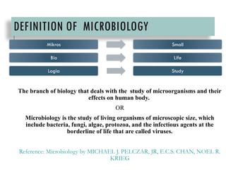
Thebranch of biology that deals with the study of microorganisms and their
effects on human body.
OR
Microbiology is the study of living organisms of microscopic size, which
include bacteria, fungi, algae, protozoa, and the infectious agents at the
borderline of life that are called viruses.
Reference: Microbiology by MICHAEL J. PELCZAR, JR, E.C.S. CHAN, NOEL R.
KRIEG
Mikros Small
Bio Life
Logia Study
HISTORICAL REVIEW OFMICROBIOLOGY
(1500’S TO 1900’S)
1500’s
Before 1500’s, most theories of diseases were based on
superstition.
No authentic knowledge of microorganisms.
People don’t know the exact cause of the disease.
Early observation and experimentation.
Several scientists put their effort in the field of microbiology.
7.
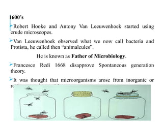
1600’s
Robert Hooke andAntony Van Leeuwenhoek started using
crude microscopes.
Van Leeuwenhoek observed what we now call bacteria and
Protista, he called then “animalcules”.
He is known as Father of Microbiology.
Francesco Redi 1668 disapprove Spontaneous generation
theory.
It was thought that microorganisms arose from inorganic or
rotting organic material.
8.
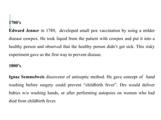
1700’s
Edward Jenner in1789, developed small pox vaccination by using a milder
disease cowpox. He took liquid from the patient with cowpox and put it into a
healthy person and observed that the healthy person didn’t get sick. This risky
experiment gave us the first way to prevent disease.
1800’s
Ignaz Semmelweis discoverer of antiseptic method. He gave concept of hand
washing before surgery could prevent “childbirth fever”. Drs would deliver
babies w/o washing hands, or after performing autopsies on women who had
died from childbirth fever.
9.
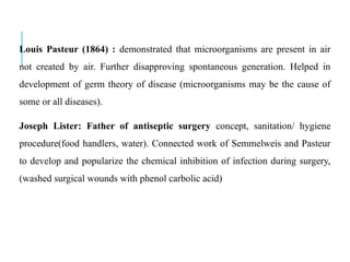
Louis Pasteur (1864): demonstrated that microorganisms are present in air
not created by air. Further disapproving spontaneous generation. Helped in
development of germ theory of disease (microorganisms may be the cause of
some or all diseases).
Joseph Lister: Father of antiseptic surgery concept, sanitation/ hygiene
procedure(food handlers, water). Connected work of Semmelweis and Pasteur
to develop and popularize the chemical inhibition of infection during surgery,
(washed surgical wounds with phenol carbolic acid)
10.
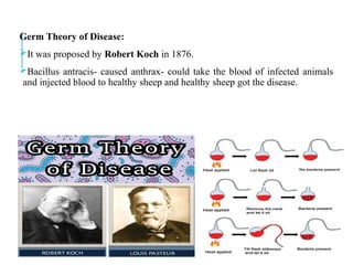
Germ Theory ofDisease:
It was proposed by Robert Koch in 1876.
Bacillus antracis- caused anthrax- could take the blood of infected animals
and injected blood to healthy sheep and healthy sheep got the disease.
11.
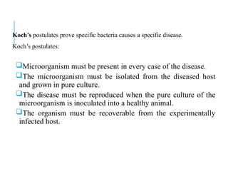
Koch’s postulates provespecific bacteria causes a specific disease.
Koch’s postulates:
Microorganism must be present in every case of the disease.
The microorganism must be isolated from the diseased host
and grown in pure culture.
The disease must be reproduced when the pure culture of the
microorganism is inoculated into a healthy animal.
The organism must be recoverable from the experimentally
infected host.
12.
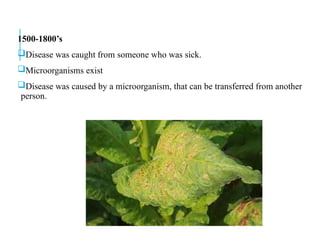
1500-1800’s
Disease was caughtfrom someone who was sick.
Microorganisms exist
Disease was caused by a microorganism, that can be transferred from another
person.
13.
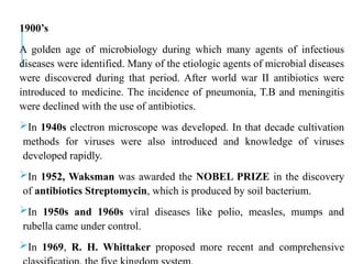
1900’s
A golden ageof microbiology during which many agents of infectious
diseases were identified. Many of the etiologic agents of microbial diseases
were discovered during that period. After world war II antibiotics were
introduced to medicine. The incidence of pneumonia, T.B and meningitis
were declined with the use of antibiotics.
In 1940s electron microscope was developed. In that decade cultivation
methods for viruses were also introduced and knowledge of viruses
developed rapidly.
In 1952, Waksman was awarded the NOBEL PRIZE in the discovery
of antibiotics Streptomycin, which is produced by soil bacterium.
In 1950s and 1960s viral diseases like polio, measles, mumps and
rubella came under control.
In 1969, R. H. Whittaker proposed more recent and comprehensive
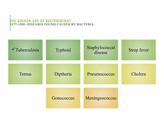
THE GOLDEN AGEOF BACTERIOLOGY
1877-1900 -DISEASES FOUND CAUSED BY BACTERIA.
Tuberculosis Typhoid
Staphylococcal
disease
Strep fever
Tetnus Diptheria Pneumococcus Cholera
Gonococcus Meningeococcus
16.
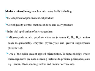
Modern microbiology reachesinto many fields including:
Development of pharmaceutical products
Use of quality control methods in food and dairy products
Industrial application of microorganism
Microorganisms also produce vitamins (vitamin C, B2, B12), amino
acids (L-glutamate), enzymes (hydrolytic) and growth supplements
(Riboflavin).
One of the major area of applied microbiology is biotechnology where
microorganisms are used as living factories to produce pharmaceuticals
e.g. insulin, blood clotting factors and number of vaccines.
17.
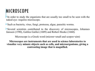
MICROSCOPE
In order tostudy the organisms that are usually too small to be seen with the
naked eye- requires microscope.
Such as bacteria, virus, fungi, protozoa, algae, parasitic worms.
Several scientists contributed to the discovery of microscopes, Johannes
Janssen (1590), Galileo Galilei (1609) and Robert Hooke (1660).
Microscope is a Greek word (micron=small and scopos=aim)
Microscopes are instruments that are used in science laboratories to
visualize very minute objects such as cells, and microorganisms, giving a
contrasting image that is magnified.
19.
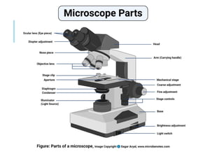
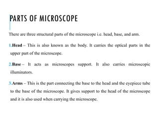
PARTS OF MICROSCOPE
Thereare three structural parts of the microscope i.e. head, base, and arm.
1.Head – This is also known as the body. It carries the optical parts in the
upper part of the microscope.
2.Base – It acts as microscopes support. It also carries microscopic
illuminators.
3.Arms – This is the part connecting the base to the head and the eyepiece tube
to the base of the microscope. It gives support to the head of the microscope
and it is also used when carrying the microscope.
20.
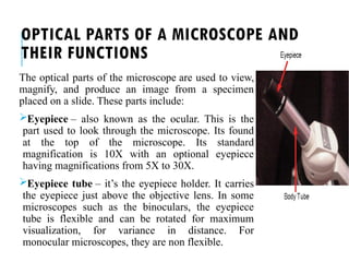
OPTICAL PARTS OFA MICROSCOPE AND
THEIR FUNCTIONS
The optical parts of the microscope are used to view,
magnify, and produce an image from a specimen
placed on a slide. These parts include:
Eyepiece – also known as the ocular. This is the
part used to look through the microscope. Its found
at the top of the microscope. Its standard
magnification is 10X with an optional eyepiece
having magnifications from 5X to 30X.
Eyepiece tube – it’s the eyepiece holder. It carries
the eyepiece just above the objective lens. In some
microscopes such as the binoculars, the eyepiece
tube is flexible and can be rotated for maximum
visualization, for variance in distance. For
monocular microscopes, they are non flexible.
21.
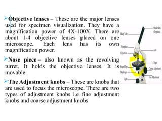
Objective lenses –These are the major lenses
used for specimen visualization. They have a
magnification power of 4X-100X. There are
about 1-4 objective lenses placed on one
microscope. Each lens has its own
magnification power.
Nose piece – also known as the revolving
turret. It holds the objective lenses. It is
movable.
The Adjustment knobs – These are knobs that
are used to focus the microscope. There are two
types of adjustment knobs i.e fine adjustment
knobs and coarse adjustment knobs.
22.
Stage – Thisis the section in which the specimen is placed for
viewing. They have stage clips that hold the specimen slides in place.
The most common stage is the mechanical stage, which allows the
control of the slides by moving the slides using the mechanical knobs
on the stage instead of moving them manually.
23.
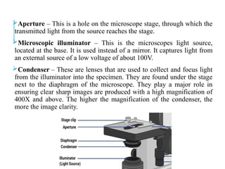
Aperture – Thisis a hole on the microscope stage, through which the
transmitted light from the source reaches the stage.
Microscopic illuminator – This is the microscopes light source,
located at the base. It is used instead of a mirror. It captures light from
an external source of a low voltage of about 100V.
Condenser – These are lenses that are used to collect and focus light
from the illuminator into the specimen. They are found under the stage
next to the diaphragm of the microscope. They play a major role in
ensuring clear sharp images are produced with a high magnification of
400X and above. The higher the magnification of the condenser, the
more the image clarity.
24.
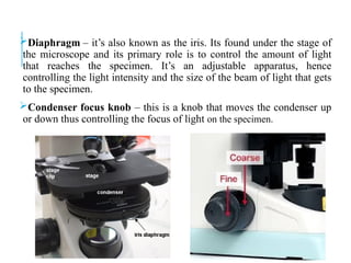
Diaphragm – it’salso known as the iris. Its found under the stage of
the microscope and its primary role is to control the amount of light
that reaches the specimen. It’s an adjustable apparatus, hence
controlling the light intensity and the size of the beam of light that gets
to the specimen.
Condenser focus knob – this is a knob that moves the condenser up
or down thus controlling the focus of light on the specimen.
25.
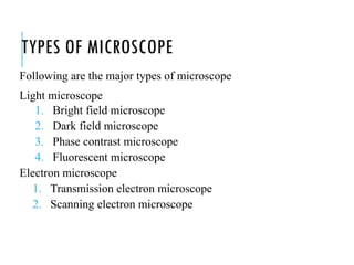
TYPES OF MICROSCOPE
Followingare the major types of microscope
Light microscope
1. Bright field microscope
2. Dark field microscope
3. Phase contrast microscope
4. Fluorescent microscope
Electron microscope
1. Transmission electron microscope
2. Scanning electron microscope
26.
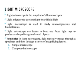
LIGHT MICROSCOPE
Lightmicroscope is the simplest of all microscopes.
Light microscope uses sunlight or artificial light
Light microscope is used to study microorganisms and
biomolecules.
Light microscope use lenses to bend and focus light rays to
produce enlarged images of small objects.
Principle: In light microscope, light typically passes through a
specimen and then through a series of magnifying lenses.
1. Simple microscope
2. Compound microscope
27.
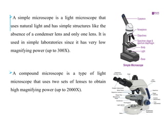
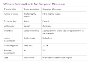
A simple microscopeis a light microscope that
uses natural light and has simple structures like the
absence of a condenser lens and only one lens. It is
used in simple laboratories since it has very low
magnifying power (up to 300X).
A compound microscope is a type of light
microscope that uses two sets of lenses to obtain
high magnifying power (up to 2000X).
29.
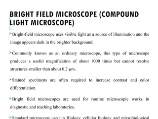
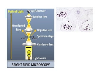
BRIGHT FIELD MICROSCOPE(COMPOUND
LIGHT MICROSCOPE)
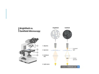
Bright-field microscope uses visible light as a source of illumination and the
image appears dark in the brighter background.
Commonly known as an ordinary microscope, this type of microscope
produces a useful magnification of about 1000 times but cannot resolve
structures smaller than about 0.2 µm.
Stained specimens are often required to increase contrast and color
differentiation.
Bright field microscopes are used for routine microscopic works in
diagnostic and teaching laboratories.
30.
PRINCIPLE
For a specimento be the focus and produce an image under the Bright field
Microscope, the specimen must pass through a uniform beam of the
illuminating light. Through differential absorption and differential refraction,
the microscope will produce a contrasting image.
32.
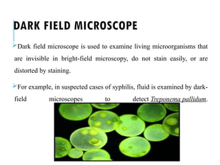
DARK FIELD MICROSCOPE
Darkfield microscope is used to examine living microorganisms that
are invisible in bright-field microscopy, do not stain easily, or are
distorted by staining.
For example, in suspected cases of syphilis, fluid is examined by dark-
field microscopes to detect Treponema pallidum.
33.
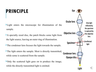
PRINCIPLE
Light enters themicroscope for illumination of the
sample.
A specially sized disc, the patch blocks some light from
the light source, leaving an outer ring of illumination.
The condenser lens focuses the light towards the sample.
The light enters the sample. Most is directly transmitted,
while some is scattered from the sample.
Only the scattered light goes on to produce the image,
while the directly transmitted light is omitted.
35.
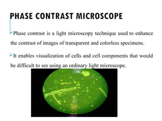
PHASE CONTRAST MICROSCOPE
Phasecontrast is a light microscopy technique used to enhance
the contrast of images of transparent and colorless specimens.
It enables visualization of cells and cell components that would
be difficult to see using an ordinary light microscope.
36.
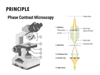
PRINCIPLE
When light passesthrough cells, small phase shifts occur, which are
invisible to the human eye. In a phase contrast microscope, these
phase shifts are converted into changes in amplitude, which can be
observed as differences in image contrast.
To study livingcells without staining. The ongoing different biological
processes in the live cells can be studied.
To study microbial motility.
To observe endospores and inclusion bodies that contain poly-
hydroxybutyrate, poly-metaphosphate, sulfur, or other substances.
39.
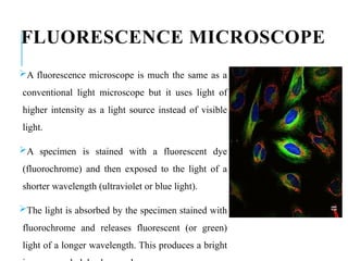
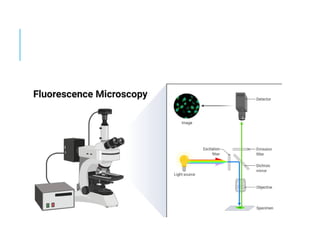
FLUORESCENCE MICROSCOPE
A fluorescencemicroscope is much the same as a
conventional light microscope but it uses light of
higher intensity as a light source instead of visible
light.
A specimen is stained with a fluorescent dye
(fluorochrome) and then exposed to the light of a
shorter wavelength (ultraviolet or blue light).
The light is absorbed by the specimen stained with
fluorochrome and releases fluorescent (or green)
light of a longer wavelength. This produces a bright
40.
The basic principleof fluorescence microscopy is to stain the components
with dyes.
Fluorescent dyes, also known as fluorophores or fluorochromes, are molecules
that absorb excitation light at a given wavelength (generally UV), and after a
short delay emit light at a longer wavelength. The delay between absorption
and emission is negligible, generally on the order of nanoseconds.
The emission light can then be filtered from the excitation light to reveal the
location of the fluorophores.
42.
USES
To identify structuresin fixed and live biological samples.
To identify different bacterial pathogens after staining them with
fluorochromes. Eg: Auramine-Rhodamine staining technique for the
detection of Mycobacterium tuberculosis.
To do ecological studies. Fluorochromes like acridine orange stain the
microorganisms. These stained organisms will fluoresce orange or
green.
To distinguish live bacteria from dead bacteria based on the color of
their fluorescence when they are treated with a special mixture of
43.
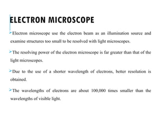
ELECTRON MICROSCOPE
Electron microscopeuse the electron beam as an illumination source and
examine structures too small to be resolved with light microscopes.
The resolving power of the electron microscope is far greater than that of the
light microscopes.
Due to the use of a shorter wavelength of electrons, better resolution is
obtained.
The wavelengths of electrons are about 100,000 times smaller than the
wavelengths of visible light.
44.
The electron travelsin a vacuum, and the magnet focuses the beam on the
sample.
On the monitor, an image is created, always black and white and can be
colored artificially.
45.
USES
To study smallerobjects such as viruses or objects or molecules having sizes
smaller than 0.2 µm.
To study the details of the internal structure of the cells.
To observe the ultrastructure of microorganisms, large molecules, biopsy
samples, metals, and crystals.
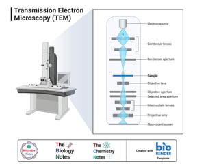
TRANSMISSION ELECTRON MICROSCOPE(TEM)
The transmission electron microscope is used to examine cells and cell
structure (even individual protein and nucleic acid molecules can be
visualized) at very high magnification and resolution.
The resolving power of a high-quality TEM is about 0.2 nanometers.
A special thin sectioning technique is needed to observe a bacterial cell by
transmission electron microscope.
A bacterial cell is cut into thin (20-60 nm) slices and treated with heavy metal
stains (such as osmic acid, permanganate, and uranium) to obtain sufficient
contrast.
49.
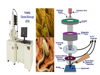
SCANNING ELECTRON MICROSCOPE(SEM)
A scanning electron microscope (SEM) is used to observe the external
features of an organism.
The specimen is coated with a thin film of a heavy metal such as gold. An
electron beam then scans back and forth across the specimen.
Electrons scattered from the metal coating are collected and activate a
viewing screen to produce an image.
SEM can obtain magnification of as low as 15X to as high as 100,000X.
50.
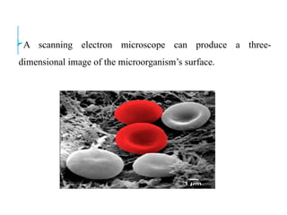
A scanning electronmicroscope can produce a three-
dimensional image of the microorganism’s surface.
52.
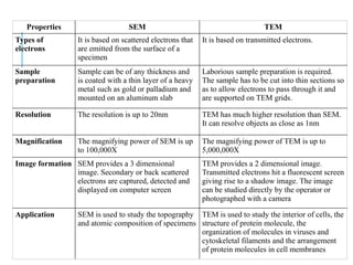
Properties SEM TEM
Typesof
electrons
It is based on scattered electrons that
are emitted from the surface of a
specimen
It is based on transmitted electrons.
Sample
preparation
Sample can be of any thickness and
is coated with a thin layer of a heavy
metal such as gold or palladium and
mounted on an aluminum slab
Laborious sample preparation is required.
The sample has to be cut into thin sections so
as to allow electrons to pass through it and
are supported on TEM grids.
Resolution The resolution is up to 20nm TEM has much higher resolution than SEM.
It can resolve objects as close as 1nm
Magnification The magnifying power of SEM is up
to 100,000X
The magnifying power of TEM is up to
5,000,000X
Image formation SEM provides a 3 dimensional
image. Secondary or back scattered
electrons are captured, detected and
displayed on computer screen
TEM provides a 2 dimensional image.
Transmitted electrons hit a fluorescent screen
giving rise to a shadow image. The image
can be studied directly by the operator or
photographed with a camera
Application SEM is used to study the topography
and atomic composition of specimens
TEM is used to study the interior of cells, the
structure of protein molecule, the
organization of molecules in viruses and
cytoskeletal filaments and the arrangement
of protein molecules in cell membranes
53.
SCOPE OF MICROBIOLOGYWITH REFERENCE TO
PHARMACEUTICAL SCIENCE
Pharmaceutical microbiology:
“Pharmaceutical microbiology is the applied branch of microbiology
which allow pharmacist to manufacture pharmaceuticals from
microorganisms either directly or with the use of some products
produced by them.”
54.
SCOPE OF MICROBIOLOGY
Criteriaand standards for the microbiological quality of
medicines depend upon the route of administration of the
medicine.
For example: The vast majority of medicines that are given by
mouth or placed on the skin are non-sterile, i.e. they may contain
some microorganisms (within limits on type and concentration),
whereas all injections and ophthalmic products must be sterile,
i.e. they contain no living organisms.
For a sterile product the criterion of quality is simple; there
should be no detectable microorganisms whatsoever.
The product should, therefore, be able to pass a test for
sterility, and a knowledge of the procedures and interpretation
of results of such tests is an important aspect of pharmaceutical
microbiology.
55.
Injections are alsosubject to a test for pyrogens; these are
substances that cause a rise in body temperature when introduced
into the body. Strictly speaking, any substance which causes
fever following injection is a pyrogen, but in reality the vast
majority are of bacterial origin, and it is for this reason that the
detection, assay and removal of bacterial pyrogens (endotoxins)
are considered.
Sterile medicines may be manufactured by two different
strategies.
The most preferred option is to make the product, pack it in its final container and sterilize it by heat, radiation or other
means.
The alternative is to manufacture the product from sterile ingredients under conditions that do not permit the entry of
contaminating organisms.
56.
Those responsible forthe manufacture of sterile products must be familiar
with the sterilization or aseptic manufacturing procedures available for
different product types, and those who have cause to open, use or dispense
sterile products (in a hospital pharmacy, for example) should be aware of the
aseptic handling procedures to be adopted in order to minimize the risk of
product contamination.
Microorganisms are valuable in the maintenance of our ecosystems. Their
role and benefits in the carbon and nitrogen cycles in terms of recycling dead
plant and animal material and in the fixation of atmospheric nitrogen.
57.
Apart from thesemajor applications, however, the uses of microorganisms in
the manufacture of medicines prior to 1980 were very limited. Enzymes were
developed for use in cancer chemotherapy (asparaginase) and to digest blood
clots (streptokinase), and polysaccharides also found therapeutical
applications (e.g. dextran—used as a plasma expander).
There is a large range of antimicrobial drugs used to prevent and treat
microbial infections. Because of this range and diversity of products,
pharmacists are now far more commonly called upon to advise on the relative
merits of the antibiotics available to treat particular categories of infection.
58.
Another major advancementof microbiology is recombinant
DNA technology in the 1970s. This technology permitted human
genes to be inserted into microorganisms, which were thus able
to manufacture the gene products far more efficiently than
traditional methods of extraction from animal or human tissues.
Insulin
Human growth hormone
Interferon
Blood clotting factors
Vaccines e.g., Hepatitis B vaccine.
59.
All these developments,together with miscellaneous applications in the
detection of mutagenic and carcinogenic activity in drugs and chemicals and
in the assay of antibiotics, vitamins and amino acids have ensured that the role
of microorganisms in the manufacture of medicines is now well recognized,
and that a basic knowledge of immunology , gene cloning is an integral part of
pharmaceutical microbiology.
60.
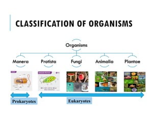
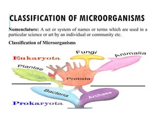
CLASSIFICATION OF MICROORGANISMS
Nomenclature:A set or system of names or terms which are used in a
particular science or art by an individual or community etc.
Classification of Microorganisms
61.
NOMENCLATURE OF MICROORGANISMS
TheGreek philosopher Aristotle attempted to classify all living
things as either Plant or Animal. He grouped animals into:
Land Dwellers
Water Dwellers
Air Dwellers
Although this system made sense to Aristotle, we would have a
difficult time in grouping elephants and earthworms, whales and
water striders, flies and falcons together.
Subsequent scientists later tried to classify living creatures by
means of locomotion, grouping butterflies and bats (flying), and
barley (both rooted in place). This system of classification was
obviously flawed as well.
62.
The efforts toclassify living things saw great progress in the work of Carl
Linnaeus, a Swedish botanist. He developed his naming system in the middle
1700’s, which essentially the same one we use today. He attempted to name all
known plants, animals, and minerals using Latin and Greek names. One of his
books, Systema Naturae, meaning “The Natural Classification", was
published in 1735 and was based on his religious belief that one could
understand God by studying his creation.
63.
Today, microorganism namesoriginate from four different
sources
1. Descriptive
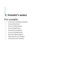
2. Scientist’s names
3. Geographic places
4. Organizations
1. Descriptive
For example:
Staphylococcus aureus (grape-like cluster of spheres, golden in color)
Streptococcus viridans (chains of spheres, green in colony color)
Proteus vulgaris (first and common)
Helicobacter pylori (spiral shaped rod at the entrance to the duodenum)
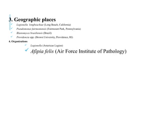
3. Geographic places
Legionella longbeachiae (Long Beach, California)
Pseudomonas fairmontensis (Fairmount Park, Pennsylvania)
Blastomyces brasiliensis (Brazil)
Providencia spp. (Brown University, Providence, RI)
4. Organizations
Legionella (American Legion)
Afipia felis (Air Force Institute of Pathology)
66.
RULES OF NOMENCLATURE
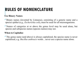
UseBinary Names
Binary names (invented by Linnaeus), consisting of a generic name and a
species epithet (e.g., Escherichia coli), must be used for all microorganisms.
Names of categories at or above the genus level may be used alone, but
species and subspecies names (species names) may not.
When to Capitalize
The genus name (and above) is always capitalized, the species name is never
capitalized, e.g. Bacillus anthracis words…never use a species name alone.
67.
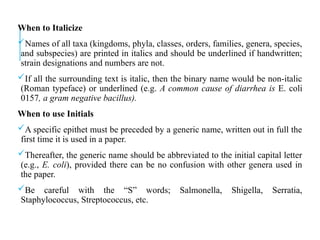
When to Italicize
Namesof all taxa (kingdoms, phyla, classes, orders, families, genera, species,
and subspecies) are printed in italics and should be underlined if handwritten;
strain designations and numbers are not.
If all the surrounding text is italic, then the binary name would be non-italic
(Roman typeface) or underlined (e.g. A common cause of diarrhea is E. coli
0157, a gram negative bacillus).
When to use Initials
A specific epithet must be preceded by a generic name, written out in full the
first time it is used in a paper.
Thereafter, the generic name should be abbreviated to the initial capital letter
(e.g., E. coli), provided there can be no confusion with other genera used in
the paper.
Be careful with the “S” words; Salmonella, Shigella, Serratia,
Staphylococcus, Streptococcus, etc.
68.
Common Names
Common namesshould be in lowercase roman type, non-italic
(e.g., streptococcus, brucella).
However when referring to the actual genus name (or above)
always capitalize and italicize.
Subspecies and Serovars
For Salmonella, genus, species, and subspecies names should be
rendered in standard form:
Salmonella enterica at first use, S. enterica thereafter;
Salmonella enterica subsp. arizonae at first use, S. enterica
subsp. arizonae thereafter.
69.
Abbreviations for Species
use“sp.” for a particular species
“spp.” for several species (“spp” stands for “species plural”).
These abbreviations are not italicized; e.g. Clostridium sp. or Clostridium
spp.
Other Abbreviations:
e.g. meaning 'for example' (it comes from the Latin, exempli gratia)
i.e. meaning 'that is' (from the Latin id est). Note that 'i.e.' specifies particular
things, whereas 'e.g.' gives examples.
etc. meaning 'and so forth' (from the Latin et cetera) [Some people, wrongly,
write ect.]
et al. meaning 'and others' (from the Latin et alia). You would use this only
when citing references.
70.
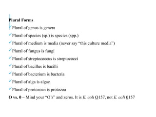
Plural Forms
Plural ofgenus is genera
Plural of species (sp.) is species (spp.)
Plural of medium is media (never say “this culture media”)
Plural of fungus is fungi
Plural of streptococcus is streptococci
Plural of bacillus is bacilli
Plural of bacterium is bacteria
Plural of alga is algae
Plural of protozoan is protozoa
O vs. 0 – Mind your “O’s” and zeros. It is E. coli O157, not E. coli 0157

![Abbreviations for Species
use “sp.” for a particular species
“spp.” for several species (“spp” stands for “species plural”).
These abbreviations are not italicized; e.g. Clostridium sp. or Clostridium
spp.
Other Abbreviations:
e.g. meaning 'for example' (it comes from the Latin, exempli gratia)
i.e. meaning 'that is' (from the Latin id est). Note that 'i.e.' specifies particular
things, whereas 'e.g.' gives examples.
etc. meaning 'and so forth' (from the Latin et cetera) [Some people, wrongly,
write ect.]
et al. meaning 'and others' (from the Latin et alia). You would use this only
when citing references.](https://image.slidesharecdn.com/microch1-250301024321-84f9b6b4/85/micro-ch-1-pptx-pharmD-Micro-biology-notes-69-320.jpg)