1) An intestinal fistula is an abnormal connection between two epithelial surfaces, most commonly the intestine and skin (enterocutaneous). The ileum is the most common site of origin.
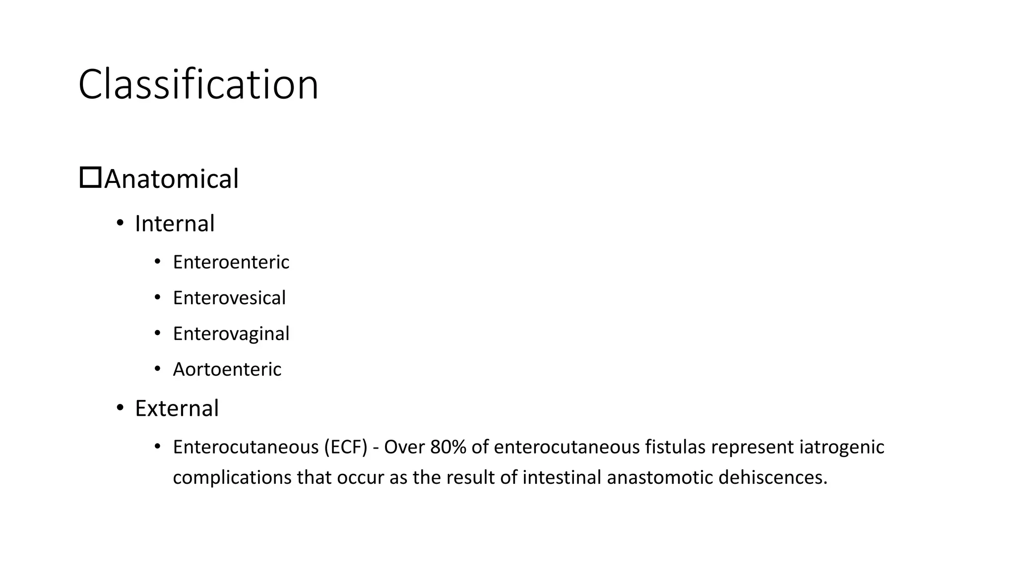
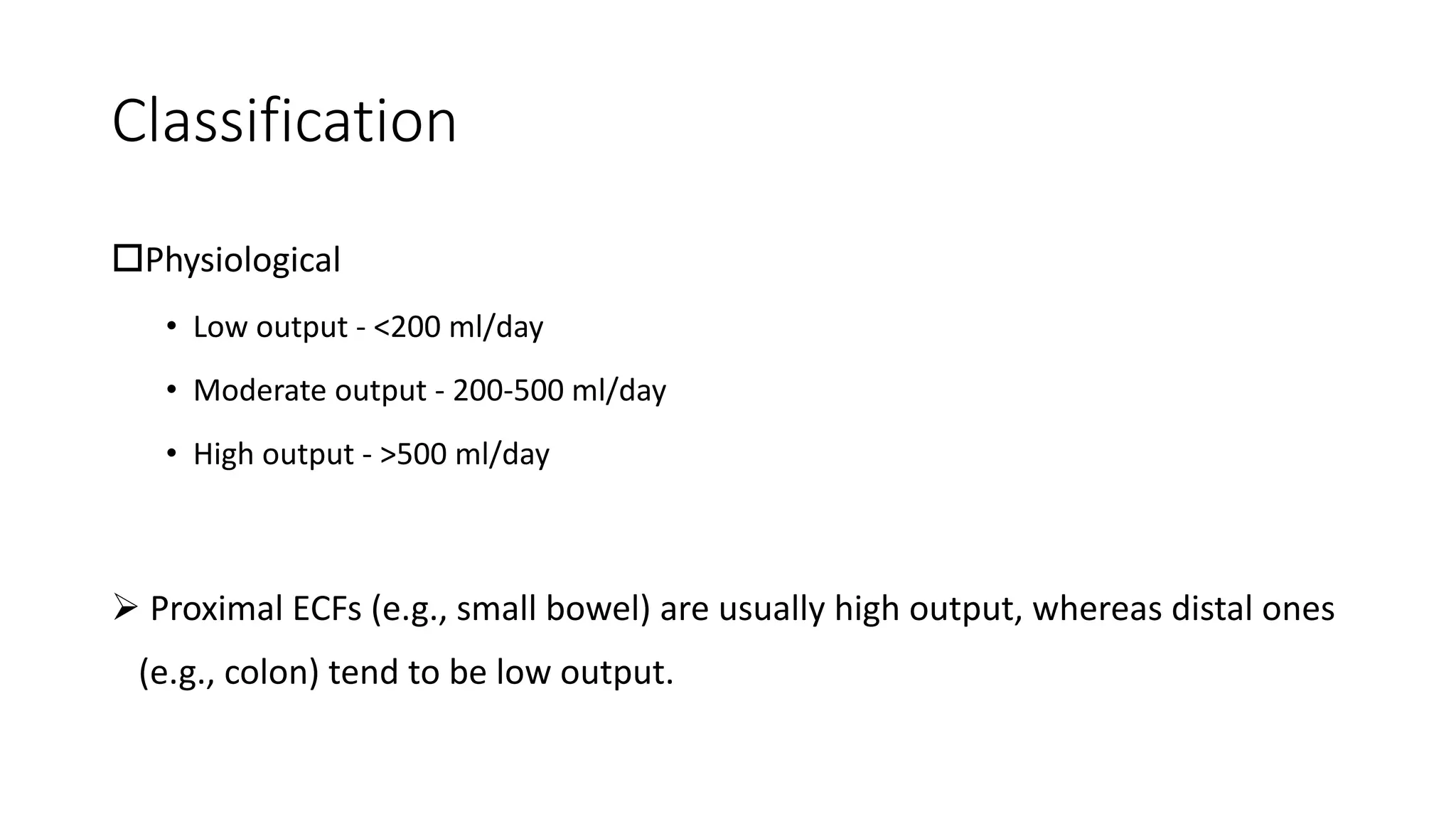
2) Fistulas can be classified anatomically by their connections or physiologically by their output. Enterocutaneous fistulas usually result from complications of intestinal surgery.
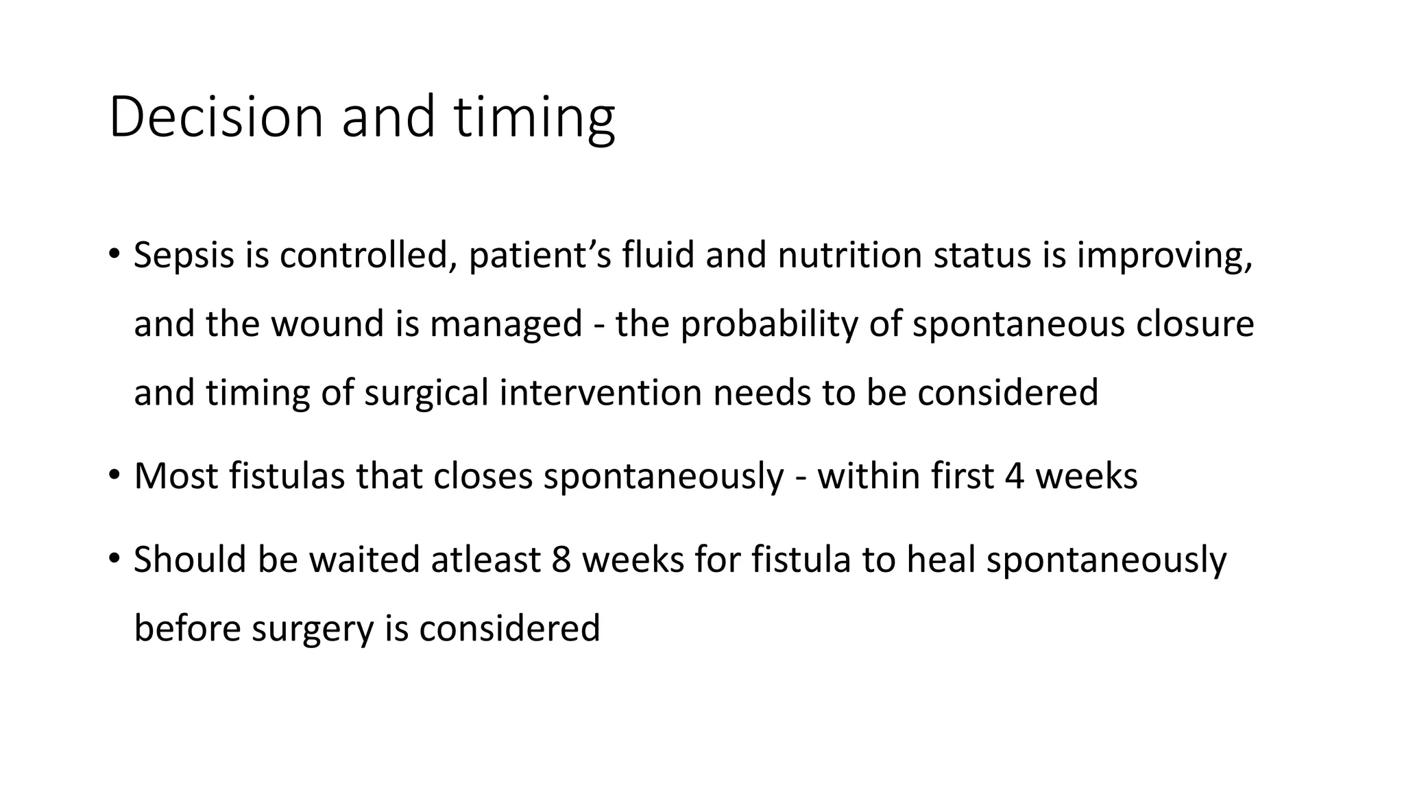
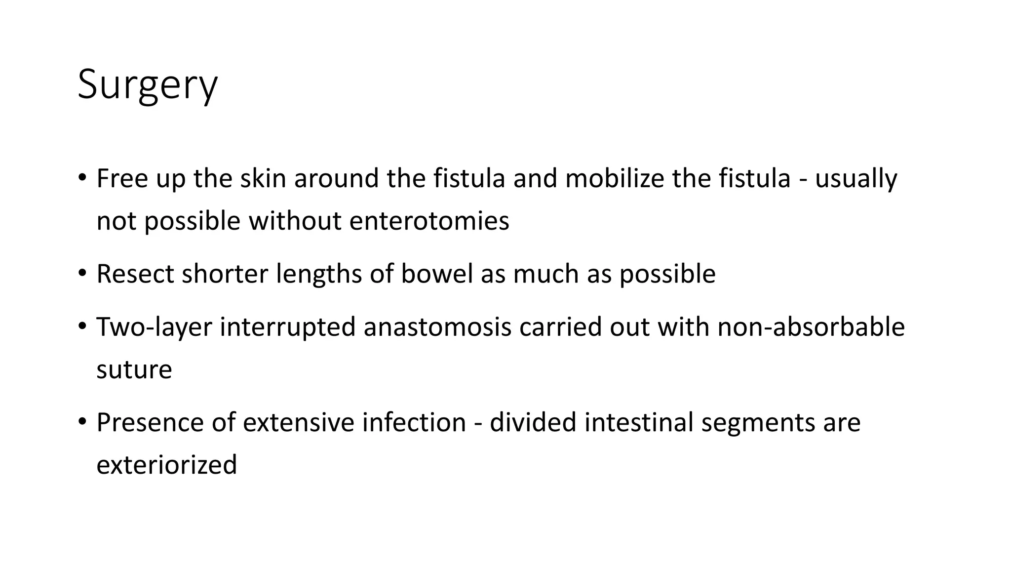
3) Management of intestinal fistulas involves stabilization of the patient through fluid resuscitation, nutritional support, and controlling sepsis before considering definitive surgical repair once the patient's condition has improved.
2. Introduction
• Fistula - defined as an abnormal communication between two
epithelialized surfaces
• Enterocutaneous fistula is the most common type of intestinal fistula
• Ilem is the most common site of origin of enterocutaneous fistula
3. Classification
Anatomical
• Internal
• Enteroenteric
• Enterovesical
• Enterovaginal
• Aortoenteric
• External
• Enterocutaneous (ECF) - Over 80% of enterocutaneous fistulas represent iatrogenic
complications that occur as the result of intestinal anastomotic dehiscences.
4. Classification
Physiological
• Low output - <200 ml/day
• Moderate output - 200-500 ml/day
• High output - >500 ml/day
Proximal ECFs (e.g., small bowel) are usually high output, whereas distal ones
(e.g., colon) tend to be low output.
5. Etiology
Congenital
• Tracheo-oesophageal fistula
Acquired
• Traumatic (75-85%)
• Iatrogenic - injury to the intestine during handling, lysis of adhesions, abdominal fascial
closure, or percutaneous drainage.
• Penetrating or blunt abdominal traumas
• Spontaneous (15-25%)
• Previous intestinal irradiation
• Intraabdominal sepsis
• Inflammatory bowel disease, especially Crohn’s disease
• Malignancy
6. Pathophysiology
• The manifestations of fistulas depend on which structures are
involved
• Low-resistance enteroenteric fistulas - malabsorption
• Enterovesicular fistulas - recurrent urinary tract infections
• Enterocutaneous fistulas
• High-output fistulas - dehydration, electrolyte imbalance and malnutrition
• Skin excoriation
8. Presentation
• Drainage of enteric material through the abdominal wound or
through existing drains
• Pronlonged ileus
• Abdominal pain
• Signs of sepsis - fever, tachycardia, leukocystosis
• Dehydration, fluid and electrolyte abnormalities and malnutrition
• Often associated with intra-abdominal abscesses
9. Diagnosis
• CT scan with oral contrast
• Leakage of contrast material from the intestinal lumen
• Demonstrate the fistula’s site of origin in the bowel
• Rule out the presence of intestinal obstruction distal to the site of origin
• Intra-abdominal abscess
• Fistulogram
• Contrast is injected under pressure through a catheter placed percutaneously
into the fistula tract
• Provide information about the length and origin of the fistula
10. Prevention
• Use of healthy bowel for anastomosis
• Preoperative mechanical bowel preparation
• Preoperative intraluminal or systemic antibiotics
• Preoperative optimization of the nutritional status
• Sound anastomotic techniques
11. Management
1. Stabilization
• Fluid and electrolyte resuscitation
• Nutritional support
• Control sepsis with antibiotics and drainage of abscess
• Skin protection and wound management
• Pharmacological measures
2. Investigation - anatomy of fistula
3. Decision and timing
4. Definitive Management
5. Rehabilitation
12. Fluid and electrolyte resuscitation
• Restoration of normovolemia, electrolyte replacement, and correcting
acid-base balance - requires accurate measurement of fistula output
and composition
• Crystalloid alone is inadequate
• Albumin and plasma (platelet rich plasma, fresh frozen plasma)
• Fresh whole blood
• Urine output should be restored to greater than 0.5 ml/kg/hr
13. Nutritional support
• Adequately nourished patient - ECF closes spontaneously
• Should begin as soon as the patient is stabilized
• Enteral or parenteral route
• Low output fistula - Enteral feeding
• High output fistula - TPN to replace fluid losses
• Advisable to provide at least a part (25%) of daily nutritional
requirement through enteral route
• Enteral feeding tube may be entered beyound the fistula -
Fistuloclysis
14. Daily nutritional requirement
Low output
• Calorie requirement: 30-35 kcal/kg/day
• Protein requirement: 1-2 gm/kg/day
High output
• Calorie requirement: 45-50 kcal/kg/day
• Protein requirement: 1.5-2.5 gm/kg/day
Vitamins, trace elements, essential fatty acids should be provided
15. Control sepsis and drainage of abscess
• Recognition of residual or ongoing sepsis - fever, tachycardia,
leukocytosis and failure to improve in general condition
• Appropriate antibiotics should be used
• Often have accompanying intraabdominal abscess
• Drainage of obvious abscesses - radiological guided aspiration
• Purulence is thick and cannot be aspirated - drainage in the operating
room
16. Skin care & wound management
• Prevent skin excoriation - stomahesive paste, aluminium paint
• Fistula output <50 mL/day can be managed with dressing and skin
barrier
• Fistula output >500 mL/day usually requires a pouch system
• NG tube placement - decompression
• NPO
• TPN
• Placement of sump drain and vacuum assisted closure (VAC) device - apply
negative pressure and help to control the drainage and consequently
minimize wound size
17. Pharmacological measures
• PPI and H2 blockers - to decrease gastric secretions
• Somatostatin analogues (octreotide) - decreased fistula output
• Infliximab (monoclonal antibody to TNF-α) may help with fistula
closure in patients with Crohn’s disease
19. Decision and timing
• Sepsis is controlled, patient’s fluid and nutrition status is improving,
and the wound is managed - the probability of spontaneous closure
and timing of surgical intervention needs to be considered
• Most fistulas that closes spontaneously - within first 4 weeks
• Should be waited atleast 8 weeks for fistula to heal spontaneously
before surgery is considered
20. Decision and timing
• Surgical therapy is inevitable in many
cases, especially when unfavorable
characteristics are present
• Surgical intervention for ECFs should
be delayed until both the
intraabdominal and systemic
conditions have been optimized
21. Aids to closure
• Keep edges protected and
clean
• Sumps with gentle suction
• Keep stool and pus away
from edges
22. Definitive management - Surgery
• Should only be attempted after an adequate trial of soft sumps,
antibiotics, nutrition, and keeping the patient’s abdomen clean
• If no closure has occurred in 60 days - show no sign of closing
• The incision should be made in a clean area away from the fistula -
keep skin edges clean
• Lysis of adhesions - if possible, avoid making enterotomies
23. Surgery
• Free up the skin around the fistula and mobilize the fistula - usually
not possible without enterotomies
• Resect shorter lengths of bowel as much as possible
• Two-layer interrupted anastomosis carried out with non-absorbable
suture
• Presence of extensive infection - divided intestinal segments are
exteriorized
24. Post-operative care and rehabilitation
• Ambulation
• Wound should be reinforced with bulky dressings
• Do not be in a rush to feed the patient
• When feeding, maintain calories and protein until bowel function is
normal
• Make certain caloric and protein intake are adequate before stopping
total parenteral nutrition
• Allow 4 to 6 months of adequate nutritional support and
rehabilitation before patients should think about returning to work
26. References
• Williams N, O’Connell PR, McCaskie AW. Bailey & Love’s Short Practice
of Surgery. 27th edition. Florida (US): CRC Press; 2017.
• Townsend CM et al. Sabiston: Textbook of Surgery: The Biological
Basis of Modern Surgical Practice. 21st edition. Missouri (US):
Elsevier; 2022.
• Brunicardi FC et al. Schwartz’s: Principles of Surgery. 11th edition.
McGraw-Hill Education; 2019.
• Cameron JL, Cameron AM. Current Surgical Therapy. 13th edition.
Elsevier; 2020.