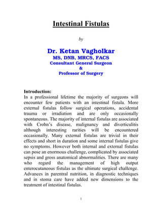

![Four Phase Approach [Sheldon et al]
Initial Phase (on presentation)
1. Restore blood volume.
2. Begin correction of fluid and electrolyte imbalance.
3. Control fistula, protect skin, collect and measure
effluent.
4. Drain abscesses and consider antibiotic therapy.
Second Phase (Up to 2 days)
1. Continue fluid and electrolyte therapy
2. Begin IV feeding.
Third Phase (Up to 5 days)
1. Institute enteral feeding if possible either orally or by
tube feeding or by jejunostomy below a high fistula.
2. Demonstrate the anatomy of the fistulas by contrast
studies and fistulography.
Fourth Phase (After 5 days)
1. Continue nutritional treatment until the fistula closes
or if it fails to close, until the patient is able to
withstand definitive surgery.
2. Operate to eliminate sepsis if recurring.
4](https://image.slidesharecdn.com/intestinalfistulas-121114073002-phpapp02/85/Intestinal-fistulas-5-320.jpg)
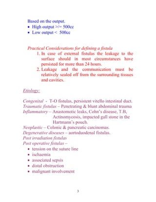
![Intravenous Treatment Regimens:
A] Resuscitation
B] Fluid & electrolyte regimens.
C] Nutritional regimens (enteral/parentral)
Common water and electrolyte problems in fistula patients:
Dehydration, hyponatremia, hypokalemia, metabolic
acidosis, metabolic alkalosis, hypernatremia &
hyperosmolar syndrome in patients fed IV or orally with
elemental diets.
Water requirements = Normal requirements + add.
Requirements resulting from the fistula- modifications
imposed by complications such as renal failure.
Daily requirements= basal requirements+additional
requirements
5% dextrose 2000cc 1250cc 3250cc
Normal saline 500cc 750cc 1250cc
KCl 80mmol 40mmol 120mmol
5](https://image.slidesharecdn.com/intestinalfistulas-121114073002-phpapp02/85/Intestinal-fistulas-6-320.jpg)


This document discusses intestinal fistulas, including: - Definitions, classifications, and etiologies of intestinal fistulas. Fistulas can be internal or external, simple or complicated, and caused by conditions like Crohn's disease or trauma. - A four phase approach to management: initial stabilization, continued support for 2 days, enteral feeding trial from 2-5 days, and definitive surgery after 5 days if needed. - Nutritional management involves IV or enteral feeding to correct deficiencies from fistula output. Output and electrolytes must be closely monitored. - Investigations help determine fistula anatomy and underlying causes. Surgical intervention aims to aid closure, correct malnutrition, or re


