SlideShare a Scribd company logo
By : Kinjal Banerjee
SU-6
DISCUSSION

INTRODUCTION
Fistula is defined as an abnormal communication between two epithelialized
surfaces.
Enterocutaneous fistula is an abnormal communication between the skin with
various parts of the gut, for example duodenum, jejunum etc.
The word fistula means a “pipe” or “flute” in Latin.
 The ileum is the most common site of origin of Enterocutaneous fistula.
Patients are usually systemically ill, with sepsis being a significant risk factor.
Spontaneous closure rate depend upon many factors like anatomy, etiology,
nutritional status, output from fistula.
Fluid and electrolyte imbalance, malnutrition, sepsis & related complications,
local wound excoriation are some of the known complications.
ETIOLOGY
Webster and Carey proposed five mechanisms of fistula formation :
1> Congenital : - Rare
- Due to the complete failure of Vitellointestinal duct to obliterate.
- Fecal matter at umbilicus after post natal slough of the stump.
2> Trauma :

- Major penetrating trauma to the abdomen may lead to ECF.
- Damage control laparotomy techniques have higher risk of
delayed ECF formation(2% to 25%).
3> Infection : - Abscess or invasive intestinal infections like amoebiasis,
coccidiomycosis, tuberculosis etc.
- Intestinal perforation in ileum from tuberculosis or enteric fever
esp in 3rd world countries.
- Actinomyces infection post appendectomy.
4> Perforation or Injury with Abscess & Operative complications :
- Intra-abdominal abscess may erode into the gut & through the
skin.
- 75-85% are iatrogenic - after surgery for bowel obstruction,
cancer, or IBD
5> Inflammation, Irradiation or Tumour :
- Crohn’s disease may cause ECF – post op more common
than spontaneous.
- ECF after anastomosis/appendicectomy/simple exploration
in Crohn’s disease.
- Irradiation for pelvic malignancies etc may lead to a chronic
non-healing ECF.
PREVENTION OF FISTULA

- Acute intra-operative perforations should be identified and closed.
- Serosal tears should be examined carefully and repaired if required.
- Aggressive interloop adhesion break-up should be avoided to prevent serosal
tears.
DIAGNOSIS OF PERFORATION AND FISTULA
-Post operatively anastomotic leaks/unrecognised perforations and
subsequent fistulae may manifest as instability or patient’s failure to improve.
-Fever, abdominal pain followed by exiting of intestinal contents from drain or
incision site.
-Occasionally,heavily purulent discharge may mask an ECF like in enteric
perforations.
-Activated charcoal or indigo carmine by mouth can help in diagnosing ECF.
- Intraluminal instillation of methylene blue and saline or direct endoscopy helps
to identify small perforations.
STAGING AND CLASSIFICATION
1. Anatomical Classification : Internal or External fistula(ECF).
- Internal fistulae are named after the structures it communicates
like gastrocolic, jejunoileal, aortoenteric fistula.
- External fistulae like gastric, duodenal, jejunal, ileal or fecal.
2. Physiologic classification : Based on output
- High-output > 500 cc/day (Difficulties in fluid management and
skin care)
- Moderate-output 200-500 cc/day
- Low-output < 200 cc/day (Usually colonic)
MANAGEMENT
S-S-N-A-P


Stabilization



Control of Sepsis and appropriate Skin care



Nutrition



Define underlying Anatomy



Plan to deal with the fistula
STABILIZATION

> The first step of management is the resuscitation and stabilization of the patient.
> Needs to be accomplished within the first 24 to 48 hours of management.
> Initial efforts directed towards intravenous fluid resuscitation, control of infection,
protection of surrounding skin & measuring and replacing ongoing losses.
> Attention should also be given to any intra-abdominal/subcutaneous abscesses
& if present they should be drained.
A. RESUSCITATION

> Restoration of normal circulating blood volume and correction of electrolyte
and acid-base imbalances are a top priority.
>Rehydration usually requires isotonic fluid(NS/RL) until the patient is euvolemic.
> Strict input and output measurements are essential and CVP monitoring
and urinary catheterisation are especially helpful.
> Ongoing fluid losses should be fully replaced and electrolyte imbalances
must be corrected.
RESUSCITATION CONTD..

> Enterocutaneous fistulae are usually associated with hypokalemia and
metabolic acidosis, which require correction.
> Electrolyte measurement of the fistula output helps in planning of
replenishment of ongoing losses.
> Urine output should be restored to greater than 0.5mL/kg/hr.
> Requirement of blood transfusion depends upon overall hemodynamic
status, patient’s oxygen carrying capacity and oxygen delivery.
B. NUTRITION

> In 1972, Roback and Nicholoff reported closure of 73% of enteric fistulae
in adequately nourished patients, as against 19% in inadequate ones.
> Nutritional support needs to begin as soon as the patient is stabilized.
> Nutrition can be given by parenteral or enteral route,based on the anatomy
of the fistula.
> Its advisable to provide atleast a part of the daily nutritional requirement
through enteral route.(Even 25% of daily requirement given enterally,
can help reap its benefits)
NUTRITION CONTD..

> Nutrition via the enteral route helps in maintaining the intestinal mucosal
barrier, more efficacious delivery of nutrients, stimulating hepatic protein
synthesis.
> WHO’s ORS (40g/L of glucose, 90 mEq/L of Na, 20 mEq/L of K) should be
given when oral intake is possible.
> In proximal fistulae, the enteral feeding tube may be entered beyond the
fistula to provide enteral nutrition(fistuloclysis).
> It is advisable to enter feeding tube beyond ligament of Treitz for a gastric
or duodenal fistula.
NUTRITION CONTD..

> Enteral feeds of highly absorbable, low-residue nutrients can be given if even
4ft of functional bowel exists in between ligament of Treitz and the proximal
end of the fistula.
> During the time the enteral nutrition is increasing, patient should be
supplemented with TPN.
> TPN is also given in patients who do not tolerate enteral feeds or have
long standing ileus or before fistulous tract is well established.
NUTRITION CONTD..
TYPE OF FISTULA

CALORIE REQ

PROTEIN REQ

Low Output

30-35 kcal/kg/day

1-2 gm/kg/day

High Output

45-50 kcal/kg/day

1.5-2.5 gm/kg/day

> Twice the daily requirement of vitamins, trace elements, zinc and upto 10 times
the daily requirement of Vitamin C should be provided.
> Short turnover proteins like retinol-binding protein, prealbumin, ferritin can be
used to monitor the weekly efficacy of protein delivery. [Alb half life is 20 days].
> Mortality rate of 42% with alb <2.5 mg/dl vs 0% if >3.5 mg/dl.
C. CONTROL OF SEPSIS AND FISTULA EFFLUENT
> Persistant fever, tachycardia and leukocytosis along with failure to improve
adequately points towards possible sepsis or abscesses.
> May require surgical drainage of abscess,if any along with adequate antibiotic
cover.
> Local skin care and prevention of skin excoriation by using stomahesive paste
or aluminium paint etc along with stoma bags.
> Excessive fistula output can be controlled by nasogastric tube placement,
withdrawal of oral feeds, initiation of parenteral nutrition or placement of
sump suction catheter as described by Suripaya and Anderson,1971.
C. CONTROL OF SEPSIS AND FISTULA EFFLUENT CONTD..

> Vacuum assisted closure [VAC] device drainage system maybe used too.
> Due to the negative pressure application, VAC device helps to control drainage,
minimizes the size of the abdominal wound, reduces frequency of dressing
and protects the skin while helping to promote fistula healing.
> For majority of ECFs, VAC devices have become the method of choice for
controlling fistula drainage and skin protection.
D. PHARMACOLOGICAL SUPPORT
> Somatostatin analogue Octreotide, at doses of 100 – 250 mcg TDS reduces
fistula output by 40 – 60% by the end of 24 hrs.
> Should be discontinued if ineffective for 48 hrs as it has side effects like
hyperglycemia, elevated cholesterol and reduced bowel motility.
> Octreotide and TPN seem to have a synergistic effect on reduction of effluent
volume and improvement in fistula closure rates.
> Proton-pump inhibitors and H2 receptor antagonists also help reduce fistula
output especially in proximal fistulas.
D. PHARMACOLOGICAL SUPPORT CONTD..
> Cyclosporine in doses of 4 mg/kg/day for 6 – 10 days followed by oral doses
of 8 mg/kg/day helps to treat refractory fistulae associated with Crohn’s disease.
> Other drugs used include Tacrolimus, Azathioprine, 6-MP, Infliximab(mAb TNFα).
> Infliximab administered at a dose of 5 mg/kg iv at 0,2,6 weeks helped in partial
resolution of 68% of multiple lesions and complete closure in 55% of patients.
> Complications include URTI, headache, fatigue etc.
INVESTIGATIONS
> Investigations are done for the next 7 to 10 days following stabilization.
> They are carried out to determine the presence and location of the fistula,
its cause and presence of comorbidities.
> Oral administration of Indigo Carmine/ Charcoal helps in diagnosing the
presence of a fistula,but not its location, cause or anatomy.
> USG abdomen helps in locating intra-abdominal abscesses, apart from
helping in performing guided aspiration of intra-abdominal collections.
> Fistulography helps in defining the length & width of the fistula, its anatomical
location, the presence of any distal obstructions etc.
> It can be performed by entering a small cathter into the fistulous tract and then
injecting the water-soluble contrast under fluoroscopy.
> Fistulogram should be performed before an upper GI series or CT scan with
oral contrast or contrast enema as it poses difficulty in interpretation.
> Fistulography should be followed up with a complete contrast study of the
GI tract.
CT scan of the abdomen with IV and oral contrast is highly sensitive and specific
for locating the fistula, defining its anatomy, commenting on the gut surrounding
fistula, presence of any intra-abdominal abscess or distal obstruction/pathology.
CT scan is highly recommended for duodenal and pancreatic fistula.
CT scan can be therapeutic by helping in CT-guided aspiration of intra-abdominal
abscesses,if any.
Endoscopy may also be used occasionally though its principal use is in internal
fistulas.
Endoscopy is usually delayed till acute inflammation gets reduced.
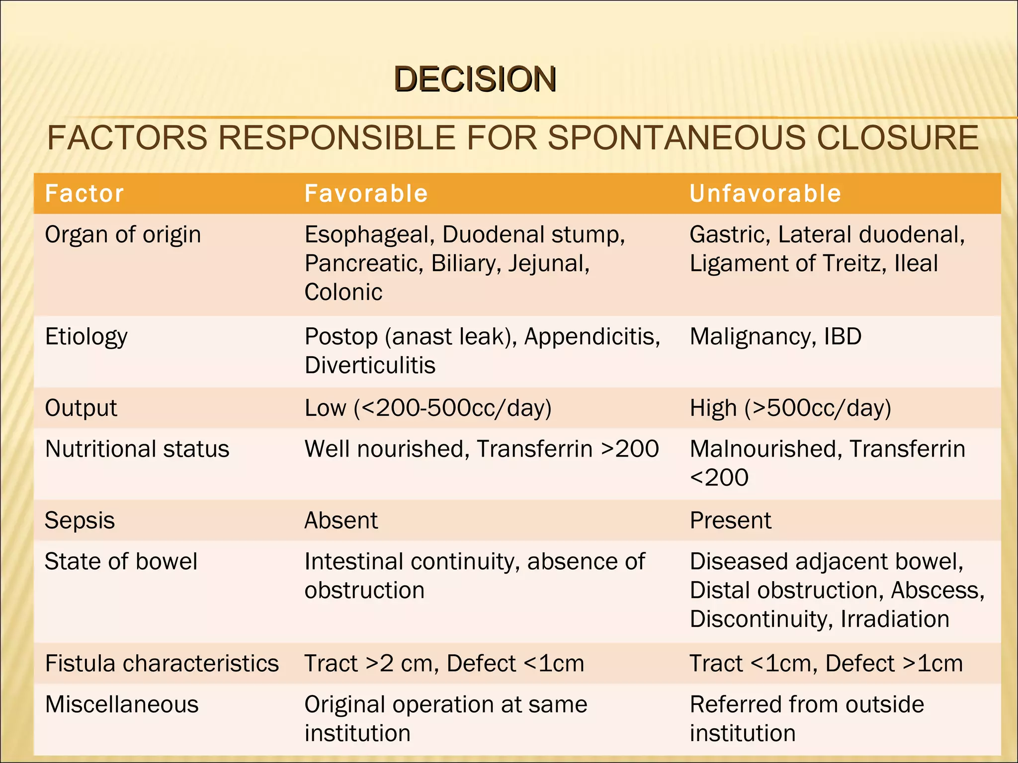
DECISION
FACTORS RESPONSIBLE FOR SPONTANEOUS CLOSURE
Factor

Favorable

Unfavorable

Organ of origin

Esophageal, Duodenal stump,
Pancreatic, Biliary, Jejunal,
Colonic

Gastric, Lateral duodenal,
Ligament of Treitz, Ileal

Etiology

Postop (anast leak), Appendicitis,
Diverticulitis

Malignancy, IBD

Output

Low (<200-500cc/day)

High (>500cc/day)

Nutritional status

Well nourished, Transferrin >200

Malnourished, Transferrin
<200

Sepsis

Absent

Present

State of bowel

Intestinal continuity, absence of
obstruction

Diseased adjacent bowel,
Distal obstruction, Abscess,
Discontinuity, Irradiation

Fistula characteristics Tract >2 cm, Defect <1cm

Tract <1cm, Defect >1cm

Miscellaneous

Referred from outside
institution

Original operation at same
institution
> Reber et al reported that more than 90% of small intestinal fistula which
closed spontaneously,did so within a month.
> Spontaneous closure rates dropped to less than 10% after 2 months and
none after 3 months.
> Factors possibly responsible for failure of spontaneous closure are:
a. Foreign Body
b. Radiation
c. Inflammation/ infection
d. Epithelialisation
[F-R-I-E-N-D-S]
e. Neoplasm
f. Distal intestinal obstruction
g. Steroids.
DEFINITIVE THERAPY
> Majority (80-90%) will close within 6 weeks with conservative management.
> Surgery between 10 days and 6 weeks post-op will encounter the worst
adhesions.
> Preferably wait upto 6 weeks before open exploration and repair of defect,
but in case of faecal fistula, due to intense inflammation, it is prudent to wait
upto 10-12 weeks.
> The patient should,by then, be nutritionally optimized, patient should not be septic
and patient should be vitally stable.
> Definitive operative correction remains the final step in the treatment of
non-healing small intestinal fistulas.
> In majority of the cases, preferred operation is resection of the involved segment
with primary end-to-end anastomosis. (Reber HA, 1978)
> However if primary anastomosis is not possible,then both the proximal and
distal ends of intestine are exteriorized.
> In case the fistula is deemed inappropriate for resection, such as when it
develops after a deep pelvic procedure, staged approach involving bypass
should be considered.
> In a staged procedure, the fistulous segment is left in-situ or the ends are
exteriorized as mucous fistula and the afferent and efferent bowel loops
are anastomosed to restore intestinal continuity.
> The staged procedure is completed when the fistula segment is removed at
a later date,although it may not always be possible.
> Enteroatmospheric fistulas, which are associated with large abdominal defects
are very difficult to manage and are associated with high mortality rates.
> Mortality rates may vary from 20% to 60%. (Schein M, Decker GA 1991)
> Enteroatmospheric fistulas usually require multiple staged operations.
ENTEROATMOSPHERIC FISTULA
> Enterocutaneous fistula treatment is complicated due to various factors like
open abdominal wound leading to desiccation of gut loops, presence of
multiple fistulous openings, dressings adhering to the gut loops etc.
> VAC devices are used initially to approximate the large abdominal defect
along with isolation of the fistula(s) by ostomy bags.
> After proper granulation tissue formation, the stabilized patient can be
considered for SSG, if his nutritional status is favourable.
> After 6 to 12 months recovery time to replenish protein and calorie stores,
resection of the fistula as definitive therapy can be provided to the patient.
> Musculocutaneous flaps, abdominal wall reconstruction by component
separation technique, use of prosthetic materials,esp biologics may also
be necessary. (human acellular dermal matrix)
> In 1995, Ho HS and Frey CF used primary closure of gastric fistula with
delayed external drainage as treatment for gastric fistulas.
> If the gastric fistula defect is too large to allow primary closure,then a
Roux-en-Y gastrojejunostomy may be done.
> Duodenal fistulas are usually treated with tube duodenostomy or Roux-en-Y
duodenojejunostomy.
> A feeding jejunostomy distal to the enteroenterostomy should always be
considered.
Roux-en-Y Duodenojejunostomy
CONCLUSION

> Enterocutaneous fistulas are abnormal communication between the gut
and skin.
> Majority of the ECF are due to iatrogenic causes (70-85%). Others include
trauma, congenital causes. Spontaneous ECF may arise due to Crohn’s
disease.
> Malnutrition, Electrolyte imbalances, acid-base imbalances and sepsis are
the major causes of mortality in ECF.
> After initial stabilization of the patient by resuscitation( fluid, electrolytes,
blood transfusions etc), the patient is subjected to various investigations
to determine the location& anatomy of fistula, presence of distal obstruction
etc.
> Enteral nutrition is always preferable to parenteral nutrition provided the
patient tolerates enteral feeds.
> Local wound care by application of ostomy bag and pastes are essential.
> Drainage of intra-abdominal abscesses, treatment of sepsis is of utmost
importance.
> After proper optimization, patient undergoes definitive therapy which
includes resection of the fistulous segment of the gut.
> ECF with large abdominal defects may require VAC devices/ biologic mesh
SSG to help close the defect.
THANK YOU

More Related Content

What's hot

Resection & anastomosis of boweL its complications PRANAYA PPT
Resection & anastomosis of boweL its complications PRANAYA PPTResection & anastomosis of boweL its complications PRANAYA PPT
Resection & anastomosis of boweL its complications PRANAYA PPT
PRANAYA PANIGRAHI
 
ENTEROCUTANEOUS FISTULA
ENTEROCUTANEOUS FISTULAENTEROCUTANEOUS FISTULA
ENTEROCUTANEOUS FISTULA
DrAnandUjjwalSingh
 
Enterocutaneous fistula
Enterocutaneous fistulaEnterocutaneous fistula
Enterocutaneous fistula
FidelSimba
 
Liver trauma
Liver traumaLiver trauma
Liver trauma
syed ubaid
 
Bowel resection and anastomosis
Bowel  resection and anastomosisBowel  resection and anastomosis
Bowel resection and anastomosis
AjayKumar4497
 
Management of enterocutaneous fistula
Management of enterocutaneous fistula Management of enterocutaneous fistula
Management of enterocutaneous fistula
Zelalem Semegnew
 
Bleeding duodenal ulcer
Bleeding duodenal ulcerBleeding duodenal ulcer
Bleeding duodenal ulcer
Drbd Soni
 
Right hemicolectomy
Right hemicolectomyRight hemicolectomy
Right hemicolectomy
Warujpong Boonkum
 
Stoma
StomaStoma
Entero cutaneous fistula
Entero cutaneous fistulaEntero cutaneous fistula
Entero cutaneous fistula
prabhanjan chakravarthy
 
Hemorrhoidectomy/ operative surgery
Hemorrhoidectomy/  operative surgeryHemorrhoidectomy/  operative surgery
Hemorrhoidectomy/ operative surgery
Selvaraj Balasubramani
 
Bile duct injuries.slideshare
Bile duct injuries.slideshareBile duct injuries.slideshare
Bile duct injuries.slidesharedrksreenath
 
mesenteric cyst
mesenteric cystmesenteric cyst
mesenteric cyst
Veeru Reddy
 
Role of Bowel preparation in elective Surgeries
Role of Bowel preparation in elective SurgeriesRole of Bowel preparation in elective Surgeries
Role of Bowel preparation in elective Surgeries
Imad Banday
 
Splenectomy
Splenectomy Splenectomy
Splenectomy
mithun benjamin
 
Types of intestinal stomas and management
Types of intestinal stomas and management Types of intestinal stomas and management
Types of intestinal stomas and management
Ankita Singh
 
Splenic trauma - Causes, Complications, Management
Splenic trauma - Causes, Complications, ManagementSplenic trauma - Causes, Complications, Management
Splenic trauma - Causes, Complications, Management
Vikas V
 
SPLENIC INJURY.pptx
SPLENIC INJURY.pptxSPLENIC INJURY.pptx
SPLENIC INJURY.pptx
Selvaraj Balasubramani
 
Open right hemicolectomy/ step by step/ operative surgery
Open right hemicolectomy/ step by step/ operative surgeryOpen right hemicolectomy/ step by step/ operative surgery
Open right hemicolectomy/ step by step/ operative surgery
Selvaraj Balasubramani
 

What's hot (20)

Resection & anastomosis of boweL its complications PRANAYA PPT
Resection & anastomosis of boweL its complications PRANAYA PPTResection & anastomosis of boweL its complications PRANAYA PPT
Resection & anastomosis of boweL its complications PRANAYA PPT
 
ENTEROCUTANEOUS FISTULA
ENTEROCUTANEOUS FISTULAENTEROCUTANEOUS FISTULA
ENTEROCUTANEOUS FISTULA
 
Enterocutaneous fistula
Enterocutaneous fistulaEnterocutaneous fistula
Enterocutaneous fistula
 
Liver trauma
Liver traumaLiver trauma
Liver trauma
 
Bowel resection and anastomosis
Bowel  resection and anastomosisBowel  resection and anastomosis
Bowel resection and anastomosis
 
Management of enterocutaneous fistula
Management of enterocutaneous fistula Management of enterocutaneous fistula
Management of enterocutaneous fistula
 
Bleeding duodenal ulcer
Bleeding duodenal ulcerBleeding duodenal ulcer
Bleeding duodenal ulcer
 
Right hemicolectomy
Right hemicolectomyRight hemicolectomy
Right hemicolectomy
 
Stoma
StomaStoma
Stoma
 
Entero cutaneous fistula
Entero cutaneous fistulaEntero cutaneous fistula
Entero cutaneous fistula
 
Hemorrhoidectomy/ operative surgery
Hemorrhoidectomy/  operative surgeryHemorrhoidectomy/  operative surgery
Hemorrhoidectomy/ operative surgery
 
Bile duct injuries.slideshare
Bile duct injuries.slideshareBile duct injuries.slideshare
Bile duct injuries.slideshare
 
mesenteric cyst
mesenteric cystmesenteric cyst
mesenteric cyst
 
Stomas
StomasStomas
Stomas
 
Role of Bowel preparation in elective Surgeries
Role of Bowel preparation in elective SurgeriesRole of Bowel preparation in elective Surgeries
Role of Bowel preparation in elective Surgeries
 
Splenectomy
Splenectomy Splenectomy
Splenectomy
 
Types of intestinal stomas and management
Types of intestinal stomas and management Types of intestinal stomas and management
Types of intestinal stomas and management
 
Splenic trauma - Causes, Complications, Management
Splenic trauma - Causes, Complications, ManagementSplenic trauma - Causes, Complications, Management
Splenic trauma - Causes, Complications, Management
 
SPLENIC INJURY.pptx
SPLENIC INJURY.pptxSPLENIC INJURY.pptx
SPLENIC INJURY.pptx
 
Open right hemicolectomy/ step by step/ operative surgery
Open right hemicolectomy/ step by step/ operative surgeryOpen right hemicolectomy/ step by step/ operative surgery
Open right hemicolectomy/ step by step/ operative surgery
 

Similar to Enterocutaneous fistulas ppt

Enterocutaneous fistulas
Enterocutaneous fistulasEnterocutaneous fistulas
Enterocutaneous fistulas
Dr. Anurag yadav
 
Enterocutanoeus fistula
Enterocutanoeus fistulaEnterocutanoeus fistula
Enterocutanoeus fistula
Preethiya Seenivasan
 
Intestinal Obstruction (1).ppt
Intestinal Obstruction (1).pptIntestinal Obstruction (1).ppt
Intestinal Obstruction (1).ppt
nagarajan740445
 
Intestinal obstruction in small animals
Intestinal obstruction in small animalsIntestinal obstruction in small animals
Intestinal obstruction in small animals
Dr Alok Bharti
 
INTESTINAL FISTULA.pdf
INTESTINAL FISTULA.pdfINTESTINAL FISTULA.pdf
INTESTINAL FISTULA.pdf
Shapi. MD
 
Cholecystitis cholelithiasis-presentation
Cholecystitis cholelithiasis-presentationCholecystitis cholelithiasis-presentation
Cholecystitis cholelithiasis-presentation
Anshu Yadav
 
Small bowel fisulas & short bowel syndrome.pptx
Small bowel fisulas & short bowel syndrome.pptxSmall bowel fisulas & short bowel syndrome.pptx
Small bowel fisulas & short bowel syndrome.pptx
mamoon_saleh
 
4. Small bowel fisulas & short bowel syndrome.pptx
4. Small bowel fisulas & short bowel syndrome.pptx4. Small bowel fisulas & short bowel syndrome.pptx
4. Small bowel fisulas & short bowel syndrome.pptx
Mamoon Saleh
 
Intestinal Fistula.pptx
Intestinal Fistula.pptxIntestinal Fistula.pptx
Intestinal Fistula.pptx
ssuser53e121
 
Intestinal Fistula (general surgery).pptx
Intestinal Fistula (general surgery).pptxIntestinal Fistula (general surgery).pptx
Intestinal Fistula (general surgery).pptx
Saujanya Jung Pandey
 
Abdominal Problems In Children
Abdominal Problems In ChildrenAbdominal Problems In Children
Abdominal Problems In Children
Robert Shirinov
 
Digestive System Procedures
Digestive System ProceduresDigestive System Procedures
Digestive System Procedurestkasprowicz
 
Intestinal Fistula
Intestinal FistulaIntestinal Fistula
Intestinal Fistula
KIST Surgery
 
Intestinal obstruction
Intestinal obstructionIntestinal obstruction
Intestinal obstruction
Osman Altohamy
 
Adhesive intestinal obstruction
Adhesive intestinal obstructionAdhesive intestinal obstruction
Adhesive intestinal obstruction
Bashir BnYunus
 
meckels, sigmoid, intussuception.pptx
meckels, sigmoid, intussuception.pptxmeckels, sigmoid, intussuception.pptx
meckels, sigmoid, intussuception.pptx
ssuser269c09
 
Meckel’s diverticulum
Meckel’s diverticulumMeckel’s diverticulum
Meckel’s diverticulum
Syed Fahad Ali Zaidi
 
CT ENTEROGRAPHY ABDOMEN CONTRAST WITH MANITTOL.pptx
CT ENTEROGRAPHY ABDOMEN CONTRAST WITH MANITTOL.pptxCT ENTEROGRAPHY ABDOMEN CONTRAST WITH MANITTOL.pptx
CT ENTEROGRAPHY ABDOMEN CONTRAST WITH MANITTOL.pptx
rempil852
 
Enterocutaneous fistula
Enterocutaneous fistulaEnterocutaneous fistula
Enterocutaneous fistula
Youttam Laudari
 

Similar to Enterocutaneous fistulas ppt (20)

Enterocutaneous fistulas
Enterocutaneous fistulasEnterocutaneous fistulas
Enterocutaneous fistulas
 
Enterocutanoeus fistula
Enterocutanoeus fistulaEnterocutanoeus fistula
Enterocutanoeus fistula
 
Intestinal Obstruction (1).ppt
Intestinal Obstruction (1).pptIntestinal Obstruction (1).ppt
Intestinal Obstruction (1).ppt
 
Intestinal obstruction in small animals
Intestinal obstruction in small animalsIntestinal obstruction in small animals
Intestinal obstruction in small animals
 
INTESTINAL FISTULA.pdf
INTESTINAL FISTULA.pdfINTESTINAL FISTULA.pdf
INTESTINAL FISTULA.pdf
 
Cholecystitis cholelithiasis-presentation
Cholecystitis cholelithiasis-presentationCholecystitis cholelithiasis-presentation
Cholecystitis cholelithiasis-presentation
 
Small bowel fisulas & short bowel syndrome.pptx
Small bowel fisulas & short bowel syndrome.pptxSmall bowel fisulas & short bowel syndrome.pptx
Small bowel fisulas & short bowel syndrome.pptx
 
4. Small bowel fisulas & short bowel syndrome.pptx
4. Small bowel fisulas & short bowel syndrome.pptx4. Small bowel fisulas & short bowel syndrome.pptx
4. Small bowel fisulas & short bowel syndrome.pptx
 
Intestinal Fistula.pptx
Intestinal Fistula.pptxIntestinal Fistula.pptx
Intestinal Fistula.pptx
 
Intestinal Fistula (general surgery).pptx
Intestinal Fistula (general surgery).pptxIntestinal Fistula (general surgery).pptx
Intestinal Fistula (general surgery).pptx
 
Abdominal Problems In Children
Abdominal Problems In ChildrenAbdominal Problems In Children
Abdominal Problems In Children
 
Digestive System Procedures
Digestive System ProceduresDigestive System Procedures
Digestive System Procedures
 
Intestinal Fistula
Intestinal FistulaIntestinal Fistula
Intestinal Fistula
 
Intestinal obstruction
Intestinal obstructionIntestinal obstruction
Intestinal obstruction
 
In tech pancreatic-ascites
In tech pancreatic-ascitesIn tech pancreatic-ascites
In tech pancreatic-ascites
 
Adhesive intestinal obstruction
Adhesive intestinal obstructionAdhesive intestinal obstruction
Adhesive intestinal obstruction
 
meckels, sigmoid, intussuception.pptx
meckels, sigmoid, intussuception.pptxmeckels, sigmoid, intussuception.pptx
meckels, sigmoid, intussuception.pptx
 
Meckel’s diverticulum
Meckel’s diverticulumMeckel’s diverticulum
Meckel’s diverticulum
 
CT ENTEROGRAPHY ABDOMEN CONTRAST WITH MANITTOL.pptx
CT ENTEROGRAPHY ABDOMEN CONTRAST WITH MANITTOL.pptxCT ENTEROGRAPHY ABDOMEN CONTRAST WITH MANITTOL.pptx
CT ENTEROGRAPHY ABDOMEN CONTRAST WITH MANITTOL.pptx
 
Enterocutaneous fistula
Enterocutaneous fistulaEnterocutaneous fistula
Enterocutaneous fistula
 

Recently uploaded

Maxilla, Mandible & Hyoid Bone & Clinical Correlations by Dr. RIG.pptx
Maxilla, Mandible & Hyoid Bone & Clinical Correlations by Dr. RIG.pptxMaxilla, Mandible & Hyoid Bone & Clinical Correlations by Dr. RIG.pptx
Maxilla, Mandible & Hyoid Bone & Clinical Correlations by Dr. RIG.pptx
Dr. Rabia Inam Gandapore
 
BRACHYTHERAPY OVERVIEW AND APPLICATORS
BRACHYTHERAPY OVERVIEW  AND  APPLICATORSBRACHYTHERAPY OVERVIEW  AND  APPLICATORS
BRACHYTHERAPY OVERVIEW AND APPLICATORS
Krishan Murari
 
NVBDCP.pptx Nation vector borne disease control program
NVBDCP.pptx Nation vector borne disease control programNVBDCP.pptx Nation vector borne disease control program
NVBDCP.pptx Nation vector borne disease control program
Sapna Thakur
 
ARTIFICIAL INTELLIGENCE IN HEALTHCARE.pdf
ARTIFICIAL INTELLIGENCE IN  HEALTHCARE.pdfARTIFICIAL INTELLIGENCE IN  HEALTHCARE.pdf
ARTIFICIAL INTELLIGENCE IN HEALTHCARE.pdf
Anujkumaranit
 
263778731218 Abortion Clinic /Pills In Harare ,
263778731218 Abortion Clinic /Pills In Harare ,263778731218 Abortion Clinic /Pills In Harare ,
263778731218 Abortion Clinic /Pills In Harare ,
sisternakatoto
 
Evaluation of antidepressant activity of clitoris ternatea in animals
Evaluation of antidepressant activity of clitoris ternatea in animalsEvaluation of antidepressant activity of clitoris ternatea in animals
Evaluation of antidepressant activity of clitoris ternatea in animals
Shweta
 
Sex determination from mandible pelvis and skull
Sex determination from mandible pelvis and skullSex determination from mandible pelvis and skull
Sex determination from mandible pelvis and skull
ShashankRoodkee
 
Physiology of Chemical Sensation of smell.pdf
Physiology of Chemical Sensation of smell.pdfPhysiology of Chemical Sensation of smell.pdf
Physiology of Chemical Sensation of smell.pdf
MedicoseAcademics
 
Top 10 Best Ayurvedic Kidney Stone Syrups in India
Top 10 Best Ayurvedic Kidney Stone Syrups in IndiaTop 10 Best Ayurvedic Kidney Stone Syrups in India
Top 10 Best Ayurvedic Kidney Stone Syrups in India
SwastikAyurveda
 
Colonic and anorectal physiology with surgical implications
Colonic and anorectal physiology with surgical implicationsColonic and anorectal physiology with surgical implications
Colonic and anorectal physiology with surgical implications
Dr Maria Tamanna
 
Pictures of Superficial & Deep Fascia.ppt.pdf
Pictures of Superficial & Deep Fascia.ppt.pdfPictures of Superficial & Deep Fascia.ppt.pdf
Pictures of Superficial & Deep Fascia.ppt.pdf
Dr. Rabia Inam Gandapore
 
planning for change nursing Management ppt
planning for change nursing Management pptplanning for change nursing Management ppt
planning for change nursing Management ppt
Thangamjayarani
 
Superficial & Deep Fascia of the NECK.pptx
Superficial & Deep Fascia of the NECK.pptxSuperficial & Deep Fascia of the NECK.pptx
Superficial & Deep Fascia of the NECK.pptx
Dr. Rabia Inam Gandapore
 
Tom Selleck Health: A Comprehensive Look at the Iconic Actor’s Wellness Journey
Tom Selleck Health: A Comprehensive Look at the Iconic Actor’s Wellness JourneyTom Selleck Health: A Comprehensive Look at the Iconic Actor’s Wellness Journey
Tom Selleck Health: A Comprehensive Look at the Iconic Actor’s Wellness Journey
greendigital
 
Thyroid Gland- Gross Anatomy by Dr. Rabia Inam Gandapore.pptx
Thyroid Gland- Gross Anatomy by Dr. Rabia Inam Gandapore.pptxThyroid Gland- Gross Anatomy by Dr. Rabia Inam Gandapore.pptx
Thyroid Gland- Gross Anatomy by Dr. Rabia Inam Gandapore.pptx
Dr. Rabia Inam Gandapore
 
KDIGO 2024 guidelines for diabetologists
KDIGO 2024 guidelines for diabetologistsKDIGO 2024 guidelines for diabetologists
KDIGO 2024 guidelines for diabetologists
د.محمود نجيب
 
heat stroke and heat exhaustion in children
heat stroke and heat exhaustion in childrenheat stroke and heat exhaustion in children
heat stroke and heat exhaustion in children
SumeraAhmad5
 
Non-respiratory Functions of the Lungs.pdf
Non-respiratory Functions of the Lungs.pdfNon-respiratory Functions of the Lungs.pdf
Non-respiratory Functions of the Lungs.pdf
MedicoseAcademics
 
Lung Cancer: Artificial Intelligence, Synergetics, Complex System Analysis, S...
Lung Cancer: Artificial Intelligence, Synergetics, Complex System Analysis, S...Lung Cancer: Artificial Intelligence, Synergetics, Complex System Analysis, S...
Lung Cancer: Artificial Intelligence, Synergetics, Complex System Analysis, S...
Oleg Kshivets
 
Vision-1.pptx, Eye structure, basics of optics
Vision-1.pptx, Eye structure, basics of opticsVision-1.pptx, Eye structure, basics of optics
Vision-1.pptx, Eye structure, basics of optics
Sai Sailesh Kumar Goothy
 

Recently uploaded (20)

Maxilla, Mandible & Hyoid Bone & Clinical Correlations by Dr. RIG.pptx
Maxilla, Mandible & Hyoid Bone & Clinical Correlations by Dr. RIG.pptxMaxilla, Mandible & Hyoid Bone & Clinical Correlations by Dr. RIG.pptx
Maxilla, Mandible & Hyoid Bone & Clinical Correlations by Dr. RIG.pptx
 
BRACHYTHERAPY OVERVIEW AND APPLICATORS
BRACHYTHERAPY OVERVIEW  AND  APPLICATORSBRACHYTHERAPY OVERVIEW  AND  APPLICATORS
BRACHYTHERAPY OVERVIEW AND APPLICATORS
 
NVBDCP.pptx Nation vector borne disease control program
NVBDCP.pptx Nation vector borne disease control programNVBDCP.pptx Nation vector borne disease control program
NVBDCP.pptx Nation vector borne disease control program
 
ARTIFICIAL INTELLIGENCE IN HEALTHCARE.pdf
ARTIFICIAL INTELLIGENCE IN  HEALTHCARE.pdfARTIFICIAL INTELLIGENCE IN  HEALTHCARE.pdf
ARTIFICIAL INTELLIGENCE IN HEALTHCARE.pdf
 
263778731218 Abortion Clinic /Pills In Harare ,
263778731218 Abortion Clinic /Pills In Harare ,263778731218 Abortion Clinic /Pills In Harare ,
263778731218 Abortion Clinic /Pills In Harare ,
 
Evaluation of antidepressant activity of clitoris ternatea in animals
Evaluation of antidepressant activity of clitoris ternatea in animalsEvaluation of antidepressant activity of clitoris ternatea in animals
Evaluation of antidepressant activity of clitoris ternatea in animals
 
Sex determination from mandible pelvis and skull
Sex determination from mandible pelvis and skullSex determination from mandible pelvis and skull
Sex determination from mandible pelvis and skull
 
Physiology of Chemical Sensation of smell.pdf
Physiology of Chemical Sensation of smell.pdfPhysiology of Chemical Sensation of smell.pdf
Physiology of Chemical Sensation of smell.pdf
 
Top 10 Best Ayurvedic Kidney Stone Syrups in India
Top 10 Best Ayurvedic Kidney Stone Syrups in IndiaTop 10 Best Ayurvedic Kidney Stone Syrups in India
Top 10 Best Ayurvedic Kidney Stone Syrups in India
 
Colonic and anorectal physiology with surgical implications
Colonic and anorectal physiology with surgical implicationsColonic and anorectal physiology with surgical implications
Colonic and anorectal physiology with surgical implications
 
Pictures of Superficial & Deep Fascia.ppt.pdf
Pictures of Superficial & Deep Fascia.ppt.pdfPictures of Superficial & Deep Fascia.ppt.pdf
Pictures of Superficial & Deep Fascia.ppt.pdf
 
planning for change nursing Management ppt
planning for change nursing Management pptplanning for change nursing Management ppt
planning for change nursing Management ppt
 
Superficial & Deep Fascia of the NECK.pptx
Superficial & Deep Fascia of the NECK.pptxSuperficial & Deep Fascia of the NECK.pptx
Superficial & Deep Fascia of the NECK.pptx
 
Tom Selleck Health: A Comprehensive Look at the Iconic Actor’s Wellness Journey
Tom Selleck Health: A Comprehensive Look at the Iconic Actor’s Wellness JourneyTom Selleck Health: A Comprehensive Look at the Iconic Actor’s Wellness Journey
Tom Selleck Health: A Comprehensive Look at the Iconic Actor’s Wellness Journey
 
Thyroid Gland- Gross Anatomy by Dr. Rabia Inam Gandapore.pptx
Thyroid Gland- Gross Anatomy by Dr. Rabia Inam Gandapore.pptxThyroid Gland- Gross Anatomy by Dr. Rabia Inam Gandapore.pptx
Thyroid Gland- Gross Anatomy by Dr. Rabia Inam Gandapore.pptx
 
KDIGO 2024 guidelines for diabetologists
KDIGO 2024 guidelines for diabetologistsKDIGO 2024 guidelines for diabetologists
KDIGO 2024 guidelines for diabetologists
 
heat stroke and heat exhaustion in children
heat stroke and heat exhaustion in childrenheat stroke and heat exhaustion in children
heat stroke and heat exhaustion in children
 
Non-respiratory Functions of the Lungs.pdf
Non-respiratory Functions of the Lungs.pdfNon-respiratory Functions of the Lungs.pdf
Non-respiratory Functions of the Lungs.pdf
 
Lung Cancer: Artificial Intelligence, Synergetics, Complex System Analysis, S...
Lung Cancer: Artificial Intelligence, Synergetics, Complex System Analysis, S...Lung Cancer: Artificial Intelligence, Synergetics, Complex System Analysis, S...
Lung Cancer: Artificial Intelligence, Synergetics, Complex System Analysis, S...
 
Vision-1.pptx, Eye structure, basics of optics
Vision-1.pptx, Eye structure, basics of opticsVision-1.pptx, Eye structure, basics of optics
Vision-1.pptx, Eye structure, basics of optics
 

Enterocutaneous fistulas ppt

  • 1. By : Kinjal Banerjee SU-6
  • 2. DISCUSSION INTRODUCTION Fistula is defined as an abnormal communication between two epithelialized surfaces. Enterocutaneous fistula is an abnormal communication between the skin with various parts of the gut, for example duodenum, jejunum etc. The word fistula means a “pipe” or “flute” in Latin.  The ileum is the most common site of origin of Enterocutaneous fistula.
  • 3. Patients are usually systemically ill, with sepsis being a significant risk factor. Spontaneous closure rate depend upon many factors like anatomy, etiology, nutritional status, output from fistula. Fluid and electrolyte imbalance, malnutrition, sepsis & related complications, local wound excoriation are some of the known complications.
  • 4. ETIOLOGY Webster and Carey proposed five mechanisms of fistula formation : 1> Congenital : - Rare - Due to the complete failure of Vitellointestinal duct to obliterate. - Fecal matter at umbilicus after post natal slough of the stump. 2> Trauma : - Major penetrating trauma to the abdomen may lead to ECF. - Damage control laparotomy techniques have higher risk of delayed ECF formation(2% to 25%).
  • 5. 3> Infection : - Abscess or invasive intestinal infections like amoebiasis, coccidiomycosis, tuberculosis etc. - Intestinal perforation in ileum from tuberculosis or enteric fever esp in 3rd world countries. - Actinomyces infection post appendectomy. 4> Perforation or Injury with Abscess & Operative complications : - Intra-abdominal abscess may erode into the gut & through the skin. - 75-85% are iatrogenic - after surgery for bowel obstruction, cancer, or IBD
  • 6. 5> Inflammation, Irradiation or Tumour : - Crohn’s disease may cause ECF – post op more common than spontaneous. - ECF after anastomosis/appendicectomy/simple exploration in Crohn’s disease. - Irradiation for pelvic malignancies etc may lead to a chronic non-healing ECF.
  • 7. PREVENTION OF FISTULA - Acute intra-operative perforations should be identified and closed. - Serosal tears should be examined carefully and repaired if required. - Aggressive interloop adhesion break-up should be avoided to prevent serosal tears.
  • 8. DIAGNOSIS OF PERFORATION AND FISTULA -Post operatively anastomotic leaks/unrecognised perforations and subsequent fistulae may manifest as instability or patient’s failure to improve. -Fever, abdominal pain followed by exiting of intestinal contents from drain or incision site. -Occasionally,heavily purulent discharge may mask an ECF like in enteric perforations. -Activated charcoal or indigo carmine by mouth can help in diagnosing ECF. - Intraluminal instillation of methylene blue and saline or direct endoscopy helps to identify small perforations.
  • 9. STAGING AND CLASSIFICATION 1. Anatomical Classification : Internal or External fistula(ECF). - Internal fistulae are named after the structures it communicates like gastrocolic, jejunoileal, aortoenteric fistula. - External fistulae like gastric, duodenal, jejunal, ileal or fecal. 2. Physiologic classification : Based on output - High-output > 500 cc/day (Difficulties in fluid management and skin care) - Moderate-output 200-500 cc/day - Low-output < 200 cc/day (Usually colonic)
  • 10. MANAGEMENT S-S-N-A-P  Stabilization  Control of Sepsis and appropriate Skin care  Nutrition  Define underlying Anatomy  Plan to deal with the fistula
  • 11. STABILIZATION > The first step of management is the resuscitation and stabilization of the patient. > Needs to be accomplished within the first 24 to 48 hours of management. > Initial efforts directed towards intravenous fluid resuscitation, control of infection, protection of surrounding skin & measuring and replacing ongoing losses. > Attention should also be given to any intra-abdominal/subcutaneous abscesses & if present they should be drained.
  • 12. A. RESUSCITATION > Restoration of normal circulating blood volume and correction of electrolyte and acid-base imbalances are a top priority. >Rehydration usually requires isotonic fluid(NS/RL) until the patient is euvolemic. > Strict input and output measurements are essential and CVP monitoring and urinary catheterisation are especially helpful. > Ongoing fluid losses should be fully replaced and electrolyte imbalances must be corrected.
  • 13. RESUSCITATION CONTD.. > Enterocutaneous fistulae are usually associated with hypokalemia and metabolic acidosis, which require correction. > Electrolyte measurement of the fistula output helps in planning of replenishment of ongoing losses. > Urine output should be restored to greater than 0.5mL/kg/hr. > Requirement of blood transfusion depends upon overall hemodynamic status, patient’s oxygen carrying capacity and oxygen delivery.
  • 14. B. NUTRITION > In 1972, Roback and Nicholoff reported closure of 73% of enteric fistulae in adequately nourished patients, as against 19% in inadequate ones. > Nutritional support needs to begin as soon as the patient is stabilized. > Nutrition can be given by parenteral or enteral route,based on the anatomy of the fistula. > Its advisable to provide atleast a part of the daily nutritional requirement through enteral route.(Even 25% of daily requirement given enterally, can help reap its benefits)
  • 15. NUTRITION CONTD.. > Nutrition via the enteral route helps in maintaining the intestinal mucosal barrier, more efficacious delivery of nutrients, stimulating hepatic protein synthesis. > WHO’s ORS (40g/L of glucose, 90 mEq/L of Na, 20 mEq/L of K) should be given when oral intake is possible. > In proximal fistulae, the enteral feeding tube may be entered beyond the fistula to provide enteral nutrition(fistuloclysis). > It is advisable to enter feeding tube beyond ligament of Treitz for a gastric or duodenal fistula.
  • 16. NUTRITION CONTD.. > Enteral feeds of highly absorbable, low-residue nutrients can be given if even 4ft of functional bowel exists in between ligament of Treitz and the proximal end of the fistula. > During the time the enteral nutrition is increasing, patient should be supplemented with TPN. > TPN is also given in patients who do not tolerate enteral feeds or have long standing ileus or before fistulous tract is well established.
  • 17. NUTRITION CONTD.. TYPE OF FISTULA CALORIE REQ PROTEIN REQ Low Output 30-35 kcal/kg/day 1-2 gm/kg/day High Output 45-50 kcal/kg/day 1.5-2.5 gm/kg/day > Twice the daily requirement of vitamins, trace elements, zinc and upto 10 times the daily requirement of Vitamin C should be provided. > Short turnover proteins like retinol-binding protein, prealbumin, ferritin can be used to monitor the weekly efficacy of protein delivery. [Alb half life is 20 days]. > Mortality rate of 42% with alb <2.5 mg/dl vs 0% if >3.5 mg/dl.
  • 18. C. CONTROL OF SEPSIS AND FISTULA EFFLUENT > Persistant fever, tachycardia and leukocytosis along with failure to improve adequately points towards possible sepsis or abscesses. > May require surgical drainage of abscess,if any along with adequate antibiotic cover. > Local skin care and prevention of skin excoriation by using stomahesive paste or aluminium paint etc along with stoma bags. > Excessive fistula output can be controlled by nasogastric tube placement, withdrawal of oral feeds, initiation of parenteral nutrition or placement of sump suction catheter as described by Suripaya and Anderson,1971.
  • 19. C. CONTROL OF SEPSIS AND FISTULA EFFLUENT CONTD.. > Vacuum assisted closure [VAC] device drainage system maybe used too. > Due to the negative pressure application, VAC device helps to control drainage, minimizes the size of the abdominal wound, reduces frequency of dressing and protects the skin while helping to promote fistula healing. > For majority of ECFs, VAC devices have become the method of choice for controlling fistula drainage and skin protection.
  • 20.
  • 21. D. PHARMACOLOGICAL SUPPORT > Somatostatin analogue Octreotide, at doses of 100 – 250 mcg TDS reduces fistula output by 40 – 60% by the end of 24 hrs. > Should be discontinued if ineffective for 48 hrs as it has side effects like hyperglycemia, elevated cholesterol and reduced bowel motility. > Octreotide and TPN seem to have a synergistic effect on reduction of effluent volume and improvement in fistula closure rates. > Proton-pump inhibitors and H2 receptor antagonists also help reduce fistula output especially in proximal fistulas.
  • 22. D. PHARMACOLOGICAL SUPPORT CONTD.. > Cyclosporine in doses of 4 mg/kg/day for 6 – 10 days followed by oral doses of 8 mg/kg/day helps to treat refractory fistulae associated with Crohn’s disease. > Other drugs used include Tacrolimus, Azathioprine, 6-MP, Infliximab(mAb TNFα). > Infliximab administered at a dose of 5 mg/kg iv at 0,2,6 weeks helped in partial resolution of 68% of multiple lesions and complete closure in 55% of patients. > Complications include URTI, headache, fatigue etc.
  • 23. INVESTIGATIONS > Investigations are done for the next 7 to 10 days following stabilization. > They are carried out to determine the presence and location of the fistula, its cause and presence of comorbidities. > Oral administration of Indigo Carmine/ Charcoal helps in diagnosing the presence of a fistula,but not its location, cause or anatomy. > USG abdomen helps in locating intra-abdominal abscesses, apart from helping in performing guided aspiration of intra-abdominal collections.
  • 24. > Fistulography helps in defining the length & width of the fistula, its anatomical location, the presence of any distal obstructions etc. > It can be performed by entering a small cathter into the fistulous tract and then injecting the water-soluble contrast under fluoroscopy. > Fistulogram should be performed before an upper GI series or CT scan with oral contrast or contrast enema as it poses difficulty in interpretation. > Fistulography should be followed up with a complete contrast study of the GI tract.
  • 25.
  • 26. CT scan of the abdomen with IV and oral contrast is highly sensitive and specific for locating the fistula, defining its anatomy, commenting on the gut surrounding fistula, presence of any intra-abdominal abscess or distal obstruction/pathology. CT scan is highly recommended for duodenal and pancreatic fistula. CT scan can be therapeutic by helping in CT-guided aspiration of intra-abdominal abscesses,if any. Endoscopy may also be used occasionally though its principal use is in internal fistulas. Endoscopy is usually delayed till acute inflammation gets reduced.
  • 27. DECISION FACTORS RESPONSIBLE FOR SPONTANEOUS CLOSURE Factor Favorable Unfavorable Organ of origin Esophageal, Duodenal stump, Pancreatic, Biliary, Jejunal, Colonic Gastric, Lateral duodenal, Ligament of Treitz, Ileal Etiology Postop (anast leak), Appendicitis, Diverticulitis Malignancy, IBD Output Low (<200-500cc/day) High (>500cc/day) Nutritional status Well nourished, Transferrin >200 Malnourished, Transferrin <200 Sepsis Absent Present State of bowel Intestinal continuity, absence of obstruction Diseased adjacent bowel, Distal obstruction, Abscess, Discontinuity, Irradiation Fistula characteristics Tract >2 cm, Defect <1cm Tract <1cm, Defect >1cm Miscellaneous Referred from outside institution Original operation at same institution
  • 28. > Reber et al reported that more than 90% of small intestinal fistula which closed spontaneously,did so within a month. > Spontaneous closure rates dropped to less than 10% after 2 months and none after 3 months. > Factors possibly responsible for failure of spontaneous closure are: a. Foreign Body b. Radiation c. Inflammation/ infection d. Epithelialisation [F-R-I-E-N-D-S] e. Neoplasm f. Distal intestinal obstruction g. Steroids.
  • 29. DEFINITIVE THERAPY > Majority (80-90%) will close within 6 weeks with conservative management. > Surgery between 10 days and 6 weeks post-op will encounter the worst adhesions. > Preferably wait upto 6 weeks before open exploration and repair of defect, but in case of faecal fistula, due to intense inflammation, it is prudent to wait upto 10-12 weeks. > The patient should,by then, be nutritionally optimized, patient should not be septic and patient should be vitally stable.
  • 30. > Definitive operative correction remains the final step in the treatment of non-healing small intestinal fistulas. > In majority of the cases, preferred operation is resection of the involved segment with primary end-to-end anastomosis. (Reber HA, 1978) > However if primary anastomosis is not possible,then both the proximal and distal ends of intestine are exteriorized. > In case the fistula is deemed inappropriate for resection, such as when it develops after a deep pelvic procedure, staged approach involving bypass should be considered.
  • 31. > In a staged procedure, the fistulous segment is left in-situ or the ends are exteriorized as mucous fistula and the afferent and efferent bowel loops are anastomosed to restore intestinal continuity. > The staged procedure is completed when the fistula segment is removed at a later date,although it may not always be possible. > Enteroatmospheric fistulas, which are associated with large abdominal defects are very difficult to manage and are associated with high mortality rates. > Mortality rates may vary from 20% to 60%. (Schein M, Decker GA 1991) > Enteroatmospheric fistulas usually require multiple staged operations.
  • 33. > Enterocutaneous fistula treatment is complicated due to various factors like open abdominal wound leading to desiccation of gut loops, presence of multiple fistulous openings, dressings adhering to the gut loops etc. > VAC devices are used initially to approximate the large abdominal defect along with isolation of the fistula(s) by ostomy bags. > After proper granulation tissue formation, the stabilized patient can be considered for SSG, if his nutritional status is favourable. > After 6 to 12 months recovery time to replenish protein and calorie stores, resection of the fistula as definitive therapy can be provided to the patient. > Musculocutaneous flaps, abdominal wall reconstruction by component separation technique, use of prosthetic materials,esp biologics may also be necessary. (human acellular dermal matrix)
  • 34.
  • 35. > In 1995, Ho HS and Frey CF used primary closure of gastric fistula with delayed external drainage as treatment for gastric fistulas. > If the gastric fistula defect is too large to allow primary closure,then a Roux-en-Y gastrojejunostomy may be done. > Duodenal fistulas are usually treated with tube duodenostomy or Roux-en-Y duodenojejunostomy. > A feeding jejunostomy distal to the enteroenterostomy should always be considered.
  • 36.
  • 38. CONCLUSION > Enterocutaneous fistulas are abnormal communication between the gut and skin. > Majority of the ECF are due to iatrogenic causes (70-85%). Others include trauma, congenital causes. Spontaneous ECF may arise due to Crohn’s disease. > Malnutrition, Electrolyte imbalances, acid-base imbalances and sepsis are the major causes of mortality in ECF. > After initial stabilization of the patient by resuscitation( fluid, electrolytes, blood transfusions etc), the patient is subjected to various investigations to determine the location& anatomy of fistula, presence of distal obstruction etc.
  • 39. > Enteral nutrition is always preferable to parenteral nutrition provided the patient tolerates enteral feeds. > Local wound care by application of ostomy bag and pastes are essential. > Drainage of intra-abdominal abscesses, treatment of sepsis is of utmost importance. > After proper optimization, patient undergoes definitive therapy which includes resection of the fistulous segment of the gut. > ECF with large abdominal defects may require VAC devices/ biologic mesh SSG to help close the defect.