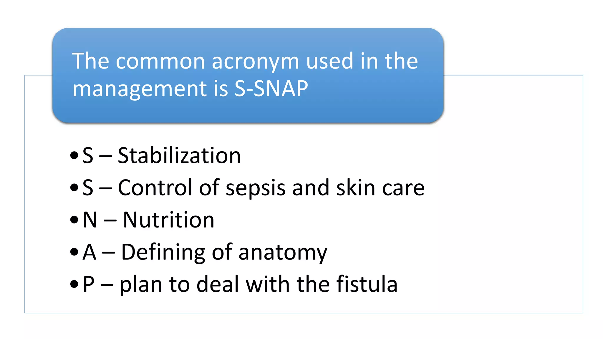
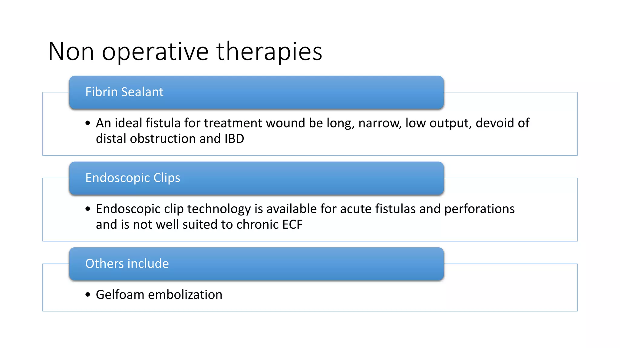
MANAGEMENT OF ENTEROCUTANOUS FISTULA
BY
DR. BARIBOTE O. S.
MBBS – NDU
30th OCTOBER, 2020
DEPARTMENT OF SURGERY
NDUTH
Outline
Introduction
History of ECF
Epidemiology
Classification
Clinical presentation
Investigation
Treatment
Conclusion
Introduction
Definition
• A fistula is an abnormal connection between two epithelized surfaces
Anatomical subdivisions:
• Internal and External
Internal fistulas are connections between two internal structures.
Examples:
External fistulas form connections between an internal structure and external
structure.
Introduction contd
An enterocutaneous fistula (ECF) is an aberrant connection between the
intra-abdominal gastrointestinal (GI) tract and skin/wound.
Conventionally, ECF is limited to those arising from the
• duodenum,
• jejunum,
• ileum,
• colon or
• rectum
although it can occur in the stomach, oesophagus and anus
History of ECF in medicine
Treatment of ECF continues to be a difficult task
• He stated “the large intestine can be sutured, not with any
certain assurance, but because this doubtful hope is preferable
to certain despair; for occasionally it heals up”
Celcus in 53 BC
• He expressed this “in such cases nothing is to be done but
dressing the wound superficially and when the contents of the
wounded viscus become less, we may hope for a cure”
John Hunter
Epidemiology
Enterocutaneous fistula mortality rates vary from 6% to 33%.
Incidence is dependent on etiology.
Infected pancreatic necrosis
• extremely high incidence of 50%.
Trauma patients
• 2% to 25% incidence,
and abdominal sepsis
• 20% to 25% incidence.
Classification
• output,
• etiology,
• and source
There are several ways in which ECF has
been classified, including
• high-output ECF - >500 mL/24 hours,
• moderate output 200 - 500 mL/24 hours
• low output <200 mL/24 hours,
According to Output:
According to etiology
• 75-85% are iatrogenic
• Typically after surgery for bowel obstruction, cancer, or IBD
• 15 -25% result from abdominal trauma or spontaneously
Berry SM, Fischer JE
• Postoperative
• Traumatic
• Spontaneous
Etiology Characterized as
Postoperative
Disruption of anastomosis
Inadvertent enterotomy
Inadvertent small bowel injury
According to etiology contd
Traumatic causes
• Iatrogenic surgical trauma to the bowel that may or may not be
recognized
• Road traffic accidents with injury to the gut
Spontaneous causes
• Malignancy
• Radiation enteritis with perforation
• Intra-abdominal sepsis
• Inflammatory bowel disease
• Crohn disease
• Ulcerative colitis
According to source
type I
type II
type III
type IV
• esophageal,
gastroduodenal,
• small bowel,
• large bowel and
• enteroatmospheric,
regardless of origin
Clinical presentation
• Postoperative abdominal pain
• Tenderness
• Distension,
• Enteric contents from the drain site and the main abdominal wound
• Tachycardia and pyrexia
Features suggestive of an ECF include
• Guarding, rigidity and rebound tenderness
Signs of localized or diffuse peritonitis
• Sepsis
• Fluid and electrolyte abnormalities
• Malnutrition
Signs of complication might be present
Investigation
Laboratory
investigations
Full blood
count +
differential
• Sepsis
• Anaemia
E/U/Cr
• Electrolyte
abnormailies
Total
proteins,
serum
albumin and
globulin
Serum
transferrin –
Low levels
(<200mg/dl)
are a
predictor of
poor healing
Imaging studies
Fistulography
• Contrast media such barium sulphate is injected into the fistulous
tract
• Conventionally performed 7-10 days
It provides
• Length of the tract
• Extent of the bowel wall disruption
• Location of the fistula
• Presence of a distal obstruction
Imaging contd
• Useful in patients with ECF involving the colon especially those
due to failure of low colorectal anastomosis
Water soluble contrast enema
• Class I: Simple, short blind ending < 2cm
• Class II: Continuous linear, long simple, >2cm
• Class III: Continuous complex, multiple linear
The following tracts can be seen
• Anterior – ventral 10 to 2 0’clock position
• Posterior – dorsal 4 to 8 0’clock position
• Lateral – right (2 to 4 0’clock) or left (8 to 10 0’clock)
Tracts positions are as follows
CT scanning and USS
•Useful in demonstrating intra-abdominal abscess cavities
Markers
•Oral administration of nonabsorbable marker eg
charcoal, congo red – help confirm the presence of ECF
•Beside administration of methylene blue diluted in
saline via NG tube can be used to confirm presence of
ECF especially those with gastrocutaneous fistula or
lateral duodenal fistula
Predictive factors for spontaneous closure and/or
mortality
Factor Favorable Unfavorable
Organ of origin Esophageal, Duodenal stump, Pancreatic,
Biliary, Jejunal, Colonic
Gastric, Lateral duodenal, Ligament of
Treitz, Ileal
Etiology Postop (anast leak), Appendicitis, Diverticulitis Malignancy, IBD
Output Low (<200-500cc/day) High (>500cc/day)
Nutritional status Well nourished, Transferrin >200 Malnourished, Transferrin <200
Sepsis Absent Present
State of bowel Intestinal continuity, absence of obstruction Diseased adjacent bowel, Distal
obstruction, Abscess, Discontinuity,
Irradiation
Fistula characteristics Tract >2 cm, Defect >1cm Tract <1cm, Defect >1cm
Miscellaneous Original operation at same institution Referred from outside institution
21
Treatment
Conservative
Surgical
• Rehydration
• Administration of antibiotics
• Correction of anemia
• Electrolyte repletion
• Drainage of obvious abscess
• Nutritional support
• Control of fistula drainage
• Skin protection
The principles of nonsurgical therapy for
ECFs include the following:
•S – Stabilization
•S – Control of sepsis and skin care
•N – Nutrition
•A – Defining of anatomy
•P – plan to deal with the fistula
The common acronym used in the
management is S-SNAP
Treatment
Resuscitation and stabilization
• 24 to 48 hours
Priorities:
• Normal circulating blood volume
• Correction of electrolyte and acid-base imbalances
Rehydration usually requires isotonic fluid (NS/RL) until the patient
is euvolemic
Strict input and output measurements are essential
Patient should be catheterized and urine output maintained at 0.5ml/kg/hr
Common problems include
• Dehydration
• Hyponatremia
• Hypokalemia
• Metabolic acidosis
Electrolyte measurement of the fistula output helps in planning of
replenishment of ongoing losses
Control of sepsis
Sepsis is responsible for 77% of mortality associated with ECF
CT or USS of the abdomen and pelvis along with percutaneous
drainage with radiographic guidance is essential to evaluate and
treat sources of infection
In cases of peritonitis and without the ability to obtain source
control with more conservative means,
• prompt fluid resuscitation,
• antibiotic administration, and
• operative control of infection are essential.
Control of sepsis contd
• infection drainage and
• exteriorization of the source in the small or large intestine,
Operative sepsis control should focus on
• in a critically ill patient or
• in the setting of significant purulence or fecal
contamination
No anastomoses
Nutrition
Nutrition is one of three necessities upon which the life and successful
treatment of a patient with ECF hinges
Fazio et al showed that mortality is 0% when serum albumin is > 3.5 mg/dL
Fistula closure rates are twice as high in those receiving adequate
supplemental nutrition as opposed to those who are not.
The goal of successful nutrition management is achieving
• an anabolic state with weight gain,
• improvement in albumin,
• prealbumin, and transferrin, and
• successful management of micronutrient needs for optimal healing
inadequate
calorie intake,
catabolism
due to
sepsis,
ongoing GI
losses
Sources of
malnutrition
Feeding
• Main stay of nutrition in ECFs
• Distal ileum
• Colon or
• Duodenum
• Elemental diets
Enteral
feeding
• Gastric fistula
• Duodenal
• Small bowel fistula
• High output fistula
Total
Parenteral
feeding
Routes of Enteral feeding
• Oral
• Feeding jejunostomy
• Nasogastric tubes
• Nasojejunal tubes
• Gastrostomy
Studies have shown that the provision of only 20%
of calories fed enterally protect the integrity of
mucosal barrier, as well as the immunologic and
hormonal function of the gut
Fistuloclysis
Nutrition needs of patient with ECF
Calorie
requiremen
t(kcal/kg/d)
Protein
requirement
(g/kg/d) Vitamin C
Other
vitamins
Elements
(zinc,
copper,
selenium)
Low-output
fistula
20–30 1–1.5 5–10 times
normal
At least
normal
At least
normal
High-output
fistula
25–35 1.5–2.5 10 times
normal
2 times
normal
2 times
trace
elements
• 75g glucose
• 20g amino acids
• 30 g lipids per 1000ml
A standard general purpose formula for TPN consists of
the following:
Monitoring of level of nutrition is essentially
• albumin,
• prealbumin,
• weights,
• and transferrin
One measure of tracking success weekly is checking
• triceps skin fold thickness (approximates body fat reserves) and
• midarm muscle circumference (approximates muscle mass)
In addition, anthropometric assessment based on
• For high output fistula, use of a pouch to collect the
effluent is preferred
• For a low output fistula, a skin barrier with
dressing/pouch is advocated
Skin care
• Restrict hypo-osmolar fluids
• Anti secretory agents
• Antimotility agents
Methods of reducing fistula output
Surgical intervention
• Foreign body
• Radiation
• Inflammation, Infection
• Epithelialization
• Neoplasm
• Distal obstruction
• High output fistula
• Low serum transferrin (<200mg/dl)
In the face of unfavorable factors that prevent spontaneous
closure:
• Fistulas from esophagus to duodenum: 2~4 weeks.
• Colonic fistulas: 30~40 days.
• Small bowel fistulas: 40~60 days.
Exceeding of expected time period for spontaneous closure:
Definitive repair of the ECF should be planned if no
spontaneous closure occurs by 12 weeks after
• sepsis control,
• nutritional optimization,
• and establishing wound cares.
Pre-operation preparation includes
• intra-luminal antibiotics,
• mechanical bowel preparation,
• discontinuation of enteral nutrition,
• meticulous skin care,
• control fistula drainage
Intraoperative details
Skin incision
• Fresh skin incision
• since there is a possibility of the gut being adherent to the site of the incision of
the index operation
Once the peritoneal cavity is assessed, the entire bowel is made free of all adhesions
Fistulous site is dissected free from the surrounding structures and excised
Restoration of bowel continuity should be done with a
• double layered anastomosis
• involving a healthy and well vascularized bowel segment
If the patient is sick and cannot withstand a resectional procedure,
• exteriorized via ileostomy or colostomy
Non operative therapies
• An ideal fistula for treatment wound be long, narrow, low output, devoid of
distal obstruction and IBD
Fibrin Sealant
• Endoscopic clip technology is available for acute fistulas and perforations
and is not well suited to chronic ECF
Endoscopic Clips
• Gelfoam embolization
Others include
Conclusion
• ECF remains a complex problem that is optimally management using a
careful and interdisciplinary approach.
• In addition to primary management of sepsis,
• conservative treatment remains the treatment mainstay,
• including the combination of wound management,
• nutritional support with EN or PN sometimes in combination, and
social support.
• Surgical treatment with resection should be carefully planned and is
used in cases that fail conservative treatment.
Reference
• Sitges-Serra A, Jaurrieta E, Sitges-Creus A. Management of
postoperative enterocutaneous fistulas: the roles of parenteral
nutrition and surgery. Br J Surg. 1982;69(3):147–150.
• Schein M, Decker G A. Postoperative external alimentary tract
fistulas. Am J Surg. 1991;161(4):435–438
• Berry S M, Fischer J E. Classification and pathophysiology of
enterocutaneous fistulas. SurgClin North Am. 1996;76(5):1009–1018.
• Lloyd D A, Gabe S M, Windsor A C. Nutrition and management of
enterocutaneous fistula. Br J Surg.2006;93(9):1045–1055.

Enterocutaneous fistula.pptx

  • 1.
    MANAGEMENT OF ENTEROCUTANOUSFISTULA BY DR. BARIBOTE O. S. MBBS – NDU 30th OCTOBER, 2020 DEPARTMENT OF SURGERY NDUTH
  • 2.
    Outline Introduction History of ECF Epidemiology Classification Clinicalpresentation Investigation Treatment Conclusion
  • 3.
    Introduction Definition • A fistulais an abnormal connection between two epithelized surfaces Anatomical subdivisions: • Internal and External Internal fistulas are connections between two internal structures. Examples: External fistulas form connections between an internal structure and external structure.
  • 4.
    Introduction contd An enterocutaneousfistula (ECF) is an aberrant connection between the intra-abdominal gastrointestinal (GI) tract and skin/wound. Conventionally, ECF is limited to those arising from the • duodenum, • jejunum, • ileum, • colon or • rectum although it can occur in the stomach, oesophagus and anus
  • 5.
    History of ECFin medicine Treatment of ECF continues to be a difficult task • He stated “the large intestine can be sutured, not with any certain assurance, but because this doubtful hope is preferable to certain despair; for occasionally it heals up” Celcus in 53 BC • He expressed this “in such cases nothing is to be done but dressing the wound superficially and when the contents of the wounded viscus become less, we may hope for a cure” John Hunter
  • 6.
    Epidemiology Enterocutaneous fistula mortalityrates vary from 6% to 33%. Incidence is dependent on etiology. Infected pancreatic necrosis • extremely high incidence of 50%. Trauma patients • 2% to 25% incidence, and abdominal sepsis • 20% to 25% incidence.
  • 7.
    Classification • output, • etiology, •and source There are several ways in which ECF has been classified, including • high-output ECF - >500 mL/24 hours, • moderate output 200 - 500 mL/24 hours • low output <200 mL/24 hours, According to Output:
  • 8.
    According to etiology •75-85% are iatrogenic • Typically after surgery for bowel obstruction, cancer, or IBD • 15 -25% result from abdominal trauma or spontaneously Berry SM, Fischer JE • Postoperative • Traumatic • Spontaneous Etiology Characterized as
  • 9.
    Postoperative Disruption of anastomosis Inadvertententerotomy Inadvertent small bowel injury
  • 10.
    According to etiologycontd Traumatic causes • Iatrogenic surgical trauma to the bowel that may or may not be recognized • Road traffic accidents with injury to the gut Spontaneous causes • Malignancy • Radiation enteritis with perforation • Intra-abdominal sepsis • Inflammatory bowel disease • Crohn disease • Ulcerative colitis
  • 12.
    According to source typeI type II type III type IV • esophageal, gastroduodenal, • small bowel, • large bowel and • enteroatmospheric, regardless of origin
  • 14.
    Clinical presentation • Postoperativeabdominal pain • Tenderness • Distension, • Enteric contents from the drain site and the main abdominal wound • Tachycardia and pyrexia Features suggestive of an ECF include • Guarding, rigidity and rebound tenderness Signs of localized or diffuse peritonitis • Sepsis • Fluid and electrolyte abnormalities • Malnutrition Signs of complication might be present
  • 15.
    Investigation Laboratory investigations Full blood count + differential •Sepsis • Anaemia E/U/Cr • Electrolyte abnormailies Total proteins, serum albumin and globulin Serum transferrin – Low levels (<200mg/dl) are a predictor of poor healing
  • 16.
    Imaging studies Fistulography • Contrastmedia such barium sulphate is injected into the fistulous tract • Conventionally performed 7-10 days It provides • Length of the tract • Extent of the bowel wall disruption • Location of the fistula • Presence of a distal obstruction
  • 19.
    Imaging contd • Usefulin patients with ECF involving the colon especially those due to failure of low colorectal anastomosis Water soluble contrast enema • Class I: Simple, short blind ending < 2cm • Class II: Continuous linear, long simple, >2cm • Class III: Continuous complex, multiple linear The following tracts can be seen • Anterior – ventral 10 to 2 0’clock position • Posterior – dorsal 4 to 8 0’clock position • Lateral – right (2 to 4 0’clock) or left (8 to 10 0’clock) Tracts positions are as follows
  • 20.
    CT scanning andUSS •Useful in demonstrating intra-abdominal abscess cavities Markers •Oral administration of nonabsorbable marker eg charcoal, congo red – help confirm the presence of ECF •Beside administration of methylene blue diluted in saline via NG tube can be used to confirm presence of ECF especially those with gastrocutaneous fistula or lateral duodenal fistula
  • 21.
    Predictive factors forspontaneous closure and/or mortality Factor Favorable Unfavorable Organ of origin Esophageal, Duodenal stump, Pancreatic, Biliary, Jejunal, Colonic Gastric, Lateral duodenal, Ligament of Treitz, Ileal Etiology Postop (anast leak), Appendicitis, Diverticulitis Malignancy, IBD Output Low (<200-500cc/day) High (>500cc/day) Nutritional status Well nourished, Transferrin >200 Malnourished, Transferrin <200 Sepsis Absent Present State of bowel Intestinal continuity, absence of obstruction Diseased adjacent bowel, Distal obstruction, Abscess, Discontinuity, Irradiation Fistula characteristics Tract >2 cm, Defect >1cm Tract <1cm, Defect >1cm Miscellaneous Original operation at same institution Referred from outside institution 21
  • 22.
  • 23.
    • Rehydration • Administrationof antibiotics • Correction of anemia • Electrolyte repletion • Drainage of obvious abscess • Nutritional support • Control of fistula drainage • Skin protection The principles of nonsurgical therapy for ECFs include the following:
  • 24.
    •S – Stabilization •S– Control of sepsis and skin care •N – Nutrition •A – Defining of anatomy •P – plan to deal with the fistula The common acronym used in the management is S-SNAP
  • 25.
    Treatment Resuscitation and stabilization •24 to 48 hours Priorities: • Normal circulating blood volume • Correction of electrolyte and acid-base imbalances Rehydration usually requires isotonic fluid (NS/RL) until the patient is euvolemic Strict input and output measurements are essential
  • 26.
    Patient should becatheterized and urine output maintained at 0.5ml/kg/hr Common problems include • Dehydration • Hyponatremia • Hypokalemia • Metabolic acidosis Electrolyte measurement of the fistula output helps in planning of replenishment of ongoing losses
  • 27.
    Control of sepsis Sepsisis responsible for 77% of mortality associated with ECF CT or USS of the abdomen and pelvis along with percutaneous drainage with radiographic guidance is essential to evaluate and treat sources of infection In cases of peritonitis and without the ability to obtain source control with more conservative means, • prompt fluid resuscitation, • antibiotic administration, and • operative control of infection are essential.
  • 28.
    Control of sepsiscontd • infection drainage and • exteriorization of the source in the small or large intestine, Operative sepsis control should focus on • in a critically ill patient or • in the setting of significant purulence or fecal contamination No anastomoses
  • 29.
    Nutrition Nutrition is oneof three necessities upon which the life and successful treatment of a patient with ECF hinges Fazio et al showed that mortality is 0% when serum albumin is > 3.5 mg/dL Fistula closure rates are twice as high in those receiving adequate supplemental nutrition as opposed to those who are not. The goal of successful nutrition management is achieving • an anabolic state with weight gain, • improvement in albumin, • prealbumin, and transferrin, and • successful management of micronutrient needs for optimal healing
  • 30.
  • 31.
    Feeding • Main stayof nutrition in ECFs • Distal ileum • Colon or • Duodenum • Elemental diets Enteral feeding • Gastric fistula • Duodenal • Small bowel fistula • High output fistula Total Parenteral feeding
  • 32.
    Routes of Enteralfeeding • Oral • Feeding jejunostomy • Nasogastric tubes • Nasojejunal tubes • Gastrostomy Studies have shown that the provision of only 20% of calories fed enterally protect the integrity of mucosal barrier, as well as the immunologic and hormonal function of the gut
  • 33.
  • 35.
    Nutrition needs ofpatient with ECF Calorie requiremen t(kcal/kg/d) Protein requirement (g/kg/d) Vitamin C Other vitamins Elements (zinc, copper, selenium) Low-output fistula 20–30 1–1.5 5–10 times normal At least normal At least normal High-output fistula 25–35 1.5–2.5 10 times normal 2 times normal 2 times trace elements
  • 36.
    • 75g glucose •20g amino acids • 30 g lipids per 1000ml A standard general purpose formula for TPN consists of the following: Monitoring of level of nutrition is essentially • albumin, • prealbumin, • weights, • and transferrin One measure of tracking success weekly is checking • triceps skin fold thickness (approximates body fat reserves) and • midarm muscle circumference (approximates muscle mass) In addition, anthropometric assessment based on
  • 37.
    • For highoutput fistula, use of a pouch to collect the effluent is preferred • For a low output fistula, a skin barrier with dressing/pouch is advocated Skin care • Restrict hypo-osmolar fluids • Anti secretory agents • Antimotility agents Methods of reducing fistula output
  • 38.
    Surgical intervention • Foreignbody • Radiation • Inflammation, Infection • Epithelialization • Neoplasm • Distal obstruction • High output fistula • Low serum transferrin (<200mg/dl) In the face of unfavorable factors that prevent spontaneous closure: • Fistulas from esophagus to duodenum: 2~4 weeks. • Colonic fistulas: 30~40 days. • Small bowel fistulas: 40~60 days. Exceeding of expected time period for spontaneous closure:
  • 39.
    Definitive repair ofthe ECF should be planned if no spontaneous closure occurs by 12 weeks after • sepsis control, • nutritional optimization, • and establishing wound cares. Pre-operation preparation includes • intra-luminal antibiotics, • mechanical bowel preparation, • discontinuation of enteral nutrition, • meticulous skin care, • control fistula drainage
  • 40.
    Intraoperative details Skin incision •Fresh skin incision • since there is a possibility of the gut being adherent to the site of the incision of the index operation Once the peritoneal cavity is assessed, the entire bowel is made free of all adhesions Fistulous site is dissected free from the surrounding structures and excised Restoration of bowel continuity should be done with a • double layered anastomosis • involving a healthy and well vascularized bowel segment If the patient is sick and cannot withstand a resectional procedure, • exteriorized via ileostomy or colostomy
  • 41.
    Non operative therapies •An ideal fistula for treatment wound be long, narrow, low output, devoid of distal obstruction and IBD Fibrin Sealant • Endoscopic clip technology is available for acute fistulas and perforations and is not well suited to chronic ECF Endoscopic Clips • Gelfoam embolization Others include
  • 42.
    Conclusion • ECF remainsa complex problem that is optimally management using a careful and interdisciplinary approach. • In addition to primary management of sepsis, • conservative treatment remains the treatment mainstay, • including the combination of wound management, • nutritional support with EN or PN sometimes in combination, and social support. • Surgical treatment with resection should be carefully planned and is used in cases that fail conservative treatment.
  • 43.
    Reference • Sitges-Serra A,Jaurrieta E, Sitges-Creus A. Management of postoperative enterocutaneous fistulas: the roles of parenteral nutrition and surgery. Br J Surg. 1982;69(3):147–150. • Schein M, Decker G A. Postoperative external alimentary tract fistulas. Am J Surg. 1991;161(4):435–438 • Berry S M, Fischer J E. Classification and pathophysiology of enterocutaneous fistulas. SurgClin North Am. 1996;76(5):1009–1018. • Lloyd D A, Gabe S M, Windsor A C. Nutrition and management of enterocutaneous fistula. Br J Surg.2006;93(9):1045–1055.

Editor's Notes

  • #4 A fistula is an abnormal connection between two epithelized surfaces Subdivisions: internal and external. Eg of Internal Fistula Enterocolic Ileosigmoid External Fistula e.g ECF, Rectovaginal fistula
  • #5 Conventionally, ECF is limited to those arising from the duodenum, jejunum, ileum, colon or rectum
  • #6 Celcus in 53 BC mentioned the problems associated with an intestinal wound breakdown John Hunter described the difficulties in treating ECFs in the mid 19th century
  • #7 Incidence is dependent on etiology. Infected pancreatic necrosis extremely high incidence of 50%. Trauma patients 2% to 25%  incidence, and abdominal sepsis 20% to 25% incidence.
  • #8 by output, etiology, and source According to Output: high-output ECF - >500 mL/24 hours, moderate output 200 - 500 mL/24 hours low output <200 mL/24 hours,
  • #9 Berry SM, Fischer JE. Enterocutaneous Fistulas. Curr Probl Surg. 1994 Jun;31(6):469-566. The etiology of ECFs can thus be characterized as postoperative, traumatic, or spontaneous
  • #10 Disruption of anastomosis Inadequate blood flow due to improper vascular supply especially when extensive mesenteric vessels have to be ligated Tension on anastomotic lines following colonic resection Restoration of continuity without adequate mobilization Minimal leak or infection leading to perianastomotic abscess formation Inadvertent enterotomy Especially in patients with adhesions when dissections can cause multiple serosal tears and occasional full-thickness tear Inadvertent small bowel injury Occur following inadvent picking up of the bowel during abdominal closure
  • #11 Spontaneous causes Malignancy Radiation enteritis with perforation Intra-abdominal sepsis Inflammatory bowel disease Crohn disease Ulcerative colitis
  • #12  In this patient, a urachal tumor was inadvertently incised when the patient underwent an appendectomy by midline incision. The patient presented with ECF (colocutaneous fistula) as the urachal tumor that ulcerated on the abdominal wall postoperatively had also infiltrated the sigmoid colon
  • #15 Postoperative abdominal pain Tenderness Distension, Enteric contents from the drain site and the main abdominal wound Tachycardia and pyrexia Guarding, rigidity and rebound tenderness
  • #16 - These can demonstrate the presence of malnutrition-associated hypoalbuminemia
  • #20 Water Soluble enema e.g gastrografin The different types of tracts that can be seen by using a water-soluble contrast enema (WCE) in patients with ECF with failure of low colorectal anastomosis may be classified as follows Other tract features include: cavity (pool of contrast within space), strictures (narrowing of anastomosis)
  • #21 Methylene blue loses diagnostic efficacy as it becomes diluted with intestinal secretions, its role in identifying distal ECFs is limited
  • #23 The conventional therapy for an enterocutaneous fistula (ECF) in the initial phase is always conservative. Immediate surgical therapy on presentation is contraindicated, because the majority of ECFs spontaneously close as a result of conservative therapy. Surgical intervention in the presence of sepsis and poor general condition would be hazardous for the patient However, patients who have an ECF with adverse factors, such as a lateral duodenal fistula, an ileal fistula, a high-output fistula, or a fistula associated with a diseased bowel, may require early surgical intervention
  • #24 In a study of 186 patients, Reber et al found that 91% of small-bowel fistulas that closed spontaneously did so within 1 month after sepsis was cured. The remaining fistulas that closed spontaneously did so by the end of 3 months after sepsis cure, with the rest of the lesions requiring surgical closure
  • #25 S – Stabilization S – Control of sepsis and skin care N – Nutrition A – Defining of anatomy P – plan to deal with the fistula
  • #26 The first step of management is the resuscitation and stabilization of the patient This needs to be accomplished within the first 24 to 48 hours
  • #28 prompt fluid resuscitation, antibiotic administration, and operative control of infection are essential.
  • #29 infection drainage and exteriorization of the source in the small or large intestine
  • #30 Uba et al reported that the majority of ECFs in children closed spontaneously following high-protein and high-carbohydrate nutrition an anabolic state with weight gain, improvement in albumin, prealbumin, and transferrin, and successful management of micronutrient needs for optimal healing
  • #31 Basically three sources They can overlap for an individual patient
  • #32 When the fistula output is very high, discontinuance of oral intake is recommended because oral intake stimulates further losses of fluids, electrolytes, and protein via the fistula. A decrease in fistula output frequently occurs with the initiation of TPN. Elemental diets, that is, non-residue balanced diets with protein components reduced to their basic elements, are preferred
  • #33 In patients with a proximal fistula, if a nasojejunal tube can be introduced beyond the site of the fistula, then these patients can be supported with enteral nutrition, provided that there is at least 4-5 ft (1.2-1.5 m) of small bowel distal to it and no distal obstruction
  • #34 Fistuloclysis Enteral feedings administered via the fistula The fistula tract is clearly defined and a feeding tube is inserted in the distal limb of the ECF
  • #38 Antisecretory agents Protein pump inhibitors Somatostatin or octreotide Antimotility agents Loperamide Codeine