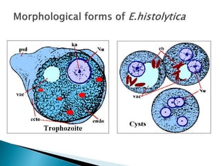
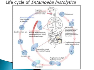
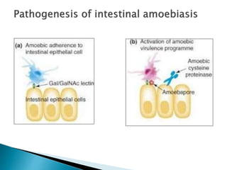
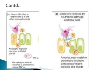
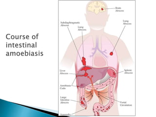
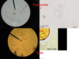
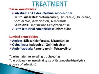
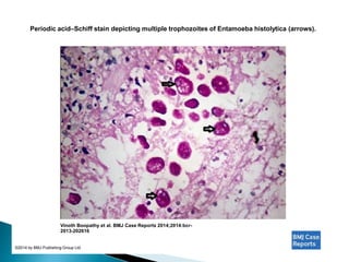
Dr.Achu Jacob Philip
Dr.Isabella
 Introduction
 Epidemiology
 Life cycle of Entamoeba histolytica
 Pathogenesis
 Pathology
 Clinical features
 Laboratory diagnosis
 Management
•Amebiasis ia an infection with the intestinal protozoan
Entamoeba histolytica.
•90% asymptomatic.
•It is the third most common cause of death from parasitic
disease.
•Asymptomatic forms are mainly caused by E.dispar.- Self
limiting (homosexual men & AIDS patients)
•Intestinal lesions mainly involve the cecum,sigmoid colon
and the rectum.
•Distant abscess occur in liver ,lung and brain.
Definition :
 Amoebiasis is an infection caused by Entamoeba
histolytica
with or without symptoms (WHO - 1969)
 Synonyms include entamoebiasis, amoebiosis,
amoebic
dysentery or bloody flux.
Global burden of the disease :
 40-50 million cases of amoebic colitis and amoebic
liver abscess
 70,000 deaths anually
 10% world population
 90% of those infected are asymptomatic, 1% may
develop invasive/extraintestinal amoebiasis.
 China, Central and South America, Indian
subcontinents
In India :
 Prevalence is 15% (3.6% to 47.4%)
 Maharashtra, Tamil Nadu, Chandigarh
Agent factors Host factors
• Virulence of organism
• Intestinal microbiota
• Sex
• Age
•Alcoholics
• Immunocompromised
(HIV) 
male homosexuals
 During amoebiasis there is a significant decrease in absolute
quantification of Bacteroides, Clostridium coccoides,
Clostridium leptum, Lactobacillus and an increase in
Bifdobacterium species.
 Lactobacillus species might be protective in the context of
protozoan infections (Preidis et al., 2011; Travers et al.,
2011).
 Thus a decrease in protective, commensal Lactobacillus
Intestinal specimen from a patient with acute
Colon (primarily
in the cecum),
sigmoid colon, and
rectum
2 types of ulcers :
nodular and irregular
Intervening
mucosal folds
may appear
normal
Submucosa : susceptible to the
lytic action of the parasite, and
produces
abundant microhemorrhages
Bowel
lumen
Amoebic ulcer with
neutrophilic infiltration
Mucosa
Submucos
a
INTESTINAL AMEBIASIS:
•Symptomatic amebic colitis develops 2-6 weeks after
ingestion of infectios cysts.
•Gradual onset of lower abdominal pain,mild
diarrhea,malaise,weight loss,back pain.
•Caecal movements may mimic acute appendicitis
•Stools will contain little fecal matter and will consist of
mainly of blood and mucus.
Fulminant intestinal infection
•Clinical features: Severe abdominal pain
High Fever
Profuse diarrhoea
Occurs predominantly in children
Also patients receiving glucocorticoids
Megacolon
•Patient will be having shock like features
•Severe bowel dilation with intramural air.
Syndrom of Postamebic colitis
•Persistent diarrhoea following documented cure of
amebiasis
AMEBIC LIVER ABSCESS:
•Febrile,Rt upper quadrent pain(dull or pleuritic) radiating to the
shoulders.
•Malaise,weight loss and hepatomegaly
•Complication
Pleuropulmonary involement (20 – 30%)
Sterile effusion
Hepatobronchial fistulae
Rupture of abscess
OTHER SITES
•Genitourinary Tract :
Direct extension
Genital ulcer,Profuse discharge
•Cerebral Involvement
Occurs in 0.1% patients.
Syptoms depends on size and site of lesion.
•Chronic granuloma arising in the large bowel.
•MC : Caecum
•Occurs in longstanding amoebic infection (with in complete treatment)
•Mistaken for carcinoma
•C/f: Pyrexia,
Mass in RIF
Blood stained mucoid diarrhoea.
Specimen collection :
Intestinal amoebiasis  Stool samples
Extraintestinal amoebiasis Aspirated pus
 Direct examination  Saline and iodine wet mounts
 Culture
 Immunodiagnosis
 Molecular methods  Polymerase chain reaction
Trophozoite
s
Cysts
Tissue amoebicides
Intestinal and Extra intestinal amoebicides
•Nitroimidazoles: Metronidazole, Tinidazole, Ornidazole,
Secnidazole, Satranidazole, Nimorazole
•Alkaloids: Emetine and Dehydroemetine
Extra intestinal amoebicides: Chloroquine
Luminal amoebicides
Amides: Diloxanide furoate, Nitazoxanide
Quinolines: Iodoquinol, Quiniodochlor
Antimicrobials: Paromomycin, Tetracyclines
To eliminate the invading trophozoites
To eradicate the intestinal cysts of Entamoeba histolytica
(source of infection)
Luminal amebicides
Tissue amebicides
Nitroimidazole –Metronidazole
Metronidazole 800 mg TDS x 7-10 days
(in severe cases 500 mg slow IV 6 hourly till oral therapy can be instituted)
OR
Tinidazole 2 g OD x 3 days
+
Luminal amoebicide
AMEBIC LIVER ABSCESS
Metronidazole 800 mg TDS x 10 days (in serious cases – IV
metronidazole x 10 days)
OR
Tinidazole 2 g oral daily x 3 - 6 days
+
Luminal amoebicide
ASPIRATION OF LIVER ABSCESS
To rule out a pyogenic abscess,mainly in multiple lesion.
No clinical response in 3 -5 days.
Threat of imminent rupture.
Left lobe abscess
29yr old male came with C/o –
•Abdominal Pain
•Fever
•Vomiting
•Patient presented with dull aching pain in the right hypochondium
for 1week.
•Associated with low grade fever, with no chills or rigor
•Vomiting four episodes – mainly food particles,non bilious.
•Patient febrile not in septic shock
•Tender hepatomegaly .
TC : 16900,DC – N 86,L 10,M3,E1.
ESR : 75
LFT Normal
•Well defined hypoechoic
lesion 11.1 x 8.2 x 7.8
•hyperechoic septation
Treatment Given
•Inj Metronidazole was started .
•Pig Tail catheter inserted under local and abscess drained.
•Patient responded to treatment
42 year old presented with acute abdomen
Soft ,
Hepatomegaly.
USG:
•Features S/o liver abscess (volume 2006cc) involving the right
lobe of liver with ? Focal subcapsular rupture.
•Moderate ascitis
Abscess of right lobe
of liver.
Sloughed out
wall of the
amebic abscess
on right lobe of
liver.
CT scan of the abdomen showing irregular wall thickening of the caecum.
Vinoth Boopathy et al. BMJ Case Reports 2014;2014:bcr-
2013-202616
©2014 by BMJ Publishing Group Ltd
Colonoscopic image showing ulcerations with yellowish white exudations in the caecum.
Vinoth Boopathy et al. BMJ Case Reports 2014;2014:bcr-
2013-202616
©2014 by BMJ Publishing Group Ltd
Periodic acid–Schiff stain depicting multiple trophozoites of Entamoeba histolytica (arrows).
Vinoth Boopathy et al. BMJ Case Reports 2014;2014:bcr-
2013-202616
©2014 by BMJ Publishing Group Ltd
Intestinal and extraintestinal amoebiasis

Intestinal and extraintestinal amoebiasis

  • 1.
  • 2.
     Introduction  Epidemiology Life cycle of Entamoeba histolytica  Pathogenesis  Pathology  Clinical features  Laboratory diagnosis  Management
  • 3.
    •Amebiasis ia aninfection with the intestinal protozoan Entamoeba histolytica. •90% asymptomatic. •It is the third most common cause of death from parasitic disease. •Asymptomatic forms are mainly caused by E.dispar.- Self limiting (homosexual men & AIDS patients) •Intestinal lesions mainly involve the cecum,sigmoid colon and the rectum. •Distant abscess occur in liver ,lung and brain.
  • 4.
    Definition :  Amoebiasisis an infection caused by Entamoeba histolytica with or without symptoms (WHO - 1969)  Synonyms include entamoebiasis, amoebiosis, amoebic dysentery or bloody flux.
  • 5.
    Global burden ofthe disease :  40-50 million cases of amoebic colitis and amoebic liver abscess  70,000 deaths anually  10% world population  90% of those infected are asymptomatic, 1% may develop invasive/extraintestinal amoebiasis.
  • 6.
     China, Centraland South America, Indian subcontinents In India :  Prevalence is 15% (3.6% to 47.4%)  Maharashtra, Tamil Nadu, Chandigarh
  • 7.
    Agent factors Hostfactors • Virulence of organism • Intestinal microbiota • Sex • Age •Alcoholics • Immunocompromised (HIV)  male homosexuals
  • 9.
     During amoebiasisthere is a significant decrease in absolute quantification of Bacteroides, Clostridium coccoides, Clostridium leptum, Lactobacillus and an increase in Bifdobacterium species.  Lactobacillus species might be protective in the context of protozoan infections (Preidis et al., 2011; Travers et al., 2011).  Thus a decrease in protective, commensal Lactobacillus
  • 15.
    Intestinal specimen froma patient with acute Colon (primarily in the cecum), sigmoid colon, and rectum 2 types of ulcers : nodular and irregular Intervening mucosal folds may appear normal
  • 16.
    Submucosa : susceptibleto the lytic action of the parasite, and produces abundant microhemorrhages Bowel lumen Amoebic ulcer with neutrophilic infiltration Mucosa Submucos a
  • 19.
    INTESTINAL AMEBIASIS: •Symptomatic amebiccolitis develops 2-6 weeks after ingestion of infectios cysts. •Gradual onset of lower abdominal pain,mild diarrhea,malaise,weight loss,back pain. •Caecal movements may mimic acute appendicitis •Stools will contain little fecal matter and will consist of mainly of blood and mucus.
  • 20.
    Fulminant intestinal infection •Clinicalfeatures: Severe abdominal pain High Fever Profuse diarrhoea Occurs predominantly in children Also patients receiving glucocorticoids Megacolon •Patient will be having shock like features •Severe bowel dilation with intramural air. Syndrom of Postamebic colitis •Persistent diarrhoea following documented cure of amebiasis
  • 21.
    AMEBIC LIVER ABSCESS: •Febrile,Rtupper quadrent pain(dull or pleuritic) radiating to the shoulders. •Malaise,weight loss and hepatomegaly •Complication Pleuropulmonary involement (20 – 30%) Sterile effusion Hepatobronchial fistulae Rupture of abscess
  • 22.
    OTHER SITES •Genitourinary Tract: Direct extension Genital ulcer,Profuse discharge •Cerebral Involvement Occurs in 0.1% patients. Syptoms depends on size and site of lesion.
  • 23.
    •Chronic granuloma arisingin the large bowel. •MC : Caecum •Occurs in longstanding amoebic infection (with in complete treatment) •Mistaken for carcinoma •C/f: Pyrexia, Mass in RIF Blood stained mucoid diarrhoea.
  • 24.
    Specimen collection : Intestinalamoebiasis  Stool samples Extraintestinal amoebiasis Aspirated pus
  • 25.
     Direct examination Saline and iodine wet mounts  Culture  Immunodiagnosis  Molecular methods  Polymerase chain reaction
  • 26.
  • 27.
    Tissue amoebicides Intestinal andExtra intestinal amoebicides •Nitroimidazoles: Metronidazole, Tinidazole, Ornidazole, Secnidazole, Satranidazole, Nimorazole •Alkaloids: Emetine and Dehydroemetine Extra intestinal amoebicides: Chloroquine Luminal amoebicides Amides: Diloxanide furoate, Nitazoxanide Quinolines: Iodoquinol, Quiniodochlor Antimicrobials: Paromomycin, Tetracyclines To eliminate the invading trophozoites To eradicate the intestinal cysts of Entamoeba histolytica (source of infection)
  • 28.
    Luminal amebicides Tissue amebicides Nitroimidazole–Metronidazole Metronidazole 800 mg TDS x 7-10 days (in severe cases 500 mg slow IV 6 hourly till oral therapy can be instituted) OR Tinidazole 2 g OD x 3 days + Luminal amoebicide
  • 29.
    AMEBIC LIVER ABSCESS Metronidazole800 mg TDS x 10 days (in serious cases – IV metronidazole x 10 days) OR Tinidazole 2 g oral daily x 3 - 6 days + Luminal amoebicide ASPIRATION OF LIVER ABSCESS To rule out a pyogenic abscess,mainly in multiple lesion. No clinical response in 3 -5 days. Threat of imminent rupture. Left lobe abscess
  • 30.
    29yr old malecame with C/o – •Abdominal Pain •Fever •Vomiting •Patient presented with dull aching pain in the right hypochondium for 1week. •Associated with low grade fever, with no chills or rigor •Vomiting four episodes – mainly food particles,non bilious. •Patient febrile not in septic shock •Tender hepatomegaly . TC : 16900,DC – N 86,L 10,M3,E1. ESR : 75 LFT Normal
  • 31.
    •Well defined hypoechoic lesion11.1 x 8.2 x 7.8 •hyperechoic septation
  • 32.
    Treatment Given •Inj Metronidazolewas started . •Pig Tail catheter inserted under local and abscess drained. •Patient responded to treatment
  • 33.
    42 year oldpresented with acute abdomen Soft , Hepatomegaly.
  • 34.
    USG: •Features S/o liverabscess (volume 2006cc) involving the right lobe of liver with ? Focal subcapsular rupture. •Moderate ascitis
  • 35.
    Abscess of rightlobe of liver.
  • 36.
    Sloughed out wall ofthe amebic abscess on right lobe of liver.
  • 38.
    CT scan ofthe abdomen showing irregular wall thickening of the caecum. Vinoth Boopathy et al. BMJ Case Reports 2014;2014:bcr- 2013-202616 ©2014 by BMJ Publishing Group Ltd
  • 39.
    Colonoscopic image showingulcerations with yellowish white exudations in the caecum. Vinoth Boopathy et al. BMJ Case Reports 2014;2014:bcr- 2013-202616 ©2014 by BMJ Publishing Group Ltd
  • 40.
    Periodic acid–Schiff staindepicting multiple trophozoites of Entamoeba histolytica (arrows). Vinoth Boopathy et al. BMJ Case Reports 2014;2014:bcr- 2013-202616 ©2014 by BMJ Publishing Group Ltd