Downloaded 380 times
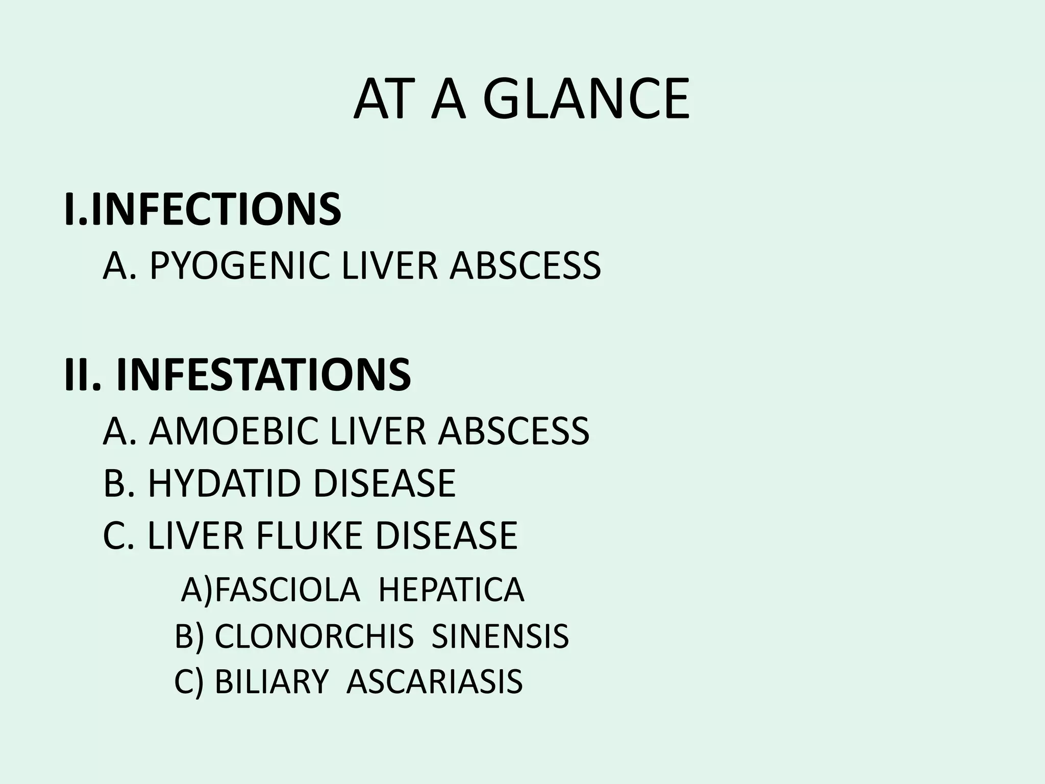


Pyogenic and amebic liver infections and infestations are described. Pyogenic liver abscesses are usually caused by bacteria spreading from another infected site via the bloodstream and are typically treated with antibiotics and drainage. Amebic liver abscesses are caused by the parasite Entamoeba histolytica and present with right upper quadrant pain, fever, and tenderness. Diagnosis involves blood tests and imaging, and treatment consists of antiparasitic medications and sometimes drainage. Hydatid cysts are caused by the tapeworm Echinococcus granulosus and seen endemic areas; they may cause liver masses, pain, or allergic reactions.


