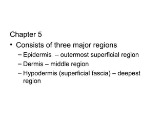
Chapter 5 Consists of three major regions Epidermis  – outermost superficial region Dermis – middle region Hypodermis (superficial fascia) – deepest region
Figure 5.1 Epidermis Dermis Hypodermis (superficial fascia) Hair root Hair shaft Pore Dermal papillae  (papillary layer of dermis) Meissner's corpuscle Free nerve ending Reticular layer of dermis  Sebaceous (oil) gland Arrector pili muscle Sensory nerve fiber Eccrine sweat gland Pacinian corpuscle Artery Vein Adipose tissue Hair follicle receptor (root hair plexus) Hair follicle Eccrine sweat gland
Epidermis – consists of stratified squamous epithelia tissue Layers of the epidermis include the following: Stratum Basale  – deepest epidermal layer; includes keratinocytes, melanocytes and Merkel cells; cells rapidly divide by mitosis; single row of cells Stratum Spinosum  – several cell layers thick; keratinocytes; cells are spiny in appearance Stratum Granulosum  – 3 to 5 cell layers thick; keratinocytes start to accumulate keratin. Stratum Lucidum  – thin translucent band; only present in thick skin (palmar and plantar skin); cells are dead Stratum Corneum  – 20 to 30 cell layers thick; cornified or horny cells; cells are dead
Layers of the Epidermis Keratinocytes – produce the fibrous protein keratin Melanocytes  – produce the brown pigment melanin Langerhans’ cells  – epidermal macrophages that help activate the immune system Merkel cells  – function as touch receptors in association with sensory nerve endings
Components of the Dermis Papillary Layer – Areolar connective tissue; dermal papillae; Meissner’s corpuscles; Reticular Layer – dense irregular connective tissue; cleavage or tension lines; collagen fibers; Skin Color Three pigments contribute to skin color: Melanin – tyrosine amino acids; found in melanocytes; all humans have the same relative number of melanocytes and skin color differences are a result of the amount of melanin made and retained; freckles and moles are local accumulations of melanin; melanocytes are stimulated to make more melanin when they are exposed to UV radiation (tan – protects against DNA damage) Carotene – yellow to orange pigment; most obvious in palms and soles; accumulates in the s. corneum Hemoglobin – pinkish hue of fair skin
Appendages of the Skin Sweat Glands Eccrine  – sweat (99% water, salts, vit. C, antibodies, metabolic waste and lactic acid) Apocrine  – axillary and anogenital areas; ducts empty into hair follicles; same as eccrine sweat plus fatty substances and protein; viscous and odorless; body odor = bacteria + sweat Ceruminous glands  – line the external ear canal; secrete cerumen (ear wax) Sebaceous Glands –  secrete sebum; function as holocrine glands; sebum softens and lubricates hair and skin; waterproofer; bactericidal Hairs and Hair Follicles  – hair has hard keratin (tougher and individual cells do not flake off); Shaft – projects from the skin (determines texture of hair); Root – imbedded in the skin; Three layers including the medulla, cortex, and cuticle (if the cuticle wears away…keratin fibrils from the medulla/cortex frizz out –”split ends”) Nails  – scalelike modification of the epidermis
Hair Filamentous strands of dead keratinized cells produced by hair follicles Contains hard keratin which is tougher and more durable than soft keratin of the skin Made up of the shaft projecting from the skin, and the root embedded in the skin Consists of a core called the medulla, a cortex, and an outermost cuticle Pigmented by melanocytes at the base of the hair Functions of hair include: Helping to maintain warmth Alerting the body to presence of insects on the skin  Guarding the scalp against physical trauma, heat loss, and sunlight
Hair Follicle Root sheath extending from the epidermal surface into the dermis Deep end is expanded forming a hair bulb A knot of sensory nerve endings (a root hair plexus) wraps around each hair bulb Bending a hair stimulates these endings, hence our hairs act as sensitive touch receptors
Hair Follicle Figure 5.5c
Structure of a Nail Scalelike modification of the epidermis on the distal, dorsal surface of fingers and toes Figure 5.6
Functions of the Integumentary System Protection – chemical, physical, and mechanical barrier Body temperature regulation is accomplished by: Dilation (cooling) and constriction (warming) of dermal vessels Increasing sweat gland secretions to cool the body  Cutaneous sensation – exoreceptors sense touch and pain Metabolic functions – synthesis of vitamin D in dermal blood vessels Blood reservoir – skin blood vessels store up to 5% of the body’s blood volume Excretion – limited amounts of nitrogenous wastes are eliminated from the body in sweat
Skin Cancer Most skin tumors are benign and do not metastasize A crucial risk factor for nonmelanoma skin cancers is the disabling of the  p53  gene Newly developed skin lotions can fix damaged DNA The three major types of skin cancer are: Basal cell carcinoma Squamous cell carcinoma Melanoma
Basal Cell Carcinoma Least malignant and most common skin cancer Stratum basale cells proliferate and invade the dermis and hypodermis Slow growing and do not often metastasize Can be cured by surgical excision in 99% of the cases http://www.healthcentral.com/mhc/top/000824.cfm
Squamous Cell Carcinoma Arises from keratinocytes of stratum spinosum Arise most often on scalp, ears, and lower lip Grows rapidly and metastasizes if not removed Prognosis is good if treated by radiation therapy or removed surgically http://www.healthcentral.com/mhc/top/000829.cfm
Melanoma Cancer of melanocytes is the most dangerous type of skin cancer because it is: Highly metastatic Resistant to chemotherapy http://www.healthcentral.com/mhc/top/000850.cfm Treated by wide surgical excision accompanied by immunotherapy Chance of survival is poor if the lesion is over 4 mm thick
Melanoma Melanomas have the following characteristics (ABCD rule) A:  Asymmetry; the two sides of the pigmented area do not match  B:  Border is irregular and exhibits indentations C:  Color (pigmented area) is black, brown, tan, and sometimes red or blue D:  Diameter is larger than 6 mm (size of a pencil eraser)
Burns http://www.healthcentral.com/mhc/top/000030.cfm Source:   http://www.healthcentral.com/mhc/top/000030.cfm
Burns First-degree – only the epidermis is damaged Symptoms include localized redness, swelling, and pain Second-degree – epidermis and upper regions of dermis are damaged Symptoms mimic first degree burns, but blisters also appear Third-degree – entire thickness of the skin is damaged Burned area appears gray-white, cherry red, or black; there is no initial edema or pain (since nerve endings are destroyed)
Rule of Nines Estimates the severity of burns Burns considered critical if: Over 25% of the body has second-degree burns Over 10% of the body has third-degree burns There are third-degree burns on face, hands, or feet
Developmental Aspects of the Integument: Fetal Epidermis develops from ectoderm Dermis and hypodermis develop from mesoderm Lanugo – downy coat of delicate hairs covering the fetus Vernix caseosa – substance produced by sebaceous glands that protects the skin of the fetus in the amnion
Developmental Aspects of the Integument: Adolescent to Adult Skin and hair become oilier and acne may appear Skin shows the effects of cumulative environmental assaults around age 30 Scaling and dermatitis become more common
Developmental Aspects of the Integument:  Old Age Epidermal replacement of cells slows and skin becomes thinner Skin becomes dry and itchy Subcutaneous fat layer diminishes, leading to intolerance of cold Decreased elasticity and loss of subcutaneous tissue leads to wrinkles Decreased numbers of melanocytes and Langerhans’ cells increase the risk of skin cancer

Chapter 5

  • 1.
    Chapter 5 Consistsof three major regions Epidermis – outermost superficial region Dermis – middle region Hypodermis (superficial fascia) – deepest region
  • 2.
    Figure 5.1 EpidermisDermis Hypodermis (superficial fascia) Hair root Hair shaft Pore Dermal papillae (papillary layer of dermis) Meissner's corpuscle Free nerve ending Reticular layer of dermis Sebaceous (oil) gland Arrector pili muscle Sensory nerve fiber Eccrine sweat gland Pacinian corpuscle Artery Vein Adipose tissue Hair follicle receptor (root hair plexus) Hair follicle Eccrine sweat gland
  • 3.
    Epidermis – consistsof stratified squamous epithelia tissue Layers of the epidermis include the following: Stratum Basale – deepest epidermal layer; includes keratinocytes, melanocytes and Merkel cells; cells rapidly divide by mitosis; single row of cells Stratum Spinosum – several cell layers thick; keratinocytes; cells are spiny in appearance Stratum Granulosum – 3 to 5 cell layers thick; keratinocytes start to accumulate keratin. Stratum Lucidum – thin translucent band; only present in thick skin (palmar and plantar skin); cells are dead Stratum Corneum – 20 to 30 cell layers thick; cornified or horny cells; cells are dead
  • 4.
    Layers of theEpidermis Keratinocytes – produce the fibrous protein keratin Melanocytes – produce the brown pigment melanin Langerhans’ cells – epidermal macrophages that help activate the immune system Merkel cells – function as touch receptors in association with sensory nerve endings
  • 5.
    Components of theDermis Papillary Layer – Areolar connective tissue; dermal papillae; Meissner’s corpuscles; Reticular Layer – dense irregular connective tissue; cleavage or tension lines; collagen fibers; Skin Color Three pigments contribute to skin color: Melanin – tyrosine amino acids; found in melanocytes; all humans have the same relative number of melanocytes and skin color differences are a result of the amount of melanin made and retained; freckles and moles are local accumulations of melanin; melanocytes are stimulated to make more melanin when they are exposed to UV radiation (tan – protects against DNA damage) Carotene – yellow to orange pigment; most obvious in palms and soles; accumulates in the s. corneum Hemoglobin – pinkish hue of fair skin
  • 6.
    Appendages of theSkin Sweat Glands Eccrine – sweat (99% water, salts, vit. C, antibodies, metabolic waste and lactic acid) Apocrine – axillary and anogenital areas; ducts empty into hair follicles; same as eccrine sweat plus fatty substances and protein; viscous and odorless; body odor = bacteria + sweat Ceruminous glands – line the external ear canal; secrete cerumen (ear wax) Sebaceous Glands – secrete sebum; function as holocrine glands; sebum softens and lubricates hair and skin; waterproofer; bactericidal Hairs and Hair Follicles – hair has hard keratin (tougher and individual cells do not flake off); Shaft – projects from the skin (determines texture of hair); Root – imbedded in the skin; Three layers including the medulla, cortex, and cuticle (if the cuticle wears away…keratin fibrils from the medulla/cortex frizz out –”split ends”) Nails – scalelike modification of the epidermis
  • 7.
    Hair Filamentous strandsof dead keratinized cells produced by hair follicles Contains hard keratin which is tougher and more durable than soft keratin of the skin Made up of the shaft projecting from the skin, and the root embedded in the skin Consists of a core called the medulla, a cortex, and an outermost cuticle Pigmented by melanocytes at the base of the hair Functions of hair include: Helping to maintain warmth Alerting the body to presence of insects on the skin Guarding the scalp against physical trauma, heat loss, and sunlight
  • 8.
    Hair Follicle Rootsheath extending from the epidermal surface into the dermis Deep end is expanded forming a hair bulb A knot of sensory nerve endings (a root hair plexus) wraps around each hair bulb Bending a hair stimulates these endings, hence our hairs act as sensitive touch receptors
  • 9.
  • 10.
    Structure of aNail Scalelike modification of the epidermis on the distal, dorsal surface of fingers and toes Figure 5.6
  • 11.
    Functions of theIntegumentary System Protection – chemical, physical, and mechanical barrier Body temperature regulation is accomplished by: Dilation (cooling) and constriction (warming) of dermal vessels Increasing sweat gland secretions to cool the body Cutaneous sensation – exoreceptors sense touch and pain Metabolic functions – synthesis of vitamin D in dermal blood vessels Blood reservoir – skin blood vessels store up to 5% of the body’s blood volume Excretion – limited amounts of nitrogenous wastes are eliminated from the body in sweat
  • 12.
    Skin Cancer Mostskin tumors are benign and do not metastasize A crucial risk factor for nonmelanoma skin cancers is the disabling of the p53 gene Newly developed skin lotions can fix damaged DNA The three major types of skin cancer are: Basal cell carcinoma Squamous cell carcinoma Melanoma
  • 13.
    Basal Cell CarcinomaLeast malignant and most common skin cancer Stratum basale cells proliferate and invade the dermis and hypodermis Slow growing and do not often metastasize Can be cured by surgical excision in 99% of the cases http://www.healthcentral.com/mhc/top/000824.cfm
  • 14.
    Squamous Cell CarcinomaArises from keratinocytes of stratum spinosum Arise most often on scalp, ears, and lower lip Grows rapidly and metastasizes if not removed Prognosis is good if treated by radiation therapy or removed surgically http://www.healthcentral.com/mhc/top/000829.cfm
  • 15.
    Melanoma Cancer ofmelanocytes is the most dangerous type of skin cancer because it is: Highly metastatic Resistant to chemotherapy http://www.healthcentral.com/mhc/top/000850.cfm Treated by wide surgical excision accompanied by immunotherapy Chance of survival is poor if the lesion is over 4 mm thick
  • 16.
    Melanoma Melanomas havethe following characteristics (ABCD rule) A: Asymmetry; the two sides of the pigmented area do not match B: Border is irregular and exhibits indentations C: Color (pigmented area) is black, brown, tan, and sometimes red or blue D: Diameter is larger than 6 mm (size of a pencil eraser)
  • 17.
    Burns http://www.healthcentral.com/mhc/top/000030.cfm Source: http://www.healthcentral.com/mhc/top/000030.cfm
  • 18.
    Burns First-degree –only the epidermis is damaged Symptoms include localized redness, swelling, and pain Second-degree – epidermis and upper regions of dermis are damaged Symptoms mimic first degree burns, but blisters also appear Third-degree – entire thickness of the skin is damaged Burned area appears gray-white, cherry red, or black; there is no initial edema or pain (since nerve endings are destroyed)
  • 19.
    Rule of NinesEstimates the severity of burns Burns considered critical if: Over 25% of the body has second-degree burns Over 10% of the body has third-degree burns There are third-degree burns on face, hands, or feet
  • 20.
    Developmental Aspects ofthe Integument: Fetal Epidermis develops from ectoderm Dermis and hypodermis develop from mesoderm Lanugo – downy coat of delicate hairs covering the fetus Vernix caseosa – substance produced by sebaceous glands that protects the skin of the fetus in the amnion
  • 21.
    Developmental Aspects ofthe Integument: Adolescent to Adult Skin and hair become oilier and acne may appear Skin shows the effects of cumulative environmental assaults around age 30 Scaling and dermatitis become more common
  • 22.
    Developmental Aspects ofthe Integument: Old Age Epidermal replacement of cells slows and skin becomes thinner Skin becomes dry and itchy Subcutaneous fat layer diminishes, leading to intolerance of cold Decreased elasticity and loss of subcutaneous tissue leads to wrinkles Decreased numbers of melanocytes and Langerhans’ cells increase the risk of skin cancer