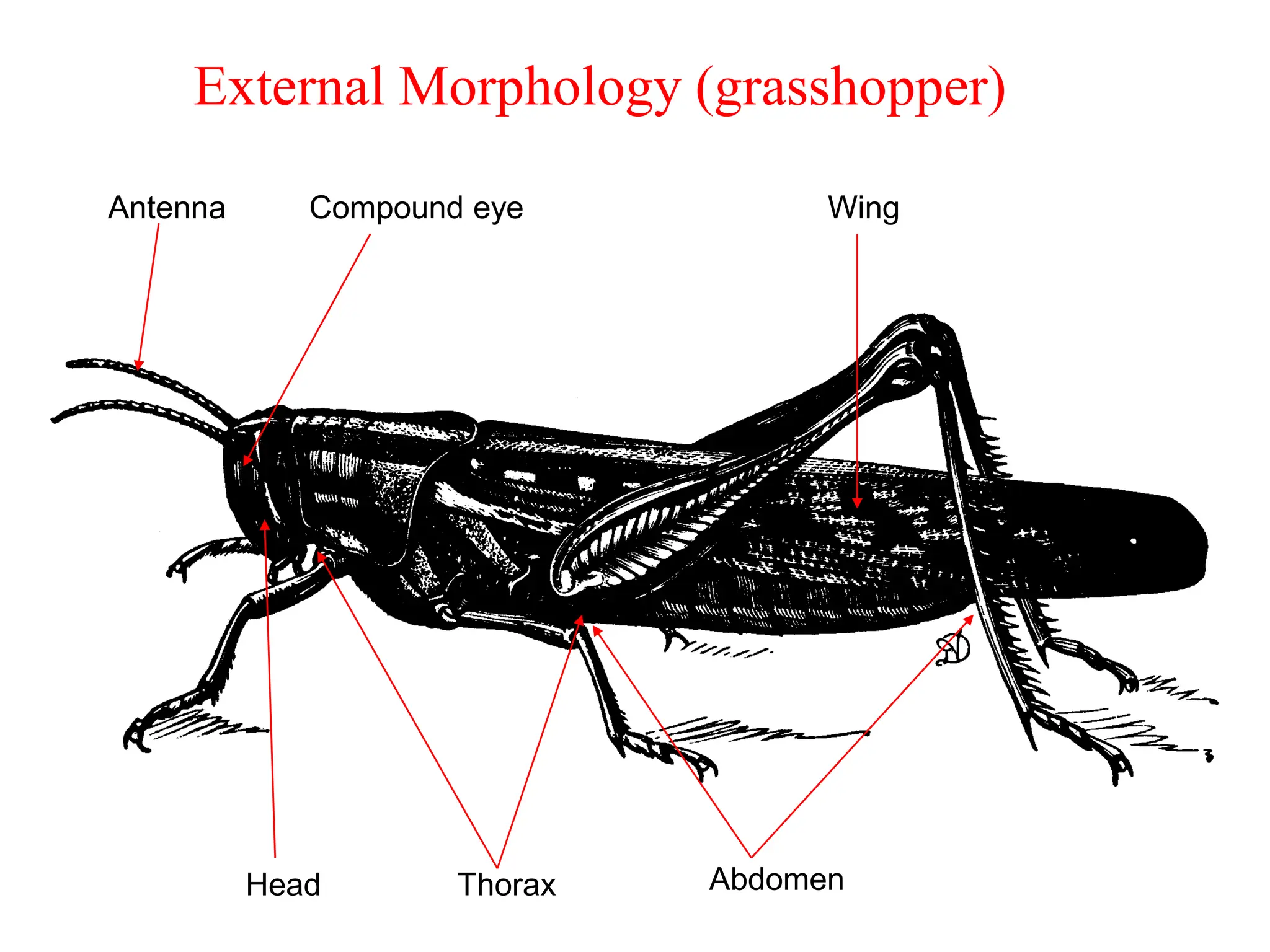
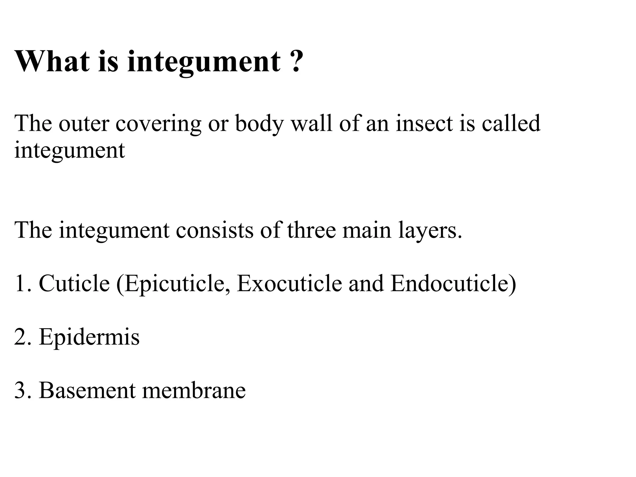
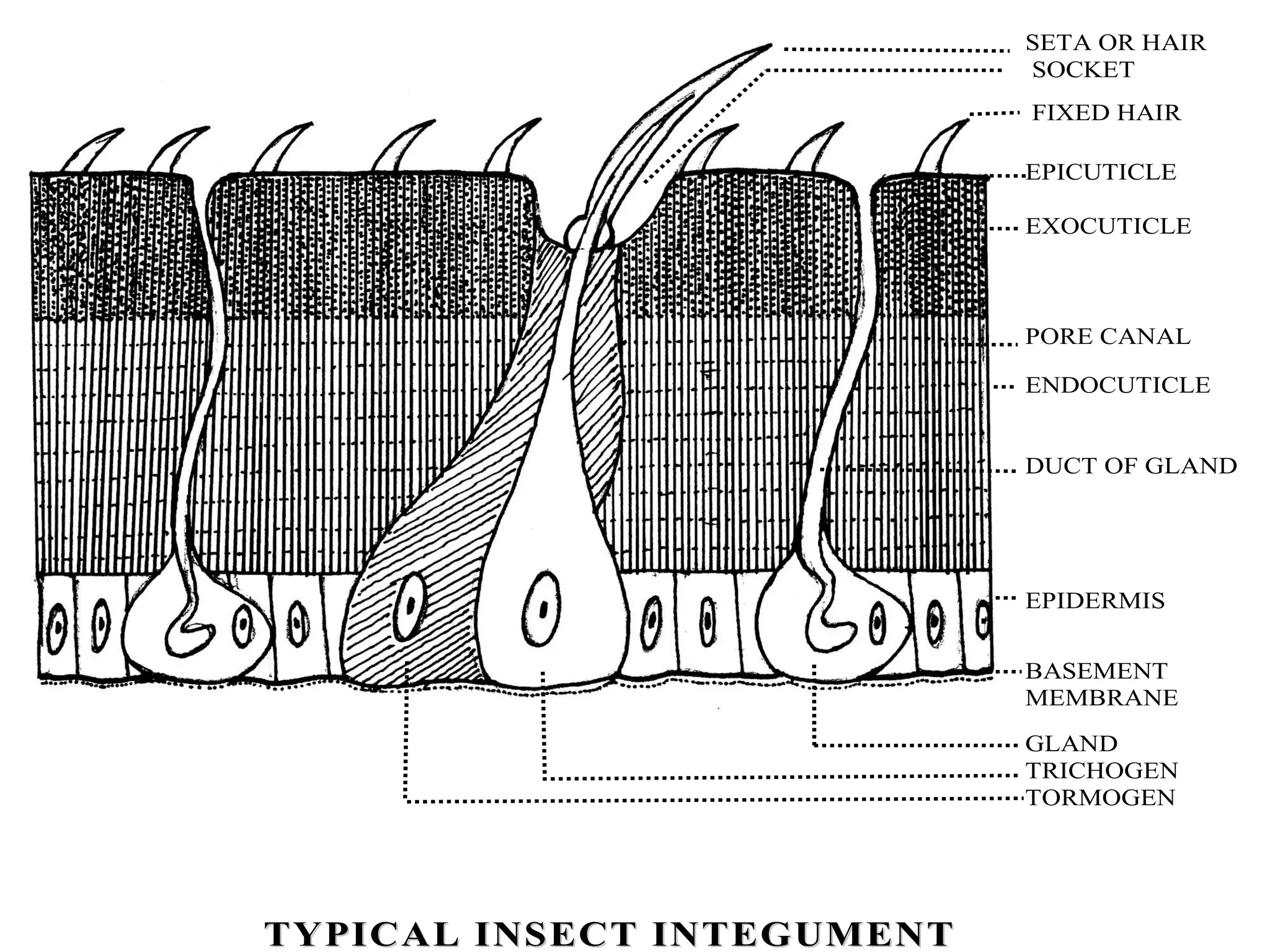
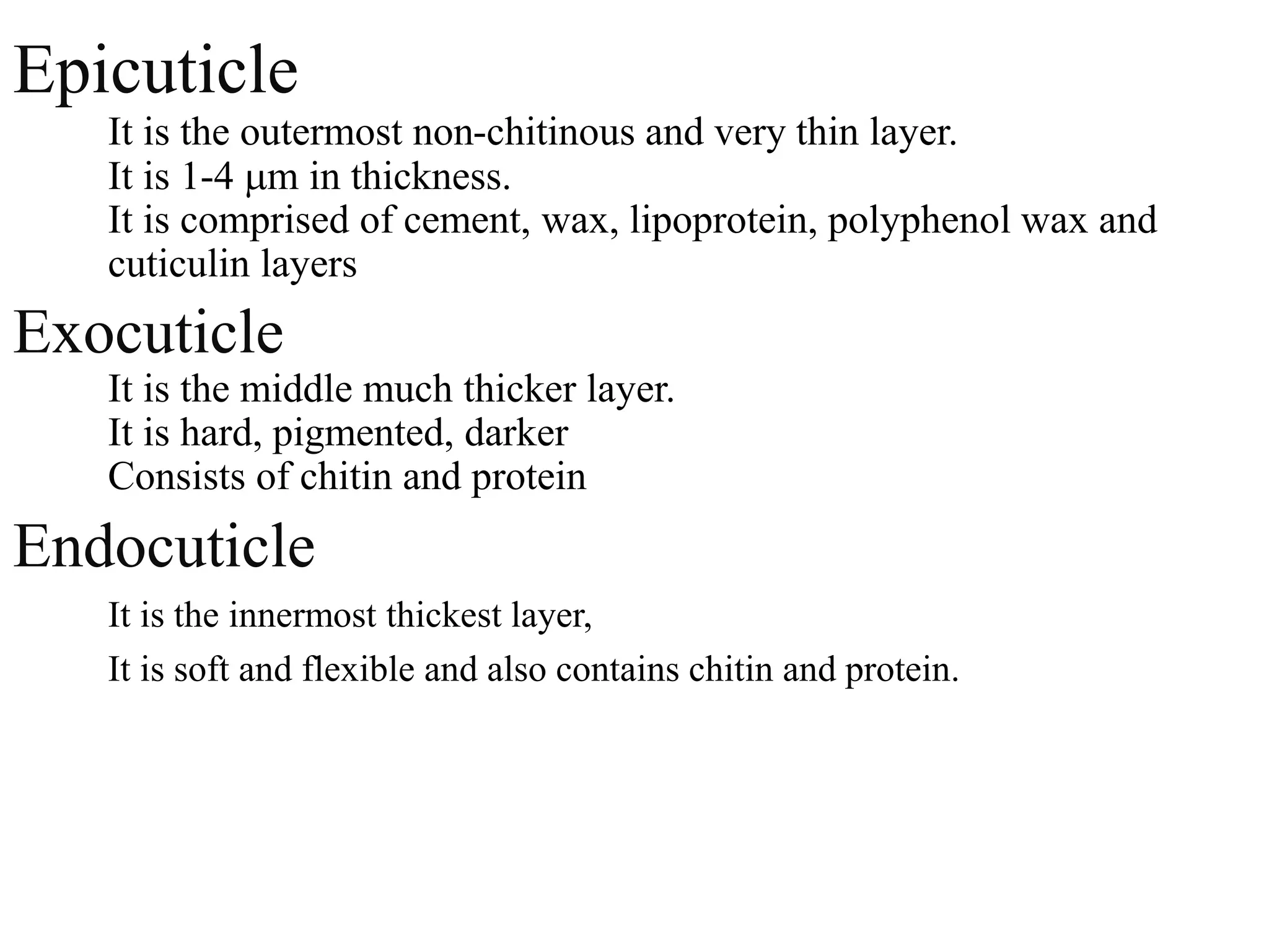
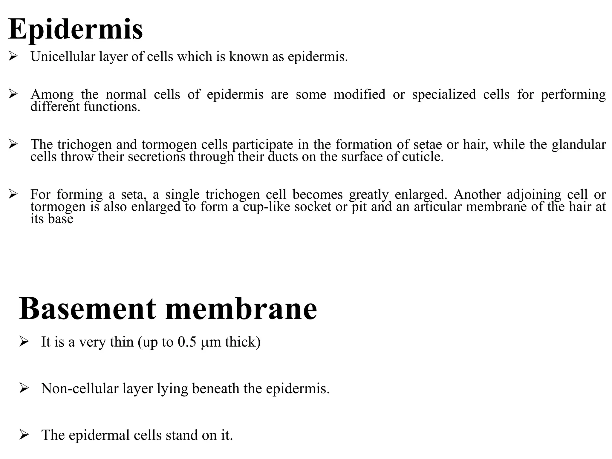
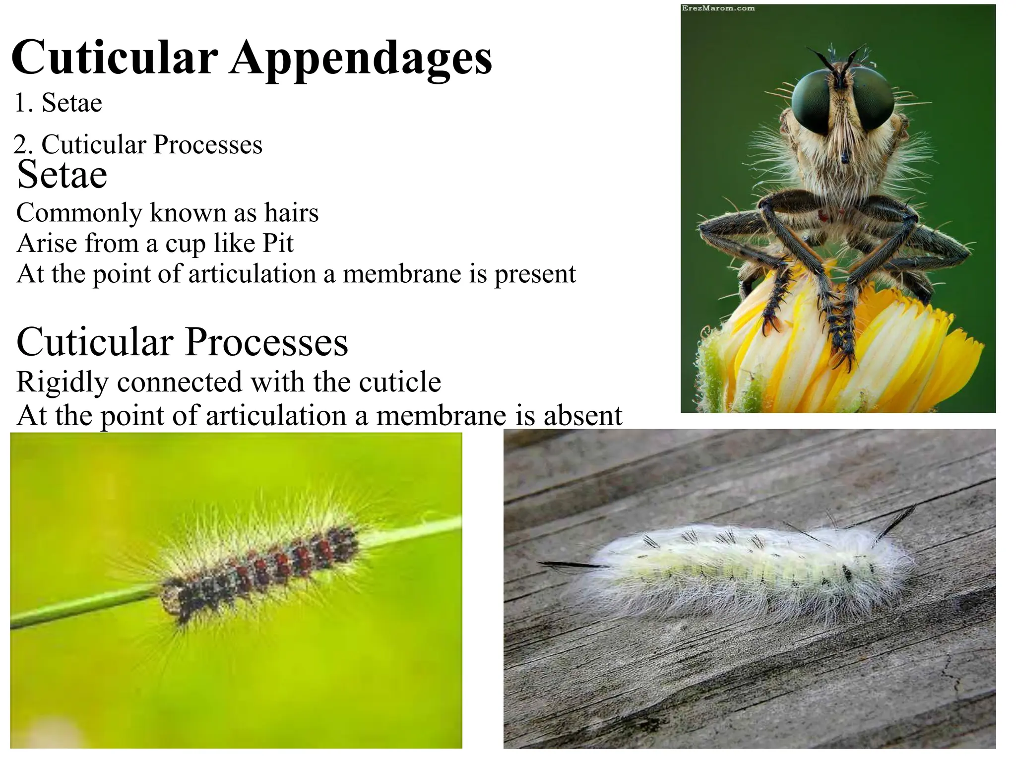
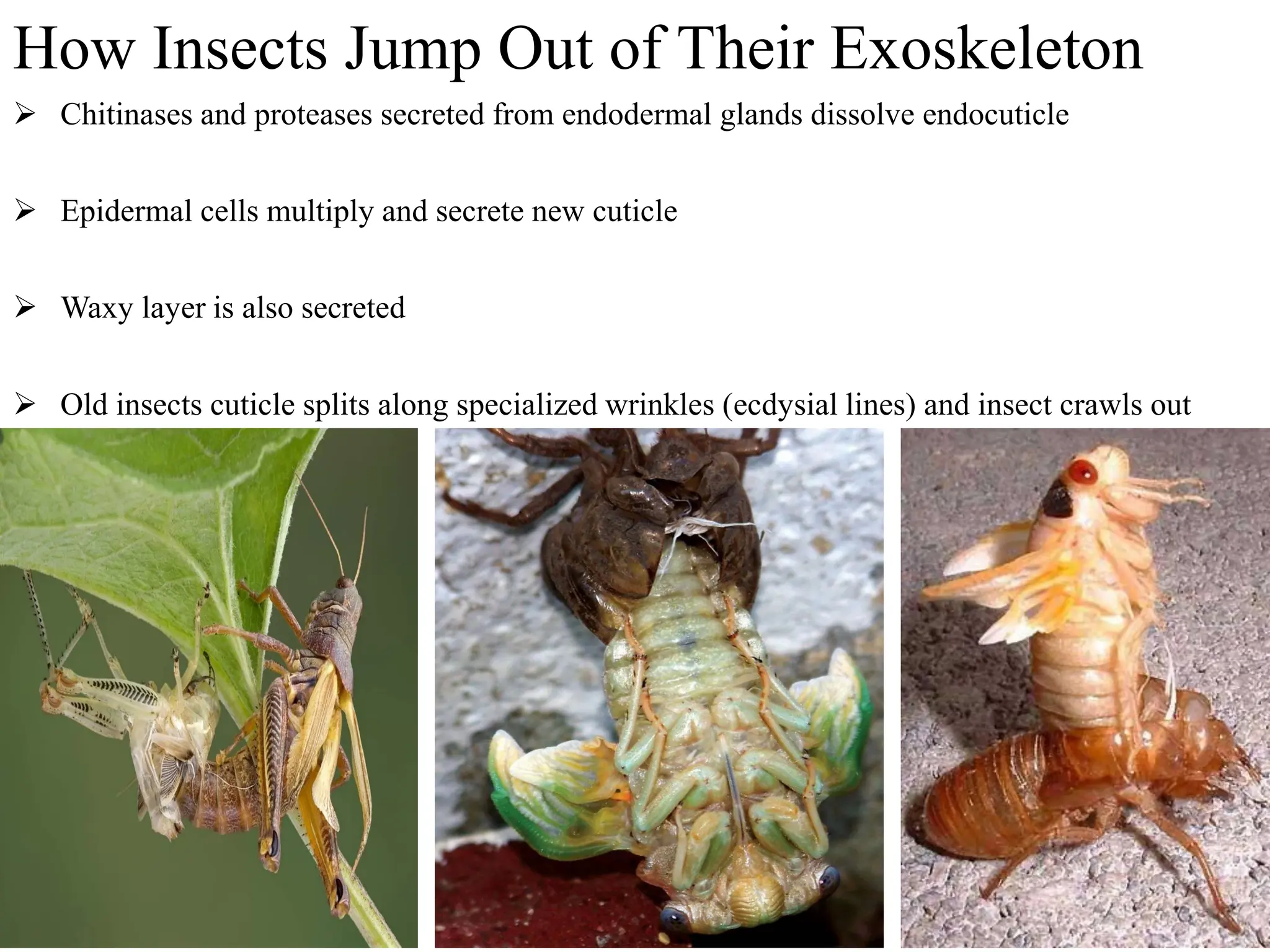
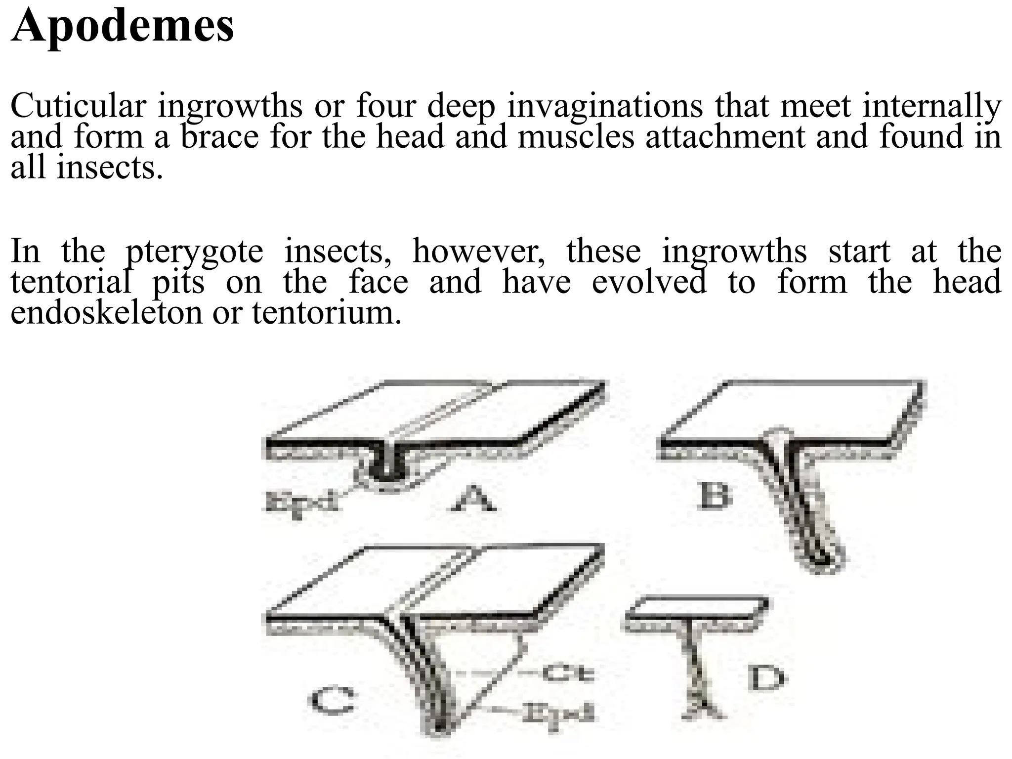
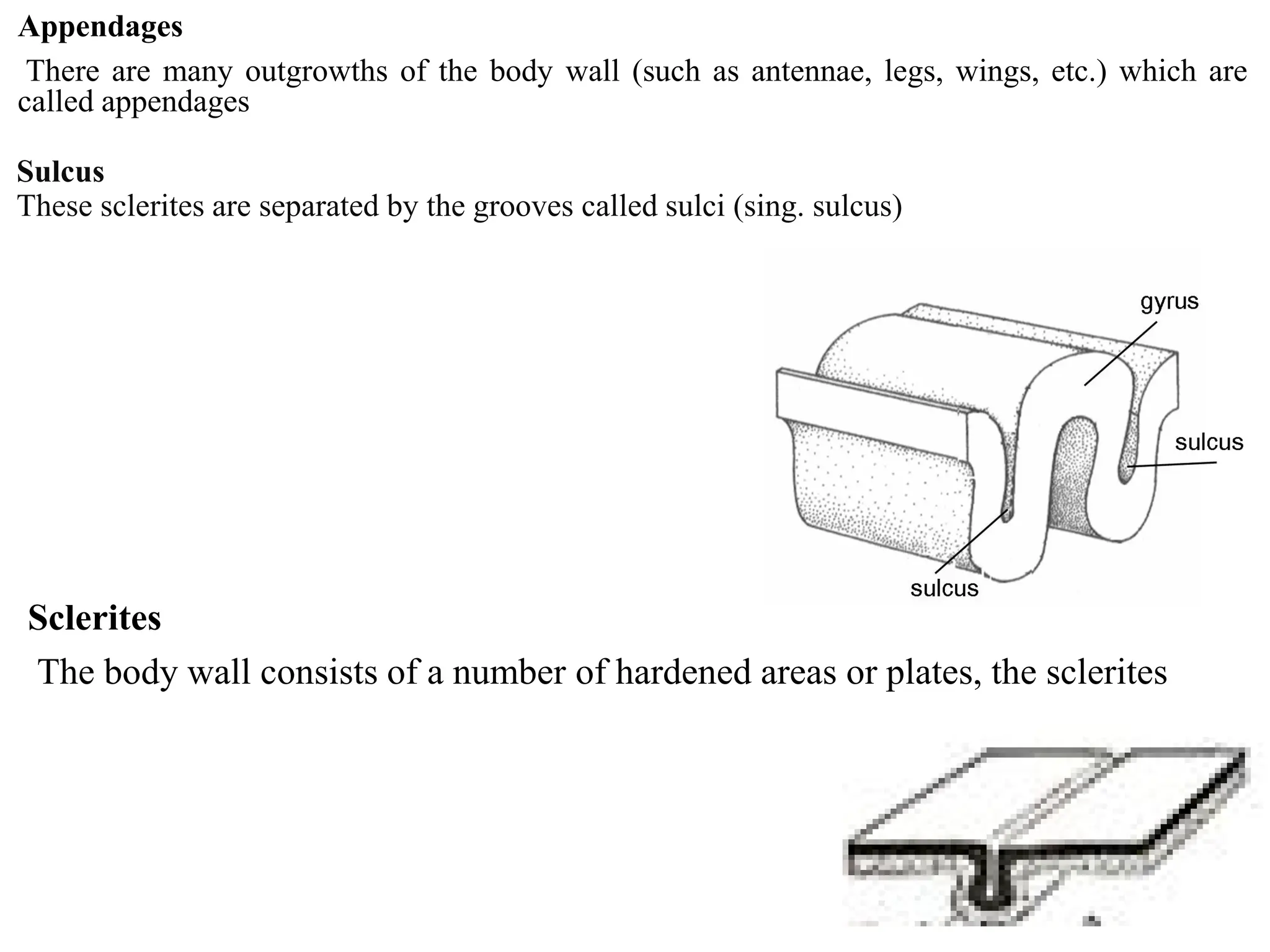
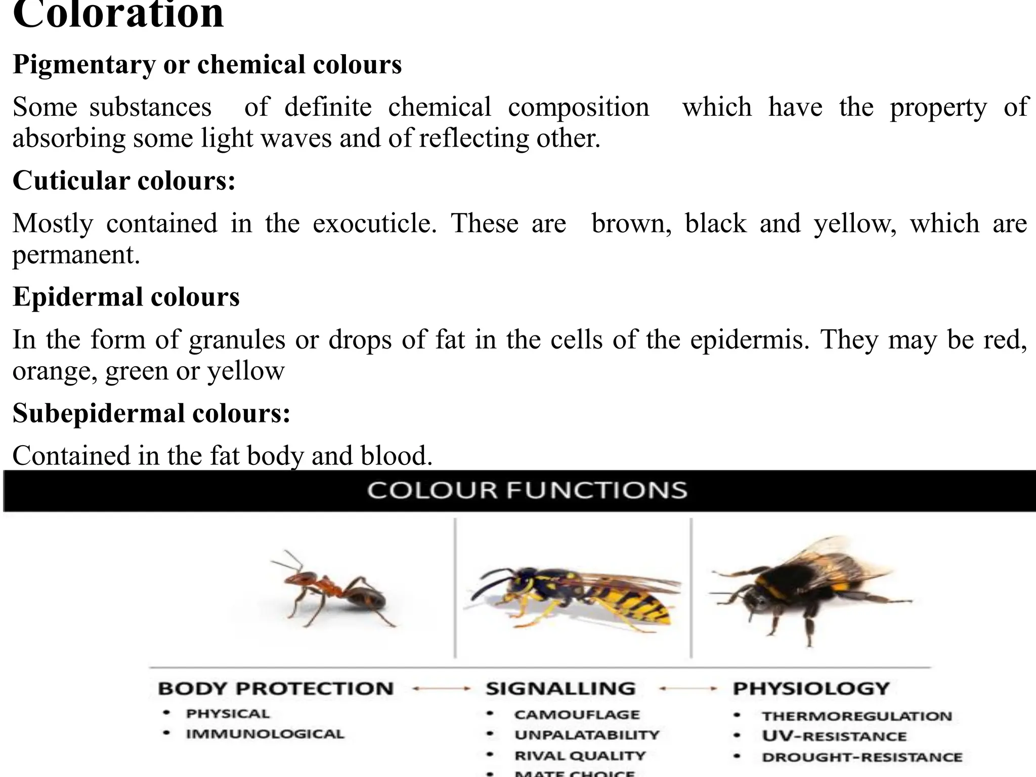
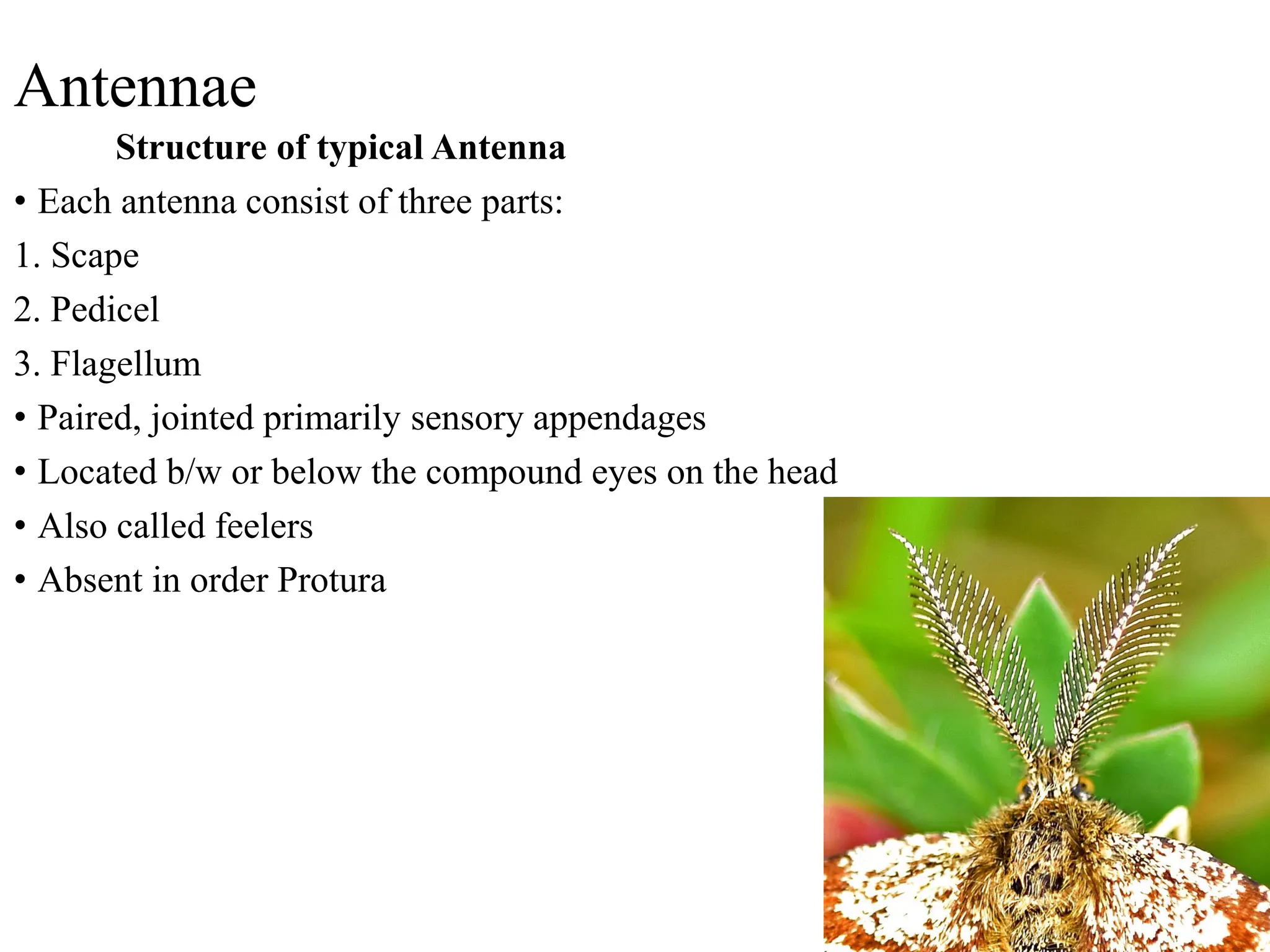
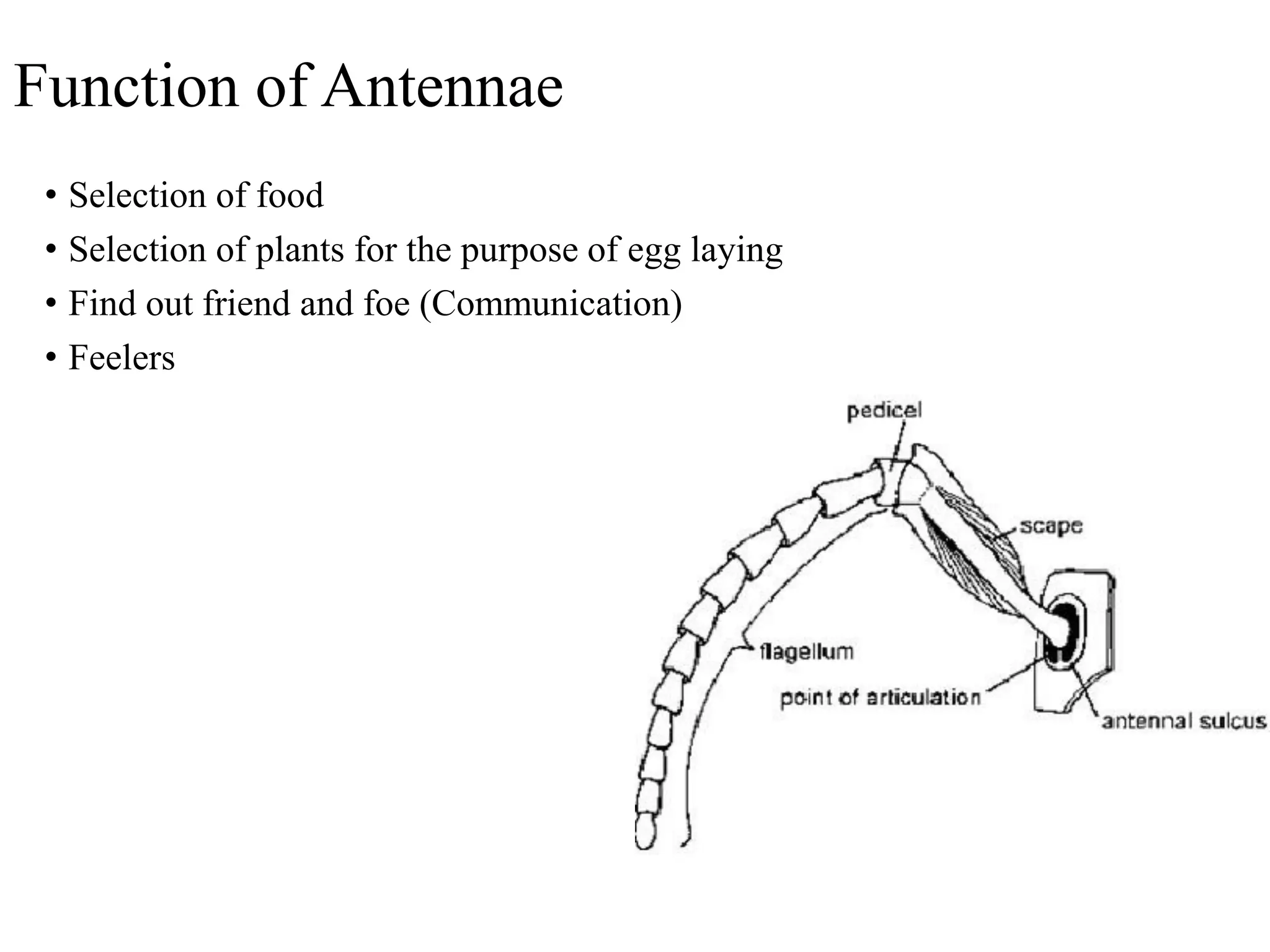
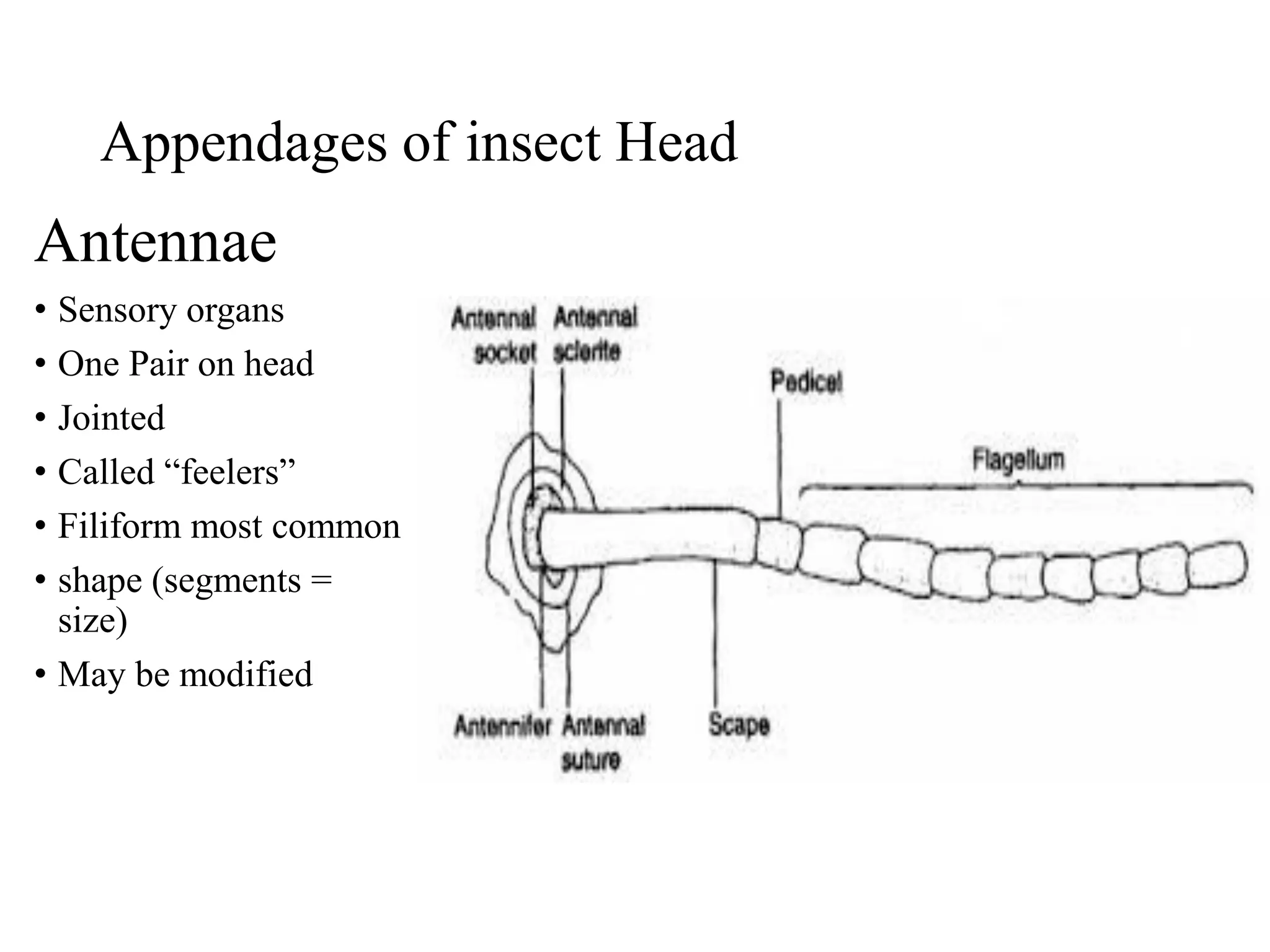
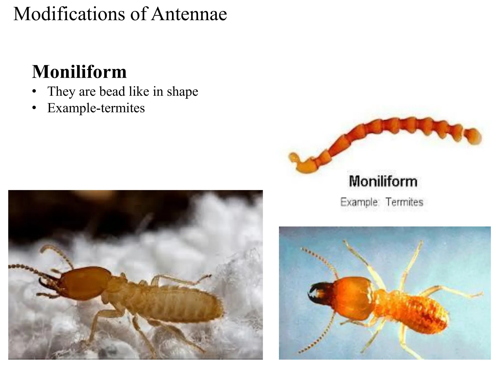
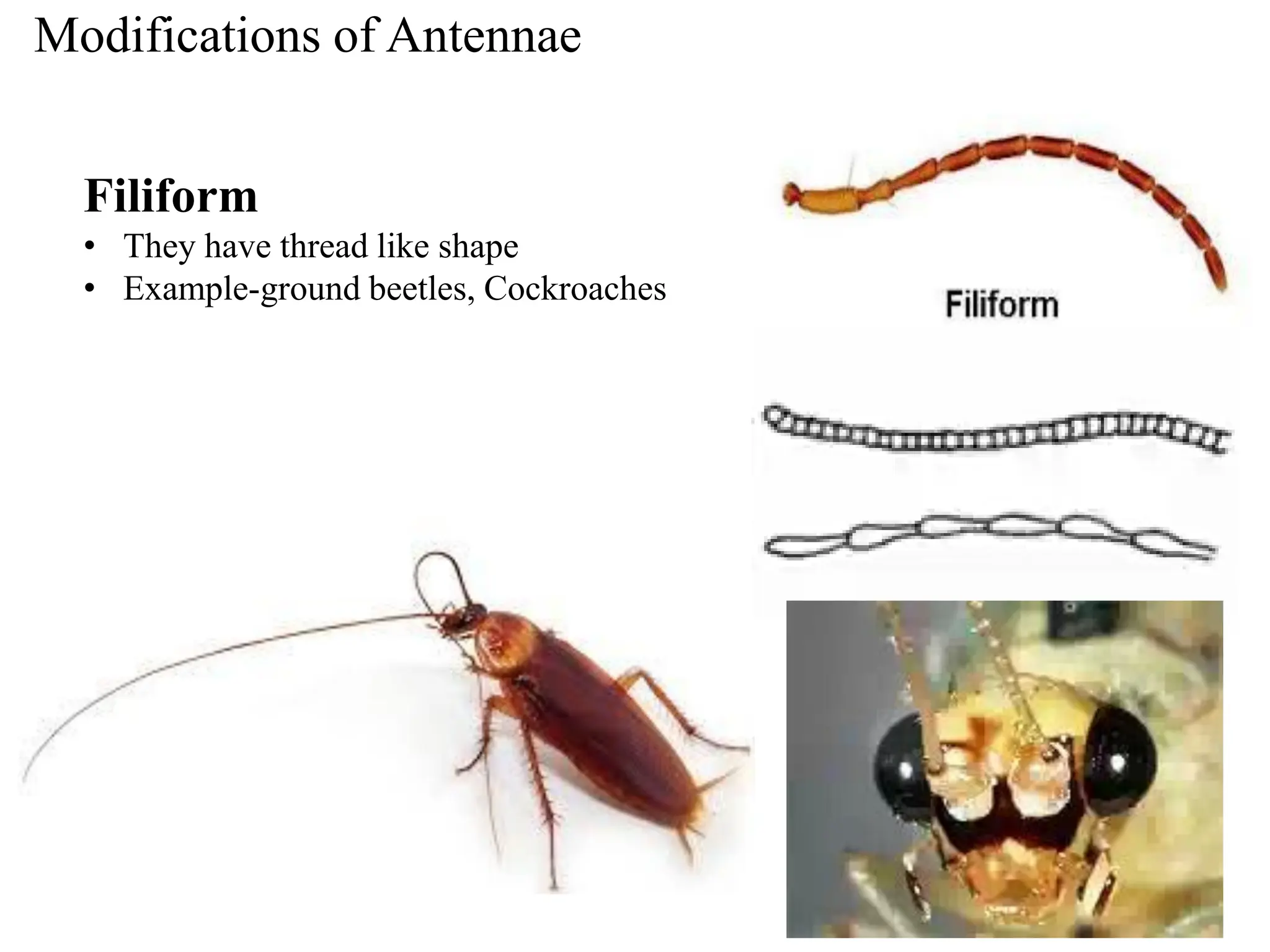
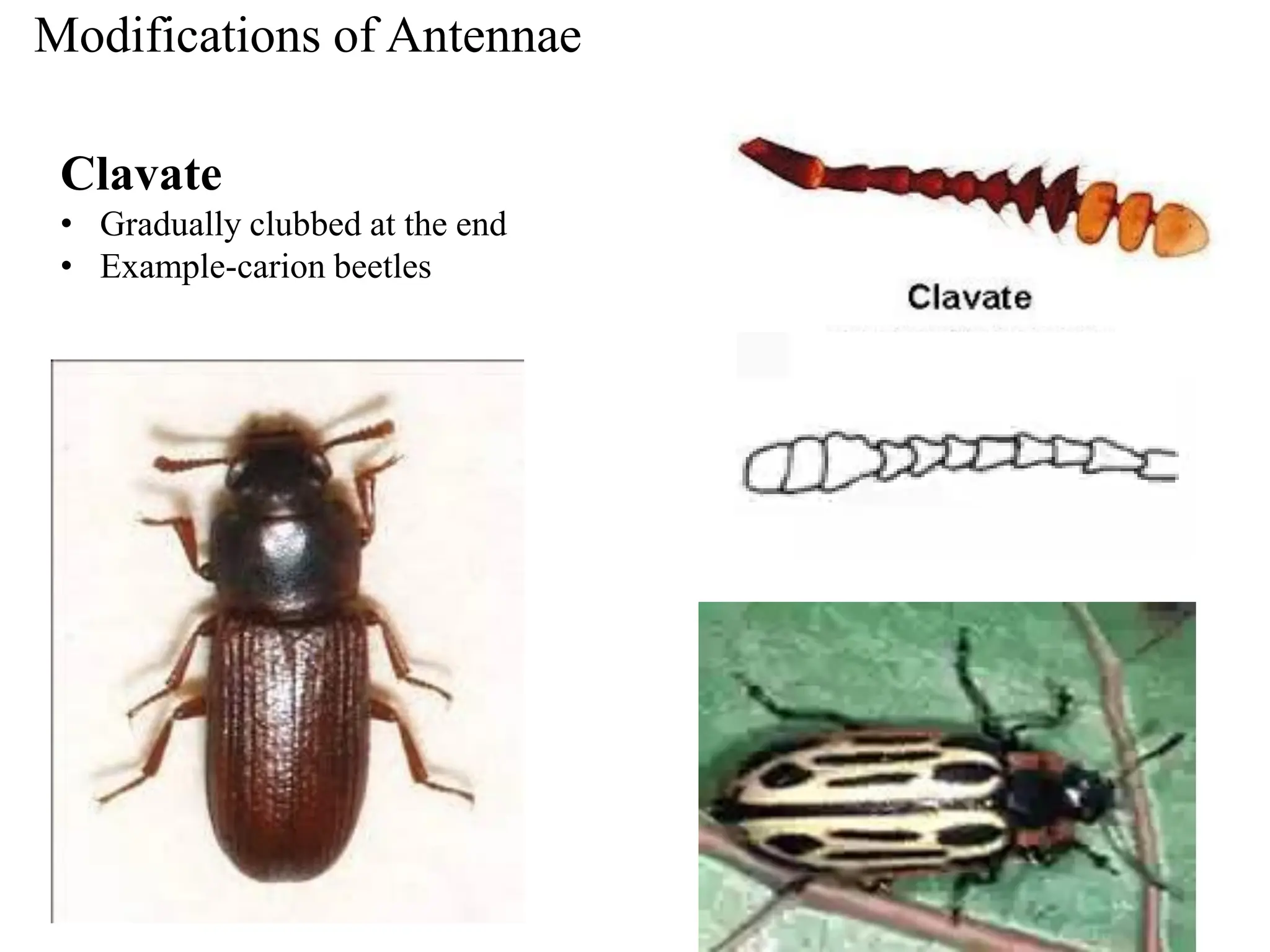
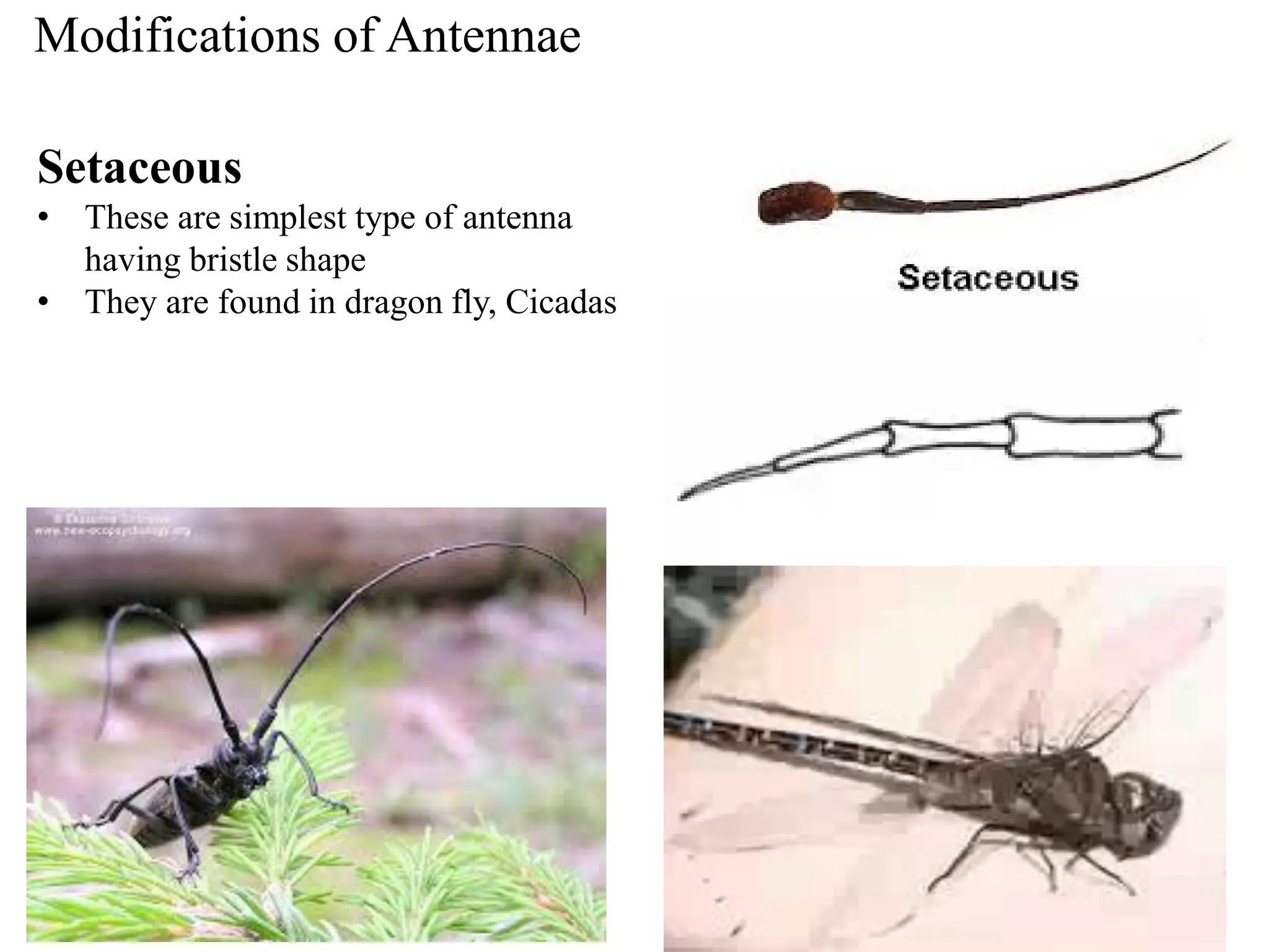
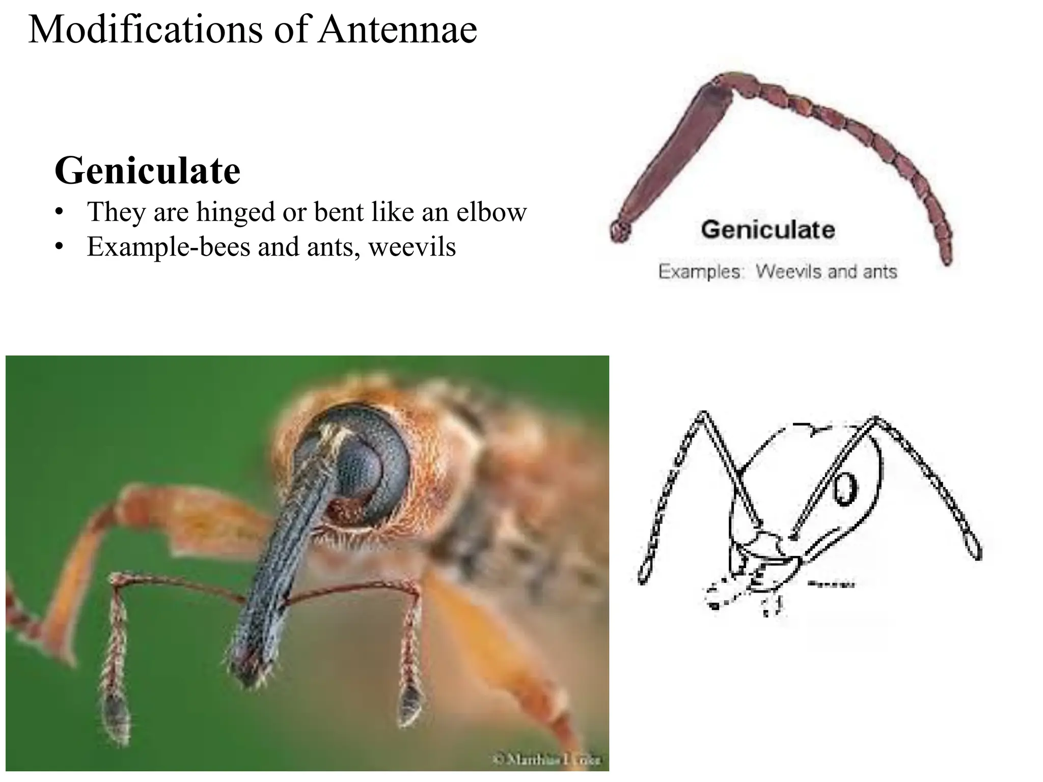
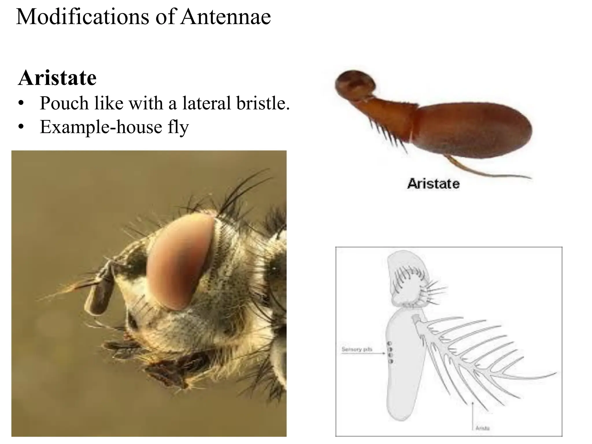
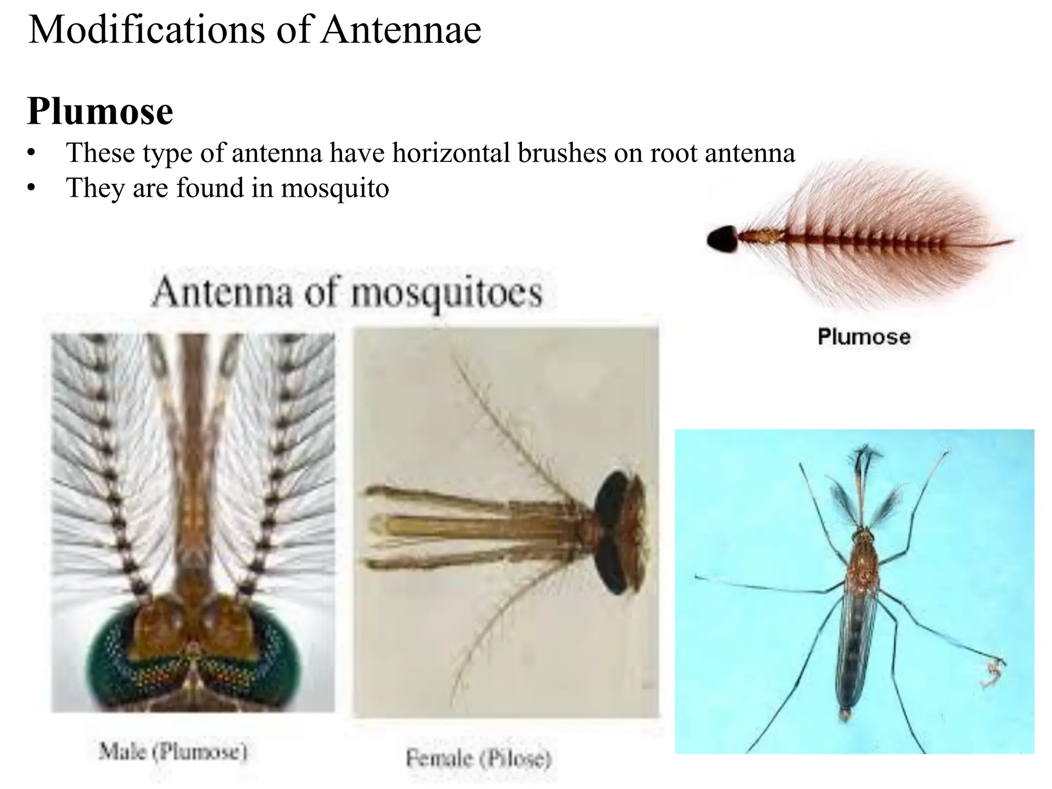
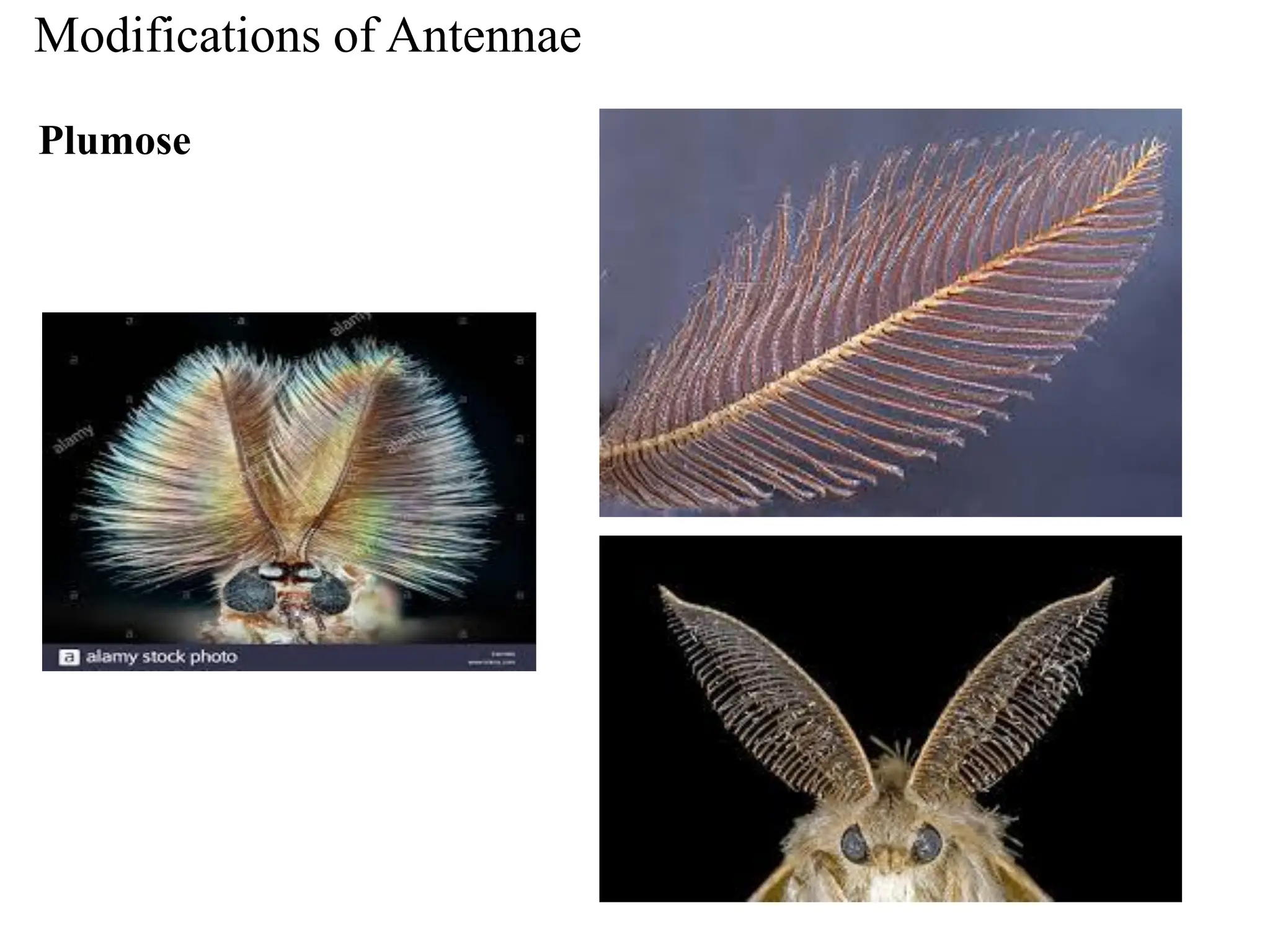
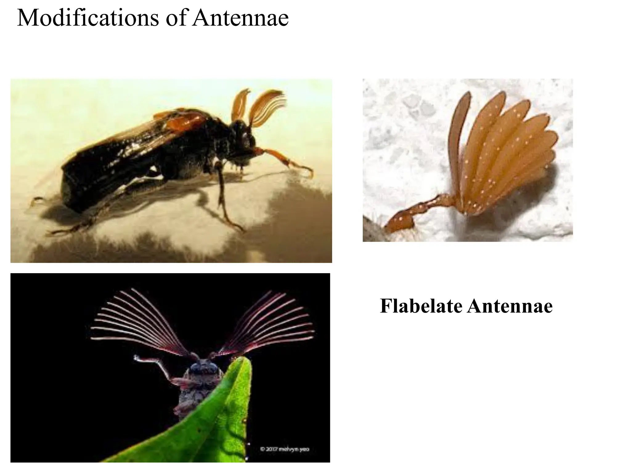
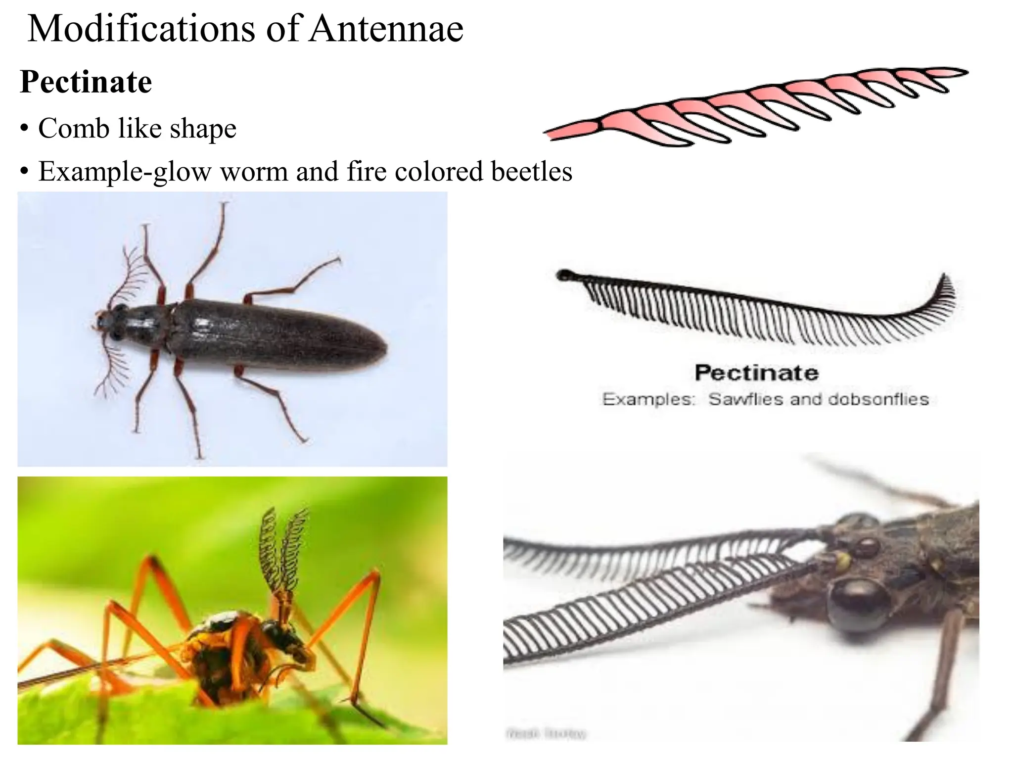
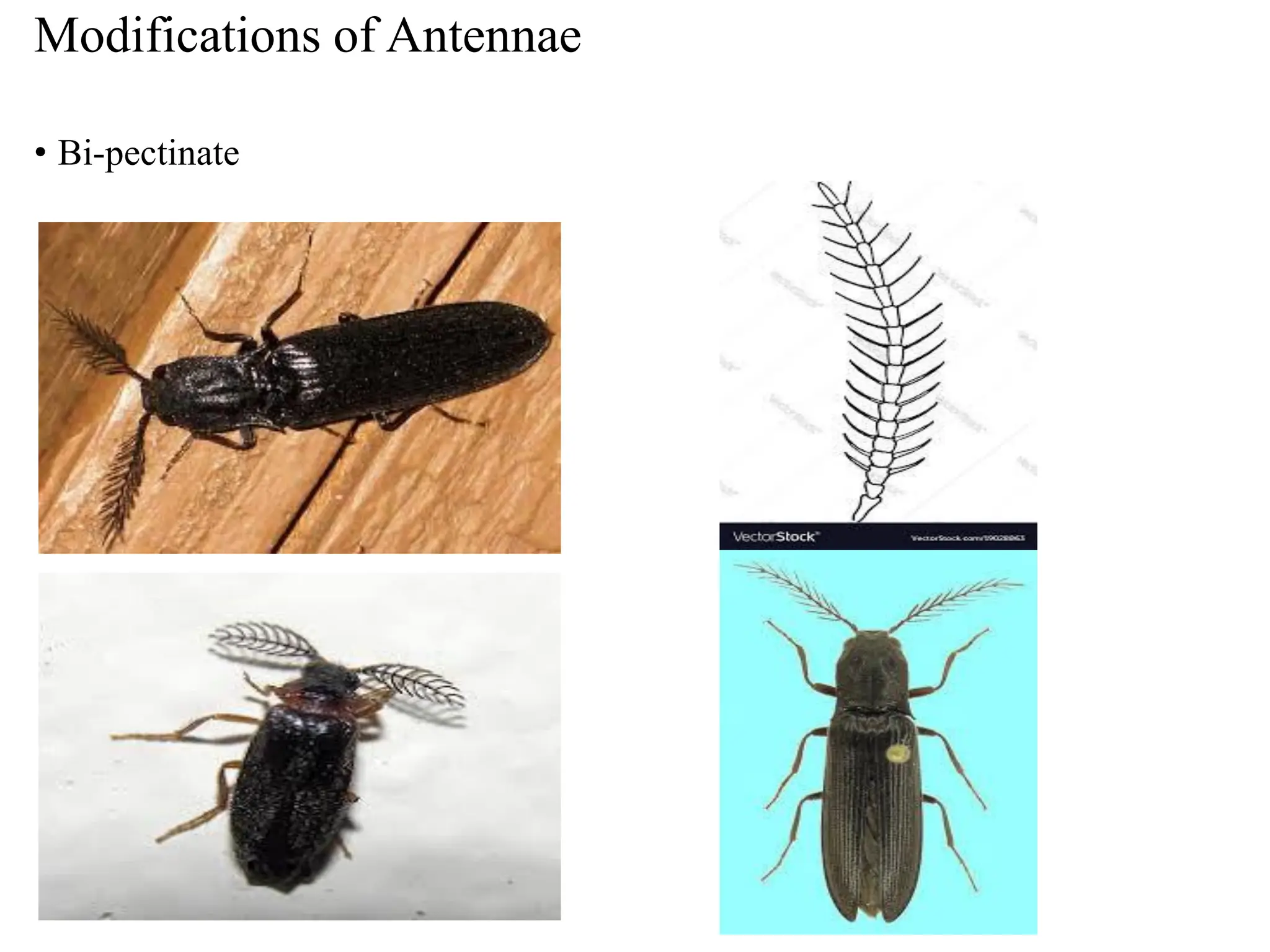
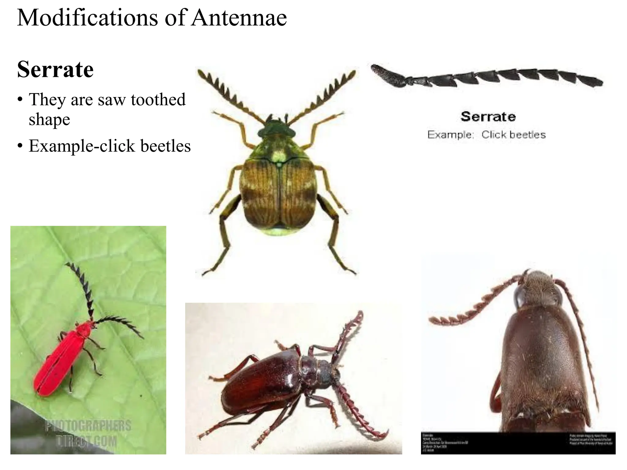
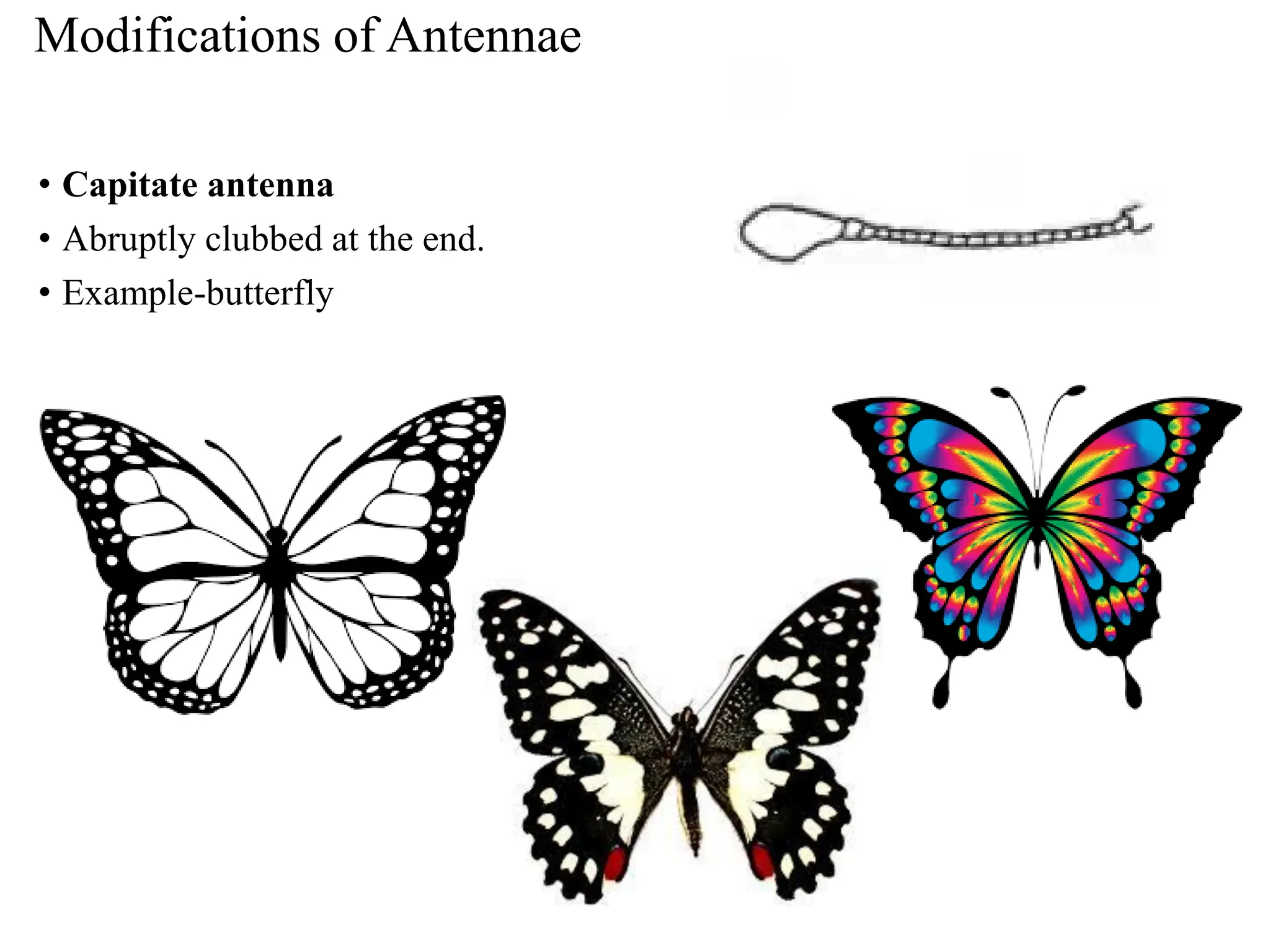
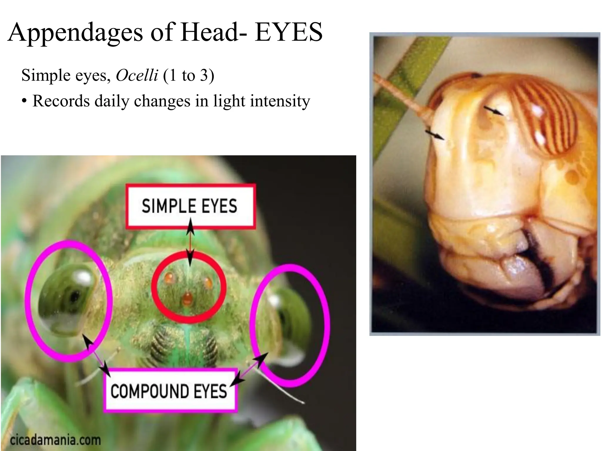
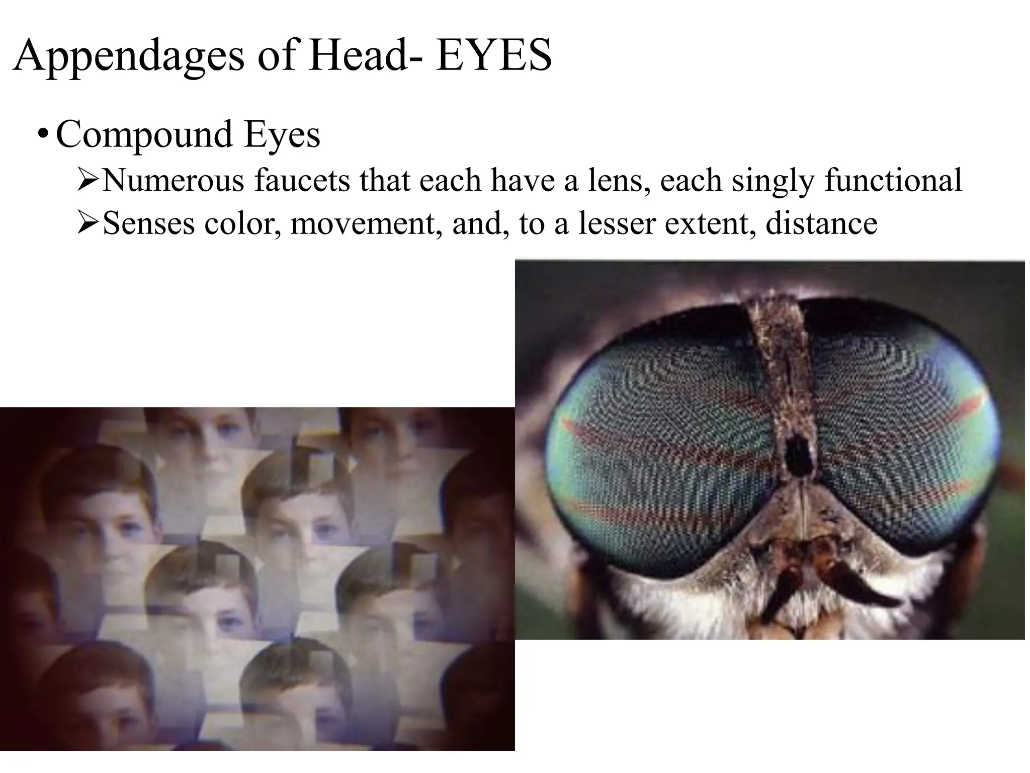
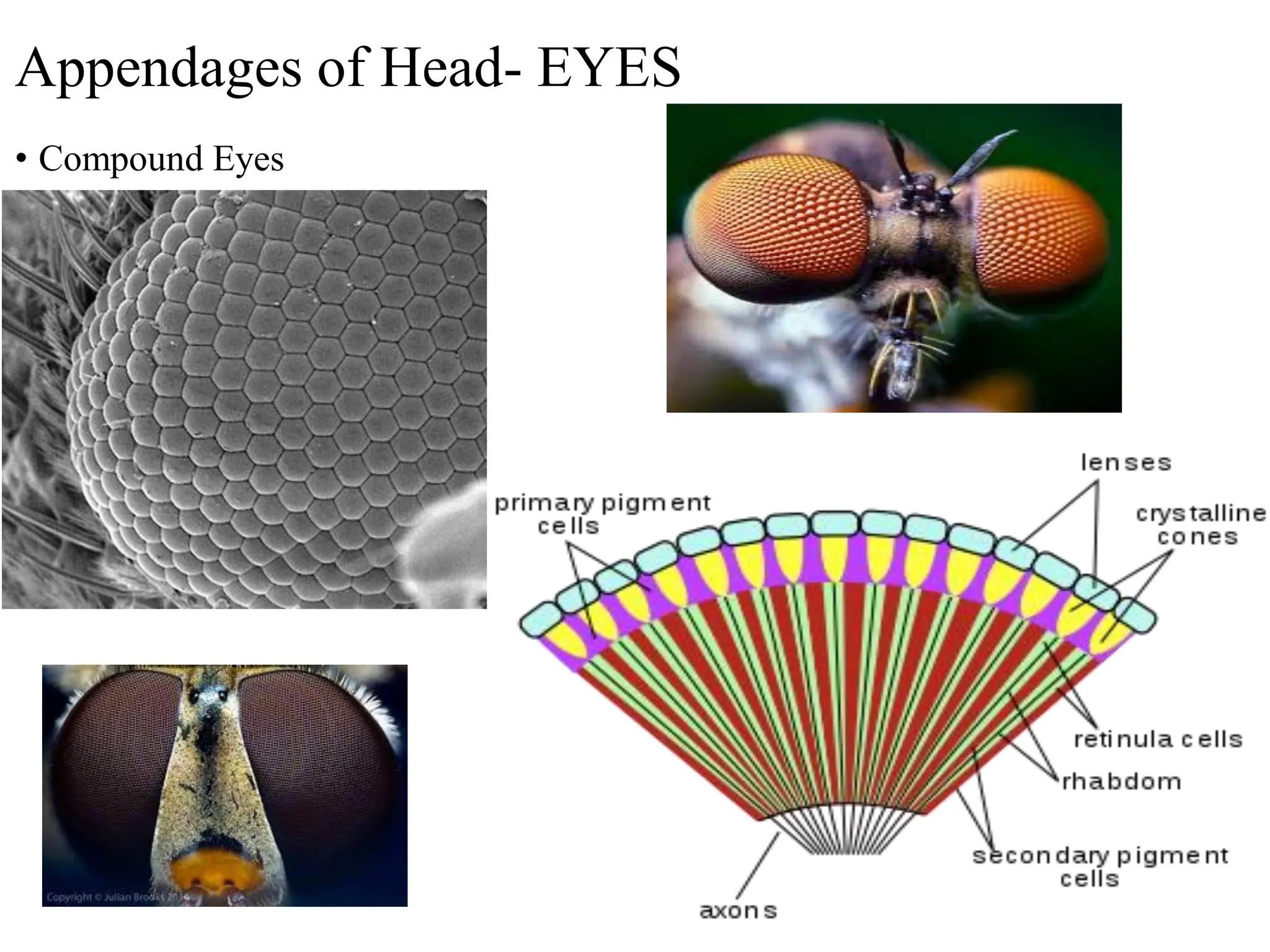
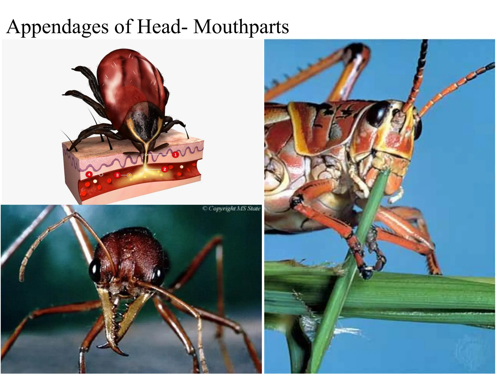
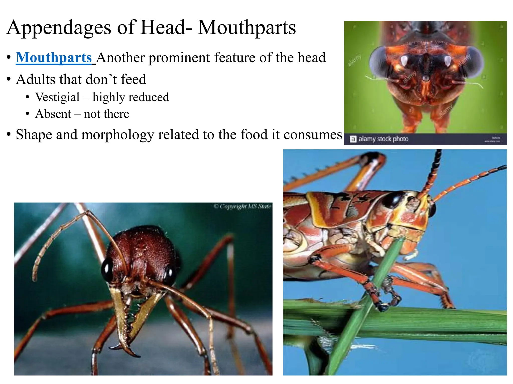
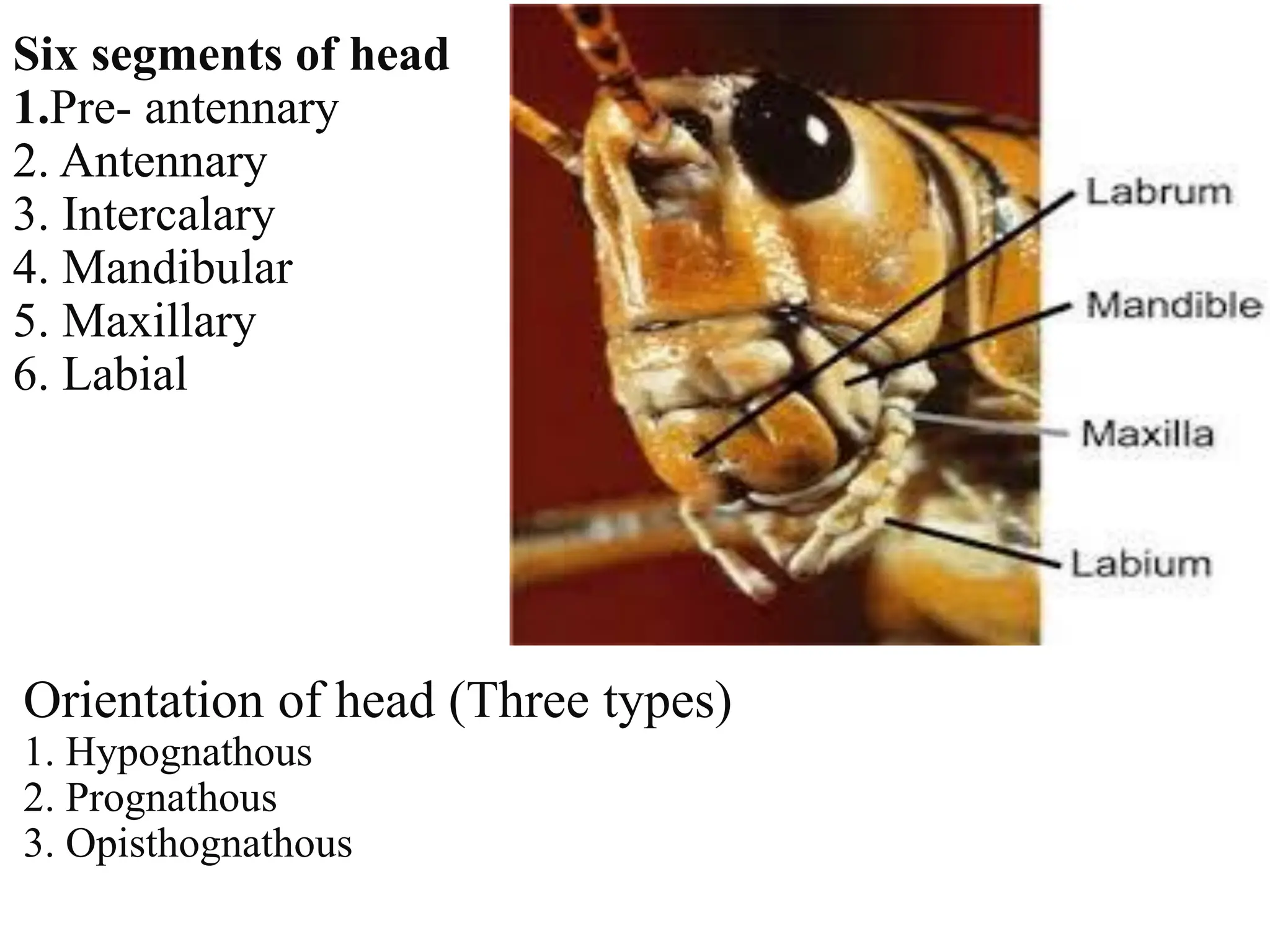
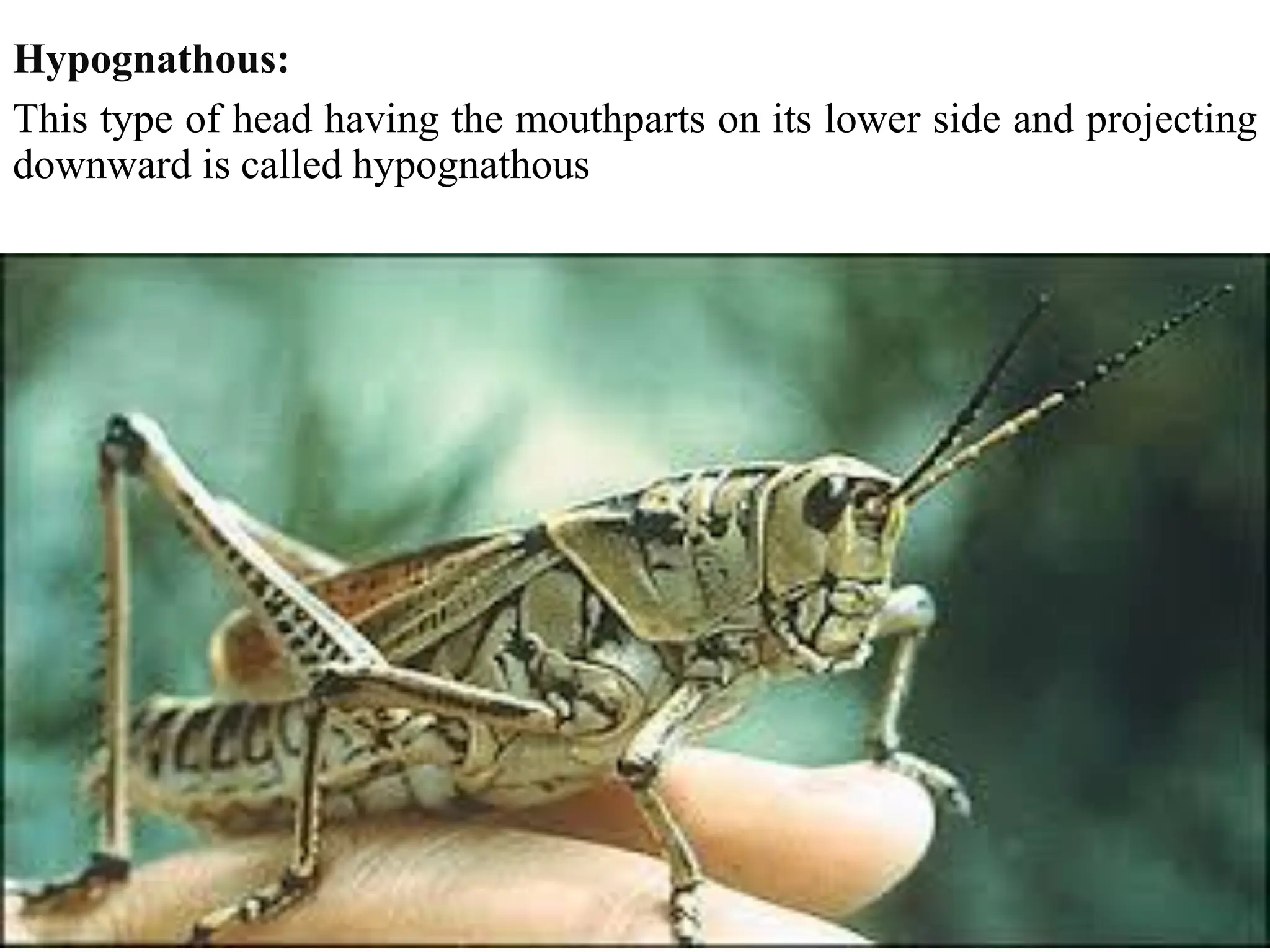
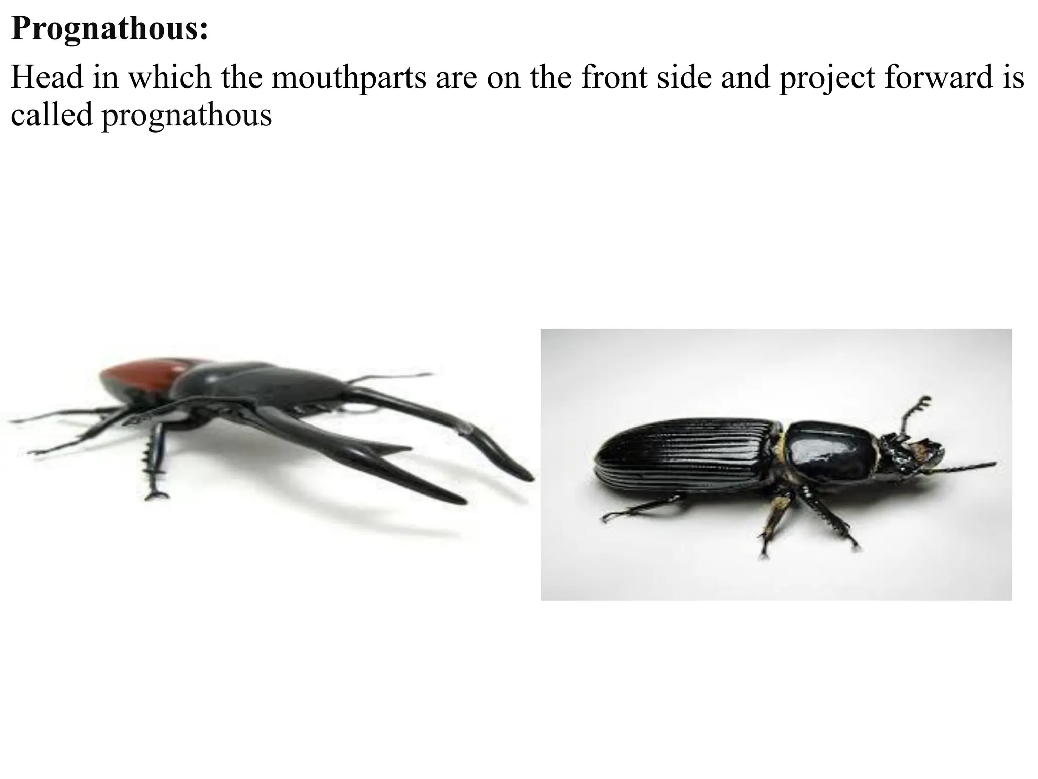
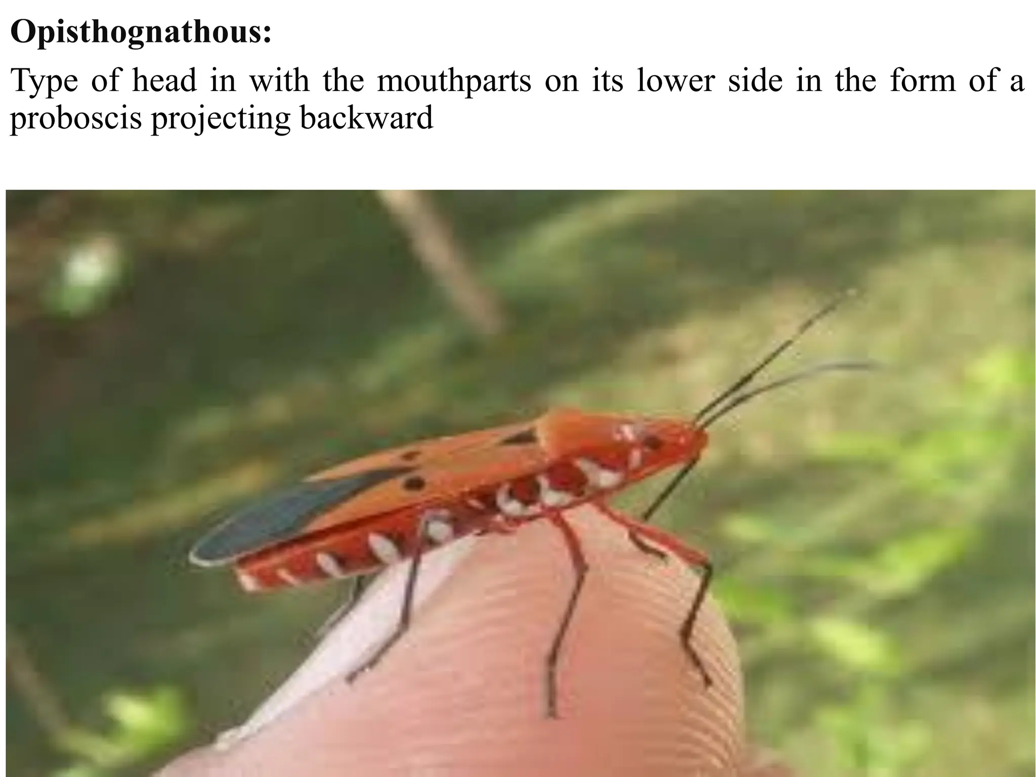
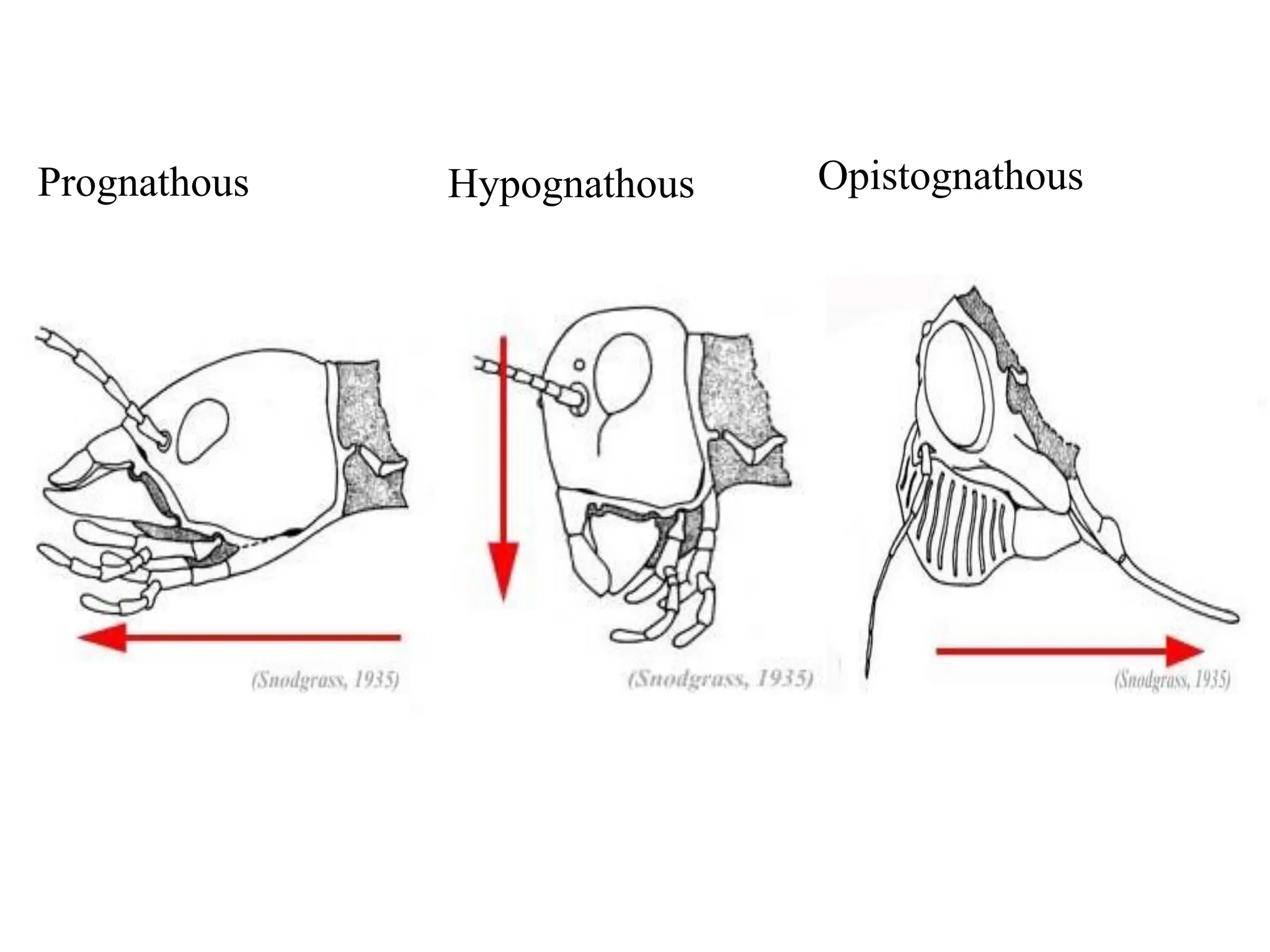
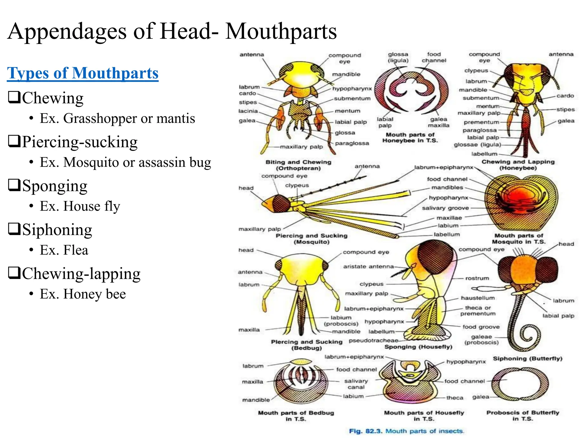
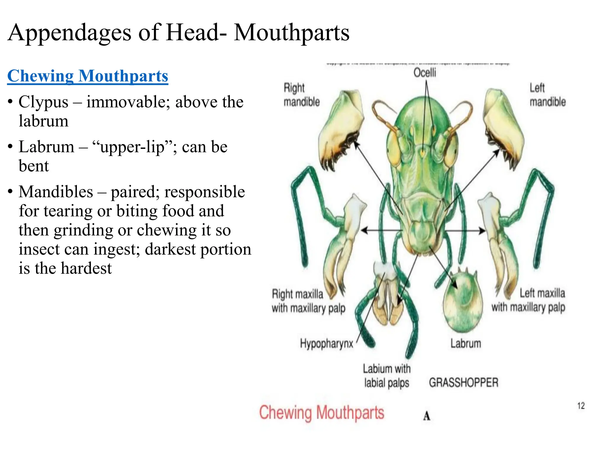
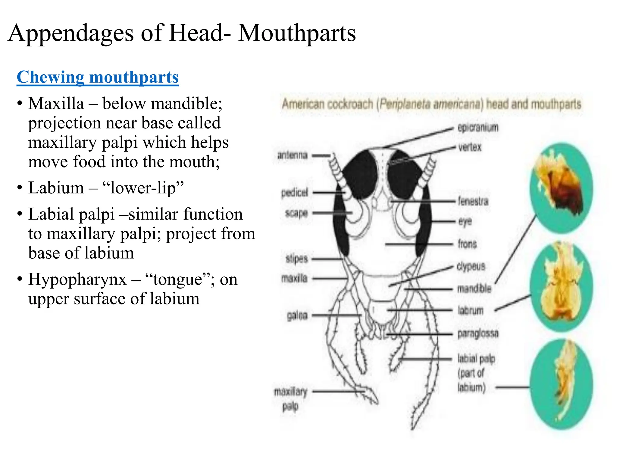
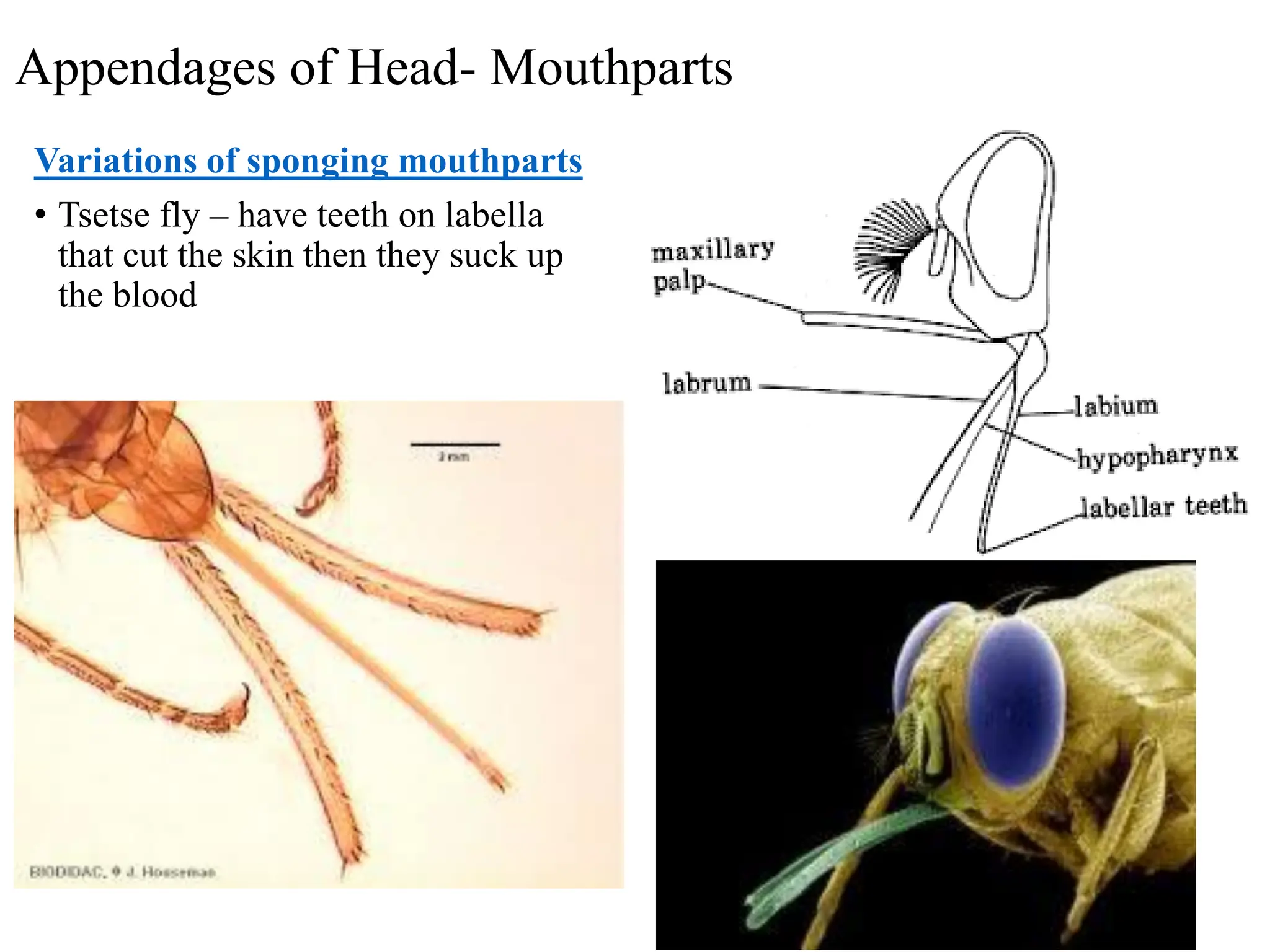
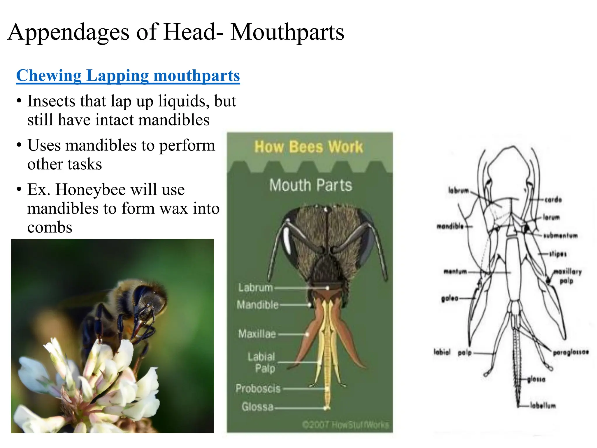
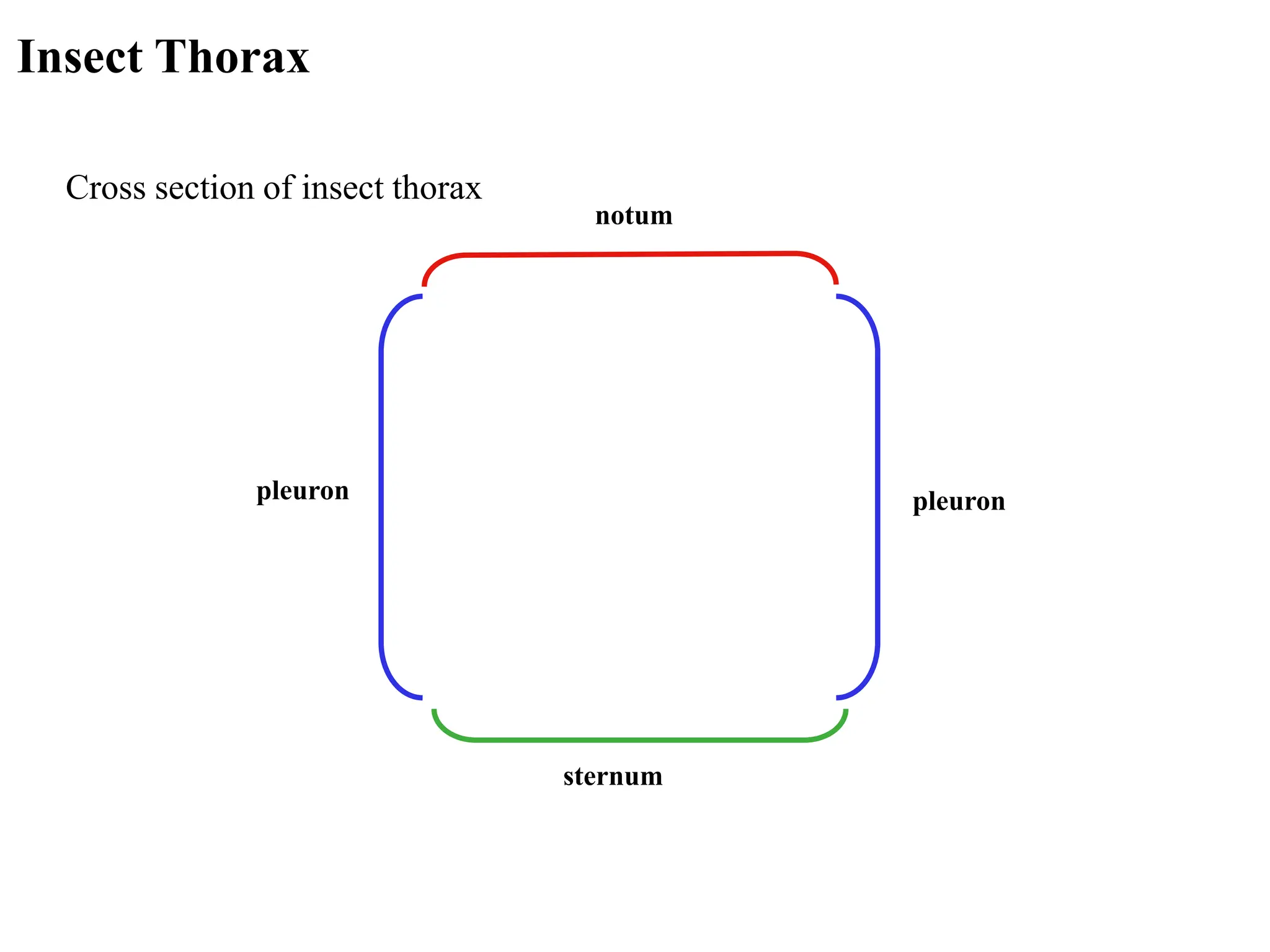
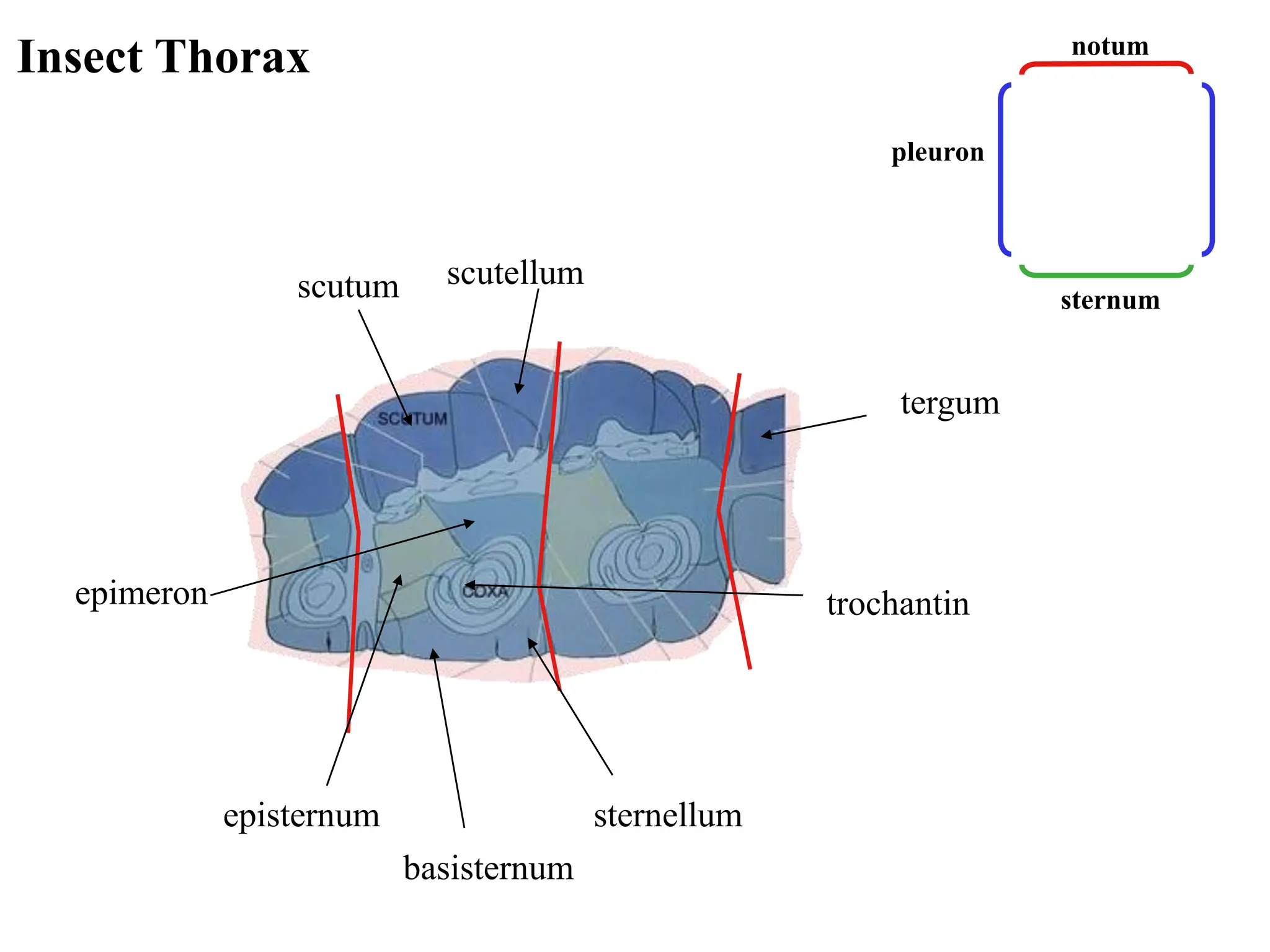
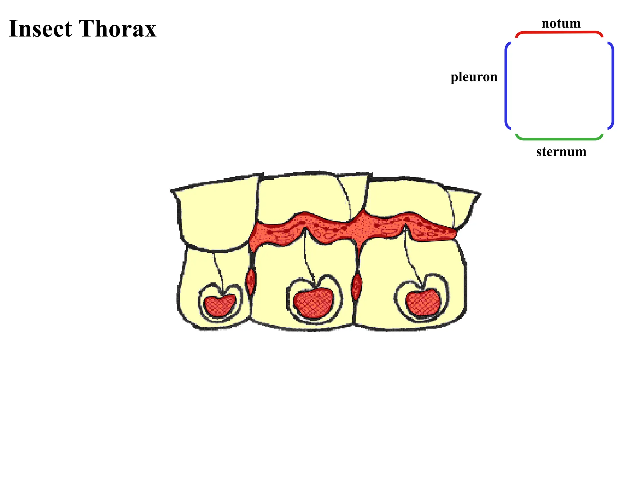
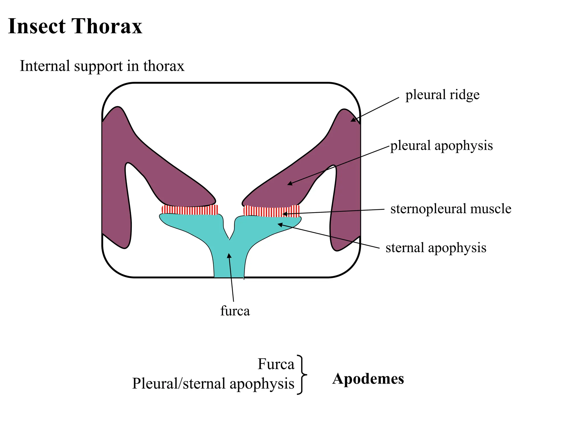
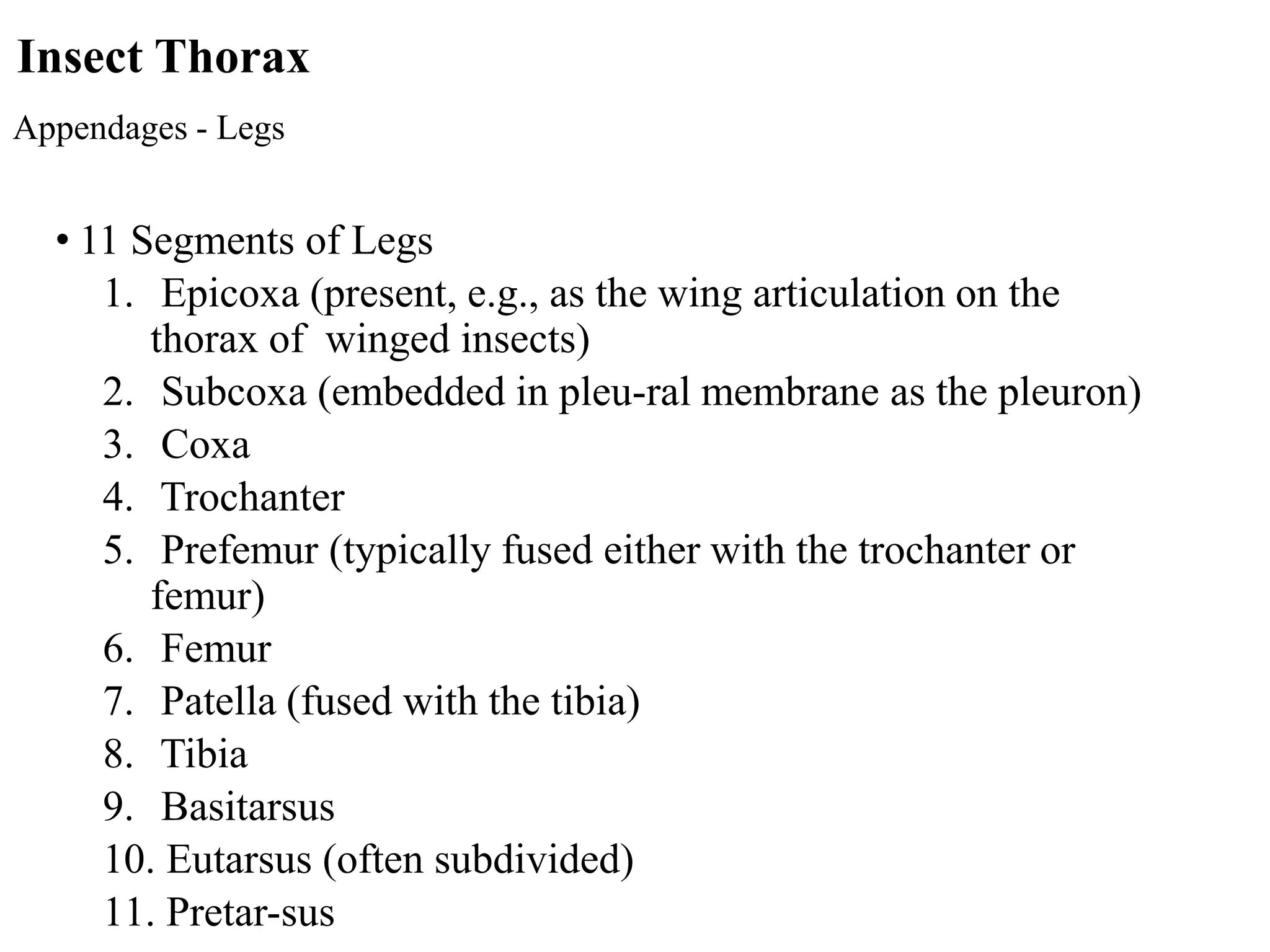
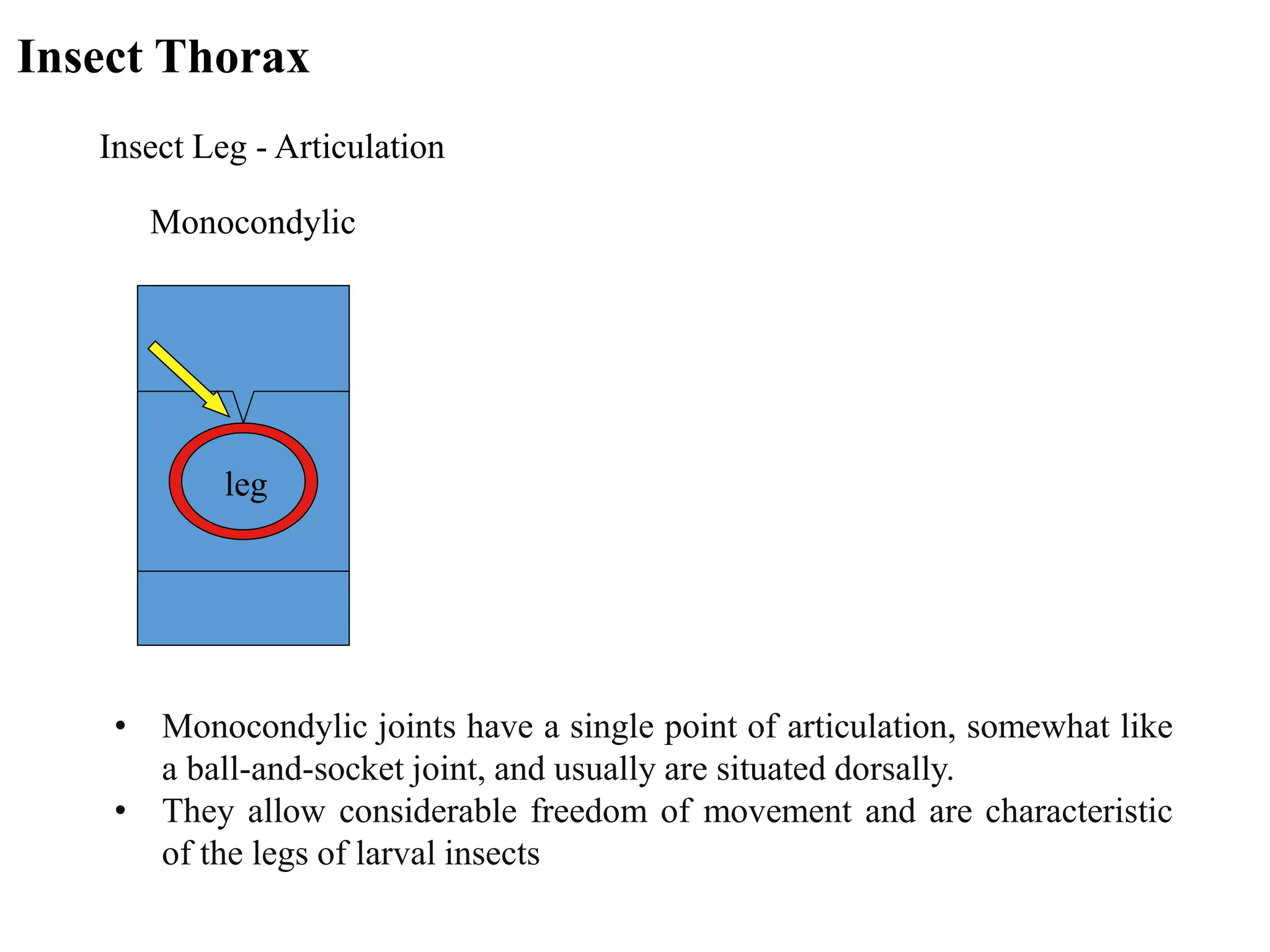
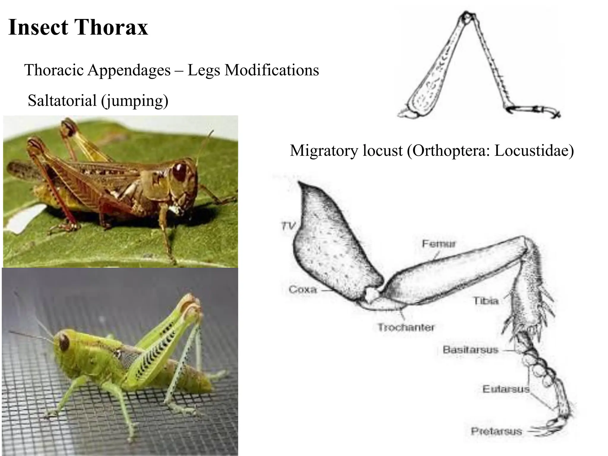
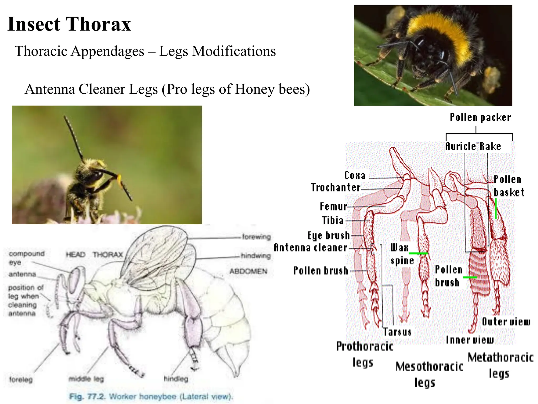
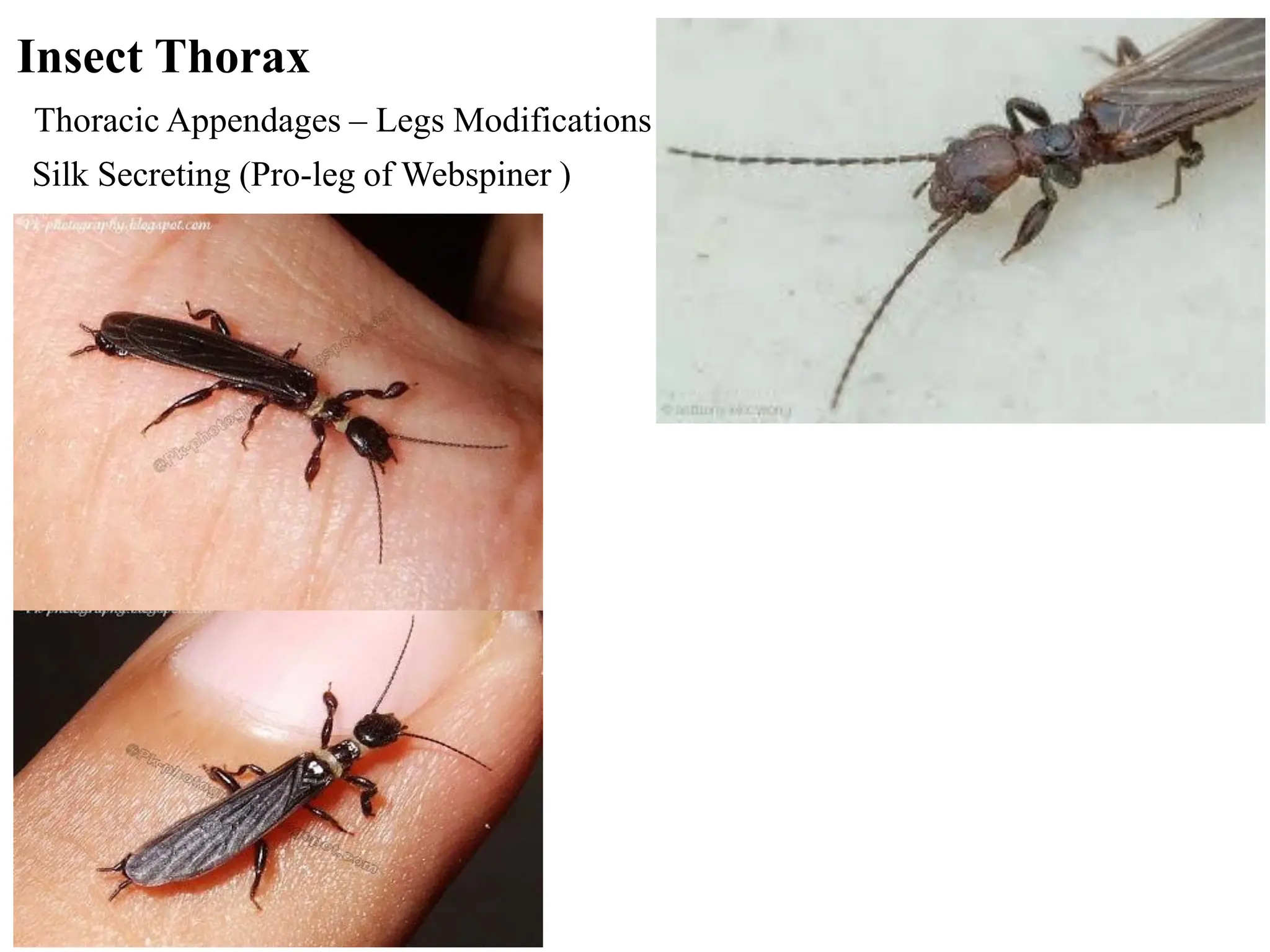
Insect Morphology
Dr. Farrukh Baig
Entomology
MNS-University of Agriculture, Multan
Outline
1. Integument
2. Head
Types of Antennae
Eyes and Types
Mouthparts and Types
3. Thorax
Legs and Types
Wings and Types
4. Abdomen
Segmentation
Body Systems
External Morphology (grasshopper)
Head Thorax Abdomen
Antenna Compound eye Wing
The integument consists of three main layers.
1. Cuticle (Epicuticle, Exocuticle and Endocuticle)
2. Epidermis
3. Basement membrane
What is integument ?
The outer covering or body wall of an insect is called
integument
Integument
Cross-section of cockroach cuticle Integument cross-section
T
TY
YP
PI
IC
CA
AL
L I
IN
NS
SE
EC
CT
T I
IN
NT
TE
EG
GU
UM
ME
EN
NT
T
SETA OR HAIR
SOCKET
FIXED HAIR
EPICUTICLE
EXOCUTICLE
PORE CANAL
ENDOCUTICLE
DUCT OF GLAND
EPIDERMIS
BASEMENT
MEMBRANE
GLAND
TRICHOGEN
TORMOGEN
Cuticle
➢ Outer non-cellular layer.
➢ It is secreted by the epidermal cells below it.
➢ When newly formed it is flexible and elastic
➢ Later on it becomes hard through the process of sclerotization.
➢ It protects the insects from water loss, penetration of insecticides
and provides ground for the attachment of muscles.
Epicuticle
It is the outermost non-chitinous and very thin layer.
It is 1-4 m in thickness.
It is comprised of cement, wax, lipoprotein, polyphenol wax and
cuticulin layers
Exocuticle
It is the middle much thicker layer.
It is hard, pigmented, darker
Consists of chitin and protein
Endocuticle
It is the innermost thickest layer,
It is soft and flexible and also contains chitin and protein.
Epidermis
➢ Unicellular layer of cells which is known as epidermis.
➢ Among the normal cells of epidermis are some modified or specialized cells for performing
different functions.
➢ The trichogen and tormogen cells participate in the formation of setae or hair, while the glandular
cells throw their secretions through their ducts on the surface of cuticle.
➢ For forming a seta, a single trichogen cell becomes greatly enlarged. Another adjoining cell or
tormogen is also enlarged to form a cup-like socket or pit and an articular membrane of the hair at
its base
Basement membrane
➢ It is a very thin (up to 0.5 m thick)
➢ Non-cellular layer lying beneath the epidermis.
➢ The epidermal cells stand on it.
Setae
Commonly known as hairs
Arise from a cup like Pit
At the point of articulation a membrane is present
Cuticular Processes
Rigidly connected with the cuticle
At the point of articulation a membrane is absent
Cuticular Appendages
1. Setae
2. Cuticular Processes
What is exoskeleton ?
The body wall having muscles on its inner side is called
exoskeleton.
Major component of exoskeleton
Chitin – 25 – 50% dry weight – mostly polysaccharide
Major Proteins
A – Sclerotins
B – Arthropodins
C – Resilin
Exoskeleton Hardening
➢Newly molted exoskeletons are soft and light colored.
➢Exposure to air and other chemicals(tyrosine) produced
by insect causes hardening.
➢Sclerotization (hardening) and later melanisation
(browning) takes place.
➢This can take several days
Exoskeleton Advantages
1. Support and strengthen the body of insects.
2. Protection of insects, especially the internal organs.
3. Protection against physical damage and abrasion
4. Provides excellent protection against many predators
5. Help for attachment of muscles
6. Retardation of water movement
7. Barrier against pesticides absorption
1. For exoskeleton it is more difficult to regulate body temperature
2. Major restriction on growth
3. Exoskeleton is the thicker and heavier
4. Once damaged the internal organisms of an exoskeleton are very
susceptible
Exoskeleton Disadvantages
How Insects Jump Out of Their Exoskeleton
➢ Chitinases and proteases secreted from endodermal glands dissolve endocuticle
➢ Epidermal cells multiply and secrete new cuticle
➢ Waxy layer is also secreted
➢ Old insects cuticle splits along specialized wrinkles (ecdysial lines) and insect crawls out
Apodemes
Cuticular ingrowths or four deep invaginations that meet internally
and form a brace for the head and muscles attachment and found in
all insects.
In the pterygote insects, however, these ingrowths start at the
tentorial pits on the face and have evolved to form the head
endoskeleton or tentorium.
Four major apodemes provide support to
an internalized ‘endoskeleton’
Tissue support and attachment sites.
For muscles.
• Tentorium---------head
• Furca---------------thorax
• Phragma----------thorax
• Apophysis--------thorax
Sulcus
These sclerites are separated by the grooves called sulci (sing. sulcus)
Appendages
There are many outgrowths of the body wall (such as antennae, legs, wings, etc.) which are
called appendages
Sclerites
The body wall consists of a number of hardened areas or plates, the sclerites
Coloration
Pigmentary or chemical colours
Some substances of definite chemical composition which have the property of
absorbing some light waves and of reflecting other.
Cuticular colours:
Mostly contained in the exocuticle. These are brown, black and yellow, which are
permanent.
Epidermal colours
In the form of granules or drops of fat in the cells of the epidermis. They may be red,
orange, green or yellow
Subepidermal colours:
Contained in the fat body and blood.
Appendages of Head
Antennae
Structure of typical Antenna
• Each antenna consist of three parts:
1. Scape
2. Pedicel
3. Flagellum
• Paired, jointed primarily sensory appendages
• Located b/w or below the compound eyes on the head
• Also called feelers
• Absent in order Protura
• Selection of food
• Selection of plants for the purpose of egg laying
• Find out friend and foe (Communication)
• Feelers
Function of Antennae
Appendages of insect Head
Antennae
• Sensory organs
• One Pair on head
• Jointed
• Called “feelers”
• Filiform most common
• shape (segments =
size)
• May be modified
Modifications of Antennae
Moniliform
• They are bead like in shape
• Example-termites
Modifications of Antennae
Filiform
• They have thread like shape
• Example-ground beetles, Cockroaches
Modifications of Antennae
Clavate
• Gradually clubbed at the end
• Example-carion beetles
Modifications of Antennae
Setaceous
• These are simplest type of antenna
having bristle shape
• They are found in dragon fly, Cicadas
Modifications of Antennae
Geniculate
• They are hinged or bent like an elbow
• Example-bees and ants, weevils
Modifications of Antennae
Aristate
• Pouch like with a lateral bristle.
• Example-house fly
Modifications of Antennae
Plumose
• These type of antenna have horizontal brushes on root antenna
• They are found in mosquito
Plumose
Modifications of Antennae
Flabelate Antennae
Pectinate
• Comb like shape
• Example-glow worm and fire colored beetles
Modifications of Antennae
• Bi-pectinate
Modifications of Antennae
Serrate
• They are saw toothed
shape
• Example-click beetles
Modifications of Antennae
• Capitate antenna
• Abruptly clubbed at the end.
• Example-butterfly
Modifications of Antennae
Appendages of Head- EYES
Simple eyes, Ocelli (1 to 3)
• Records daily changes in light intensity
•Compound Eyes
➢Numerous faucets that each have a lens, each singly functional
➢Senses color, movement, and, to a lesser extent, distance
Appendages of Head- EYES
• Compound Eyes
Appendages of Head- EYES
Appendages of Head- Mouthparts
• Mouthparts Another prominent feature of the head
• Adults that don’t feed
• Vestigial – highly reduced
• Absent – not there
• Shape and morphology related to the food it consumes
Appendages of Head- Mouthparts
Orientation of head (Three types)
1. Hypognathous
2. Prognathous
3. Opisthognathous
Six segments of head
1.Pre- antennary
2. Antennary
3. Intercalary
4. Mandibular
5. Maxillary
6. Labial
Hypognathous:
This type of head having the mouthparts on its lower side and projecting
downward is called hypognathous
Prognathous:
Head in which the mouthparts are on the front side and project forward is
called prognathous
Opisthognathous:
Type of head in with the mouthparts on its lower side in the form of a
proboscis projecting backward
Opistognathous
Prognathous Hypognathous
Types of Mouthparts
❑Chewing
• Ex. Grasshopper or mantis
❑Piercing-sucking
• Ex. Mosquito or assassin bug
❑Sponging
• Ex. House fly
❑Siphoning
• Ex. Flea
❑Chewing-lapping
• Ex. Honey bee
Appendages of Head- Mouthparts
Chewing Mouthparts
• Clypus – immovable; above the
labrum
• Labrum – “upper-lip”; can be
bent
• Mandibles – paired; responsible
for tearing or biting food and
then grinding or chewing it so
insect can ingest; darkest portion
is the hardest
Appendages of Head- Mouthparts
Chewing mouthparts
• Maxilla – below mandible;
projection near base called
maxillary palpi which helps
move food into the mouth;
• Labium – “lower-lip”
• Labial palpi –similar function
to maxillary palpi; project from
base of labium
• Hypopharynx – “tongue”; on
upper surface of labium
Appendages of Head- Mouthparts
Piercing-sucking mouthparts
• Why?
• Liquid diet
• Type of liquid diets?
• Nectar
• Plant juices
• Blood
• Mouthparts as a whole called
beak
Appendages of Head- Mouthparts
Piercing-sucking mouthparts
• Same general mouthparts as
chewing, but shaped differently
• Mandibles and maxillae are
shaped into long, slender stylets
• The stylets are housed within a
protective sheath made from the
labium
Appendages of Head- Mouthparts
Piercing-sucking mouthparts
• One or more channels
• Suck in food
• Salivary channel for secretions
• Types of secretions
• Antiesthetic
• Anti-coagluants
• Toxins
• Enzymes (break down food)
Appendages of Head- Mouthparts
Sponging Mouthparts
• Why?
• Non-invasive sucking of liquids
• Types of meals?
• Exposed food or liquid
• Blood
• Common among flies
Appendages of Head- Mouthparts
Sponging Mouthparts
• The proboscis is a
modified labium
• The end of the
proboscis are the
modified labella
• The labella are fleshy
lobes with transverse
grooves that use
capillary action to draw
food up to the mouth
Appendages of Head- Mouthparts
Variations of sponging mouthparts
• Horse fly – have blade-like part
that slices skin. Blood pools and
sponging mouthparts sponge the
blood up.
Appendages of Head- Mouthparts
Variations of sponging mouthparts
• Tsetse fly – have teeth on labella
that cut the skin then they suck up
the blood
Appendages of Head- Mouthparts
Siphoning mouthparts
• Why?
• Get liquid meals from deep within
flowers
• Types of food?
• Nectar
• Water
• Salt and minerals in liquid
• Butterflies and moths
Appendages of Head- Mouthparts
Siphoning mouthparts
• Coiled tongue is modified
maxillae
• Labial palpi are located
between head and proboscis
• Other mouthparts are either
too small to see or have been
lost evolutionarily
• Proboscis works like a straw
Appendages of Head- Mouthparts
Chewing Lapping mouthparts
• Insects that lap up liquids, but
still have intact mandibles
• Uses mandibles to perform
other tasks
• Ex. Honeybee will use
mandibles to form wax into
combs
Appendages of Head- Mouthparts
• Chewing Lapping mouthparts
Appendages of Head- Mouthparts
Other mouthparts
• Antlion larvae have huge toothed mandibles
that grab ants as they fall into their sand
traps
• Dragonfly larvae (naiads) labiums have
become a long, flat hinged structure that
shoots out and impales or hooks the prey
• Antlion larvae
• Dragonfly larvae
Appendages of Head- Mouthparts
Insect Thorax
- 3 segmented
Prothorax Mesothorax Metathorax
Pterothorax
Anterior Posterior
pleuron
notum
sternum
pleuron
Cross section of insect thorax
Insect Thorax
pleuron
notum
sternum
scutum scutellum
epimeron
basisternum
trochantin
sternellum
tergum
episternum
Insect Thorax
pleuron
notum
sternum
Insect Thorax
Internal support in thorax
pleural ridge
pleural apophysis
sternal apophysis
sternopleural muscle
furca
Furca
Pleural/sternal apophysis Apodemes
Insect Thorax
Appendages - Legs
coxa
trochanter
femur
tibia
tarsus
pretarsus
tarsomeres
coxa
Insect Thorax
• 11 Segments of Legs
1. Epicoxa (present, e.g., as the wing articulation on the
thorax of winged insects)
2. Subcoxa (embedded in pleu-ral membrane as the pleuron)
3. Coxa
4. Trochanter
5. Prefemur (typically fused either with the trochanter or
femur)
6. Femur
7. Patella (fused with the tibia)
8. Tibia
9. Basitarsus
10. Eutarsus (often subdivided)
11. Pretar-sus
Insect Thorax
Appendages - Legs
Insect Leg - Articulation
Monocondylic
leg
• Monocondylic joints have a single point of articulation, somewhat like
a ball-and-socket joint, and usually are situated dorsally.
• They allow considerable freedom of movement and are characteristic
of the legs of larval insects
Insect Thorax
Insect Leg - Articulation
Dicondylic
leg
Insect Thorax
• Dicondylic joints have an anterior and a posterior condyle, or a dorsal
and ventral condyle in the case of the trochanterofemoral joint.
• They typically limit movement to that of a hinge.
Insect Leg - Articulation
Monocondylic
Decreasing mobility
Increasing strength
Trochantin Dicondylic
leg leg leg
Insect Thorax
Modifications of insect legs
• Saltatorial (metaleg of AK grasshopper)
• Cursoroial (metaleg of cockroaches)
• Antenna cleaner (proleg of Honeybee)
• Pollen colleting (metaleg of honey bee)
• Tarsal structures (Rubber fly)
• Silk secreting (proleg of webspinner)
• Clinging (louse)
• Natatorial (metaleg of giant water bug)
• Fossorial (pro leg of mole cricket)
• Raptorial (proleg of mentid)
Insect Thorax
Saltatorial (jumping)
Migratory locust (Orthoptera: Locustidae)
Insect Thorax
Thoracic Appendages – Legs Modifications
Insect Thorax
Thoracic Appendages – Legs Modifications
Antenna Cleaner Legs (Pro legs of Honey bees)
Insect Thorax
Thoracic Appendages – Legs Modifications
Pollen collecting Legs (Meta legs of Honey bees)
Insect Thorax
Thoracic Appendages – Legs Modifications
Clinging Legs (Louse)
Insect Thorax
Thoracic Appendages – Legs Modifications
Silk Secreting (Pro-leg of Webspiner )
Insect Thorax
Thoracic Appendages – Legs Modifications
Cursorial (running) Tiger beetle, Cockroaches
Thoracic Appendages – Legs Modifications
Digging (Fossorial)
Mole cricket (Orthoptera:Gryllotalpidae)
Insect Thorax
Insect Thorax
Thoracic Appendages – Legs Modifications
Natorial (swimming) Predaceous diving beetle
Pneustonic (water surface)
Whirlig beetle (Coleoptera: Gyrinidae)
Insect Thorax
Thoracic Appendages – Legs Modifications
Raptorial (Apposition/Grasping)
Insect Thorax
Suction (mating pads)
Predaceous diving beetle (Coleoptera:Dytiscidae)
Insect Thorax
Thoracic Appendages – Legs Modifications
Thoracic Appendages - Wings
Development
cuticle
haemocoel
Evagination of body wall
1.
2.
blood(hemolymph)
trachea
nerve
cuticle
epidermis vein
The Insect Wing
costa
subcosta
radius
medial
cubitus
anal
jugal
C
Sc
R
RS
MA
MP
CuA
CuP
A
J
The Insect Wing
Wing modifications
Dragonfly (Odonata)
-archedictyon
Evolutionary Trends
1. Reduce number of cross veins = loss of weight
2. Fuse or lose longitudinal veins
3. Less sclerotization
4. Reduce fluting except on anterior
The Insect Wing
Wing folding
The Insect Wing
Wing folding
Melolontha
Wing Coupling
Paleoptera
Wing Coupling
Frenate (Lepidoptera)
frenulum retinaculum
male
female
Wing Coupling
Hamate (Hymenoptera)
Hamuli (hooks)
Wing Coupling
Jugate (Lepidoptera)
Wing Coupling
Overlap (Lepidoptera)
The Insect Wing
Wing modifications
Elytra (Several Orders) elytra
The Insect Wing
Wing modifications
Hemelytra (True Bugs - Hemiptera)
The Insect Wing
Wing modifications
Halteres
INSECT
PHYSIOLOGY
INSECT DIGESTIVE SYSTEM
Generalized Diagram (typical of grasshopper)
Absorption of digested material
Insect
Excretory
System
Malpighian
Tubules
INSECT CIRCULATORY SYSTEM
Insect
Respiratory
System
Insect Respiratory System (to Draw)
Spiracles, Tracheae, Tracheal Trunks & Air Sacs, Tracheoles
Respiratory System in Aquatic insects
Siphon
Respiratory Position of different mosquitoes
Supra-oesophageal
ganglion
Sub-oesophageal
ganglion
INSECT NERVOUS SYSTEM
Ventral Nerve Cord
Insect Abdomen
Insect Nervous System
Insect Reproductive System
Vas efferens
Testicular follicles
Ovariole
Ovary
Types of Reproduction
1- Oviparity:- Grasshoppers, butterflies
2- Viviparity:- Aphids
3- Parthenogenesis:- Wasps, Thrips
4- Paedogenesis:- Cecidomyids & Midges
(Henria sp)
5- Polyembryony:- Parasitic wasps
6- Hermaphroditism:- Phorid flies, Scale
insect
Insect Abdomen
Insect Muscular System
Insect Abdomen
Insect Exoskeleton
Caterpillar Digestive System Milkweed Bug Digestive System
Insect Abdomen
Insect Digestive System

Insect morphology insect morphology insect morphology