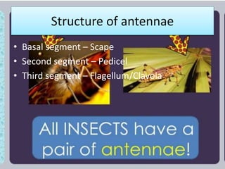
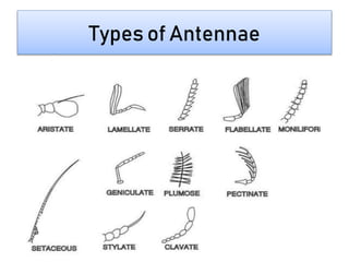
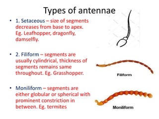
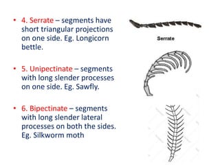
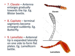
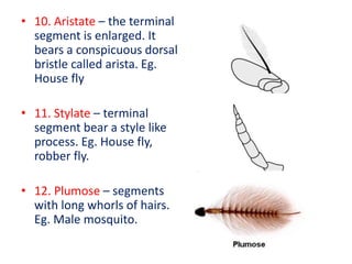
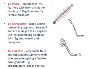
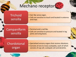
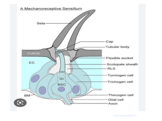
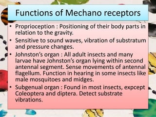
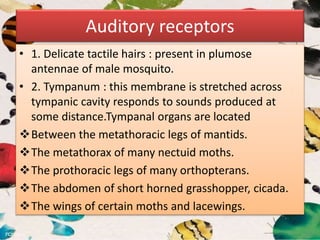
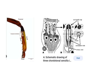
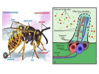
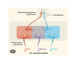
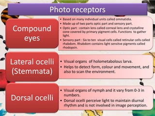
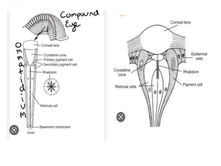
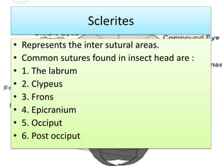
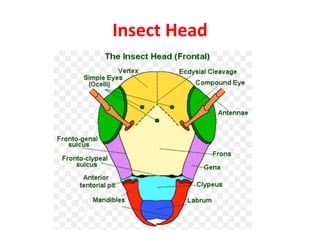
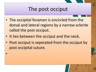
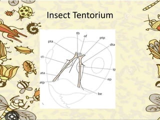
ENTO
501
Insect Morphology
By Durva Santosh Zulpe
1.Types of antennae
2.Functioning
3.Tentorium
4.Sclerite
5.Insect sense organs
Antennae
• Also called as feelers.
• Paired, mobile and segmented.
• Set in a socket of cranium called as antennal
socket.
Structure of antennae
• Basal segment – Scape
• Second segment – Pedicel
• Third segment – Flagellum/Clavola
Functions
• Detect chemicals.
• Perceives smell, humidity changes, temp.,
wind velocity, wind direction.
• Perceives the forward environment and detect
danger.
• Hearing in mosquitoes and communication in
ants.
• Useful to clasp the mate and grasp prey.
Types of Antennae
Types of antennae
• 1. Setaceous – size of segments
decreases from base to apex.
Eg. Leafhopper, dragonfly,
damselfly.
• 2. Filiform – segments are
usually cylindrical, thickness of
segments remains same
throughout. Eg. Grasshopper.
• Moniliform – segments are
either globular or spherical with
prominent constriction in
between. Eg. termites
• 4. Serrate – segments have
short triangular projections
on one side. Eg. Longicorn
bettle.
• 5. Unipectinate – segments
with long slender processes
on one side. Eg. Sawfly.
• 6. Bipectinate – segments
with long slender lateral
processes on both the sides.
Eg. Silkworm moth
• 7. Clavate – Antenna
enlarges gradually
towards the tip. Eg.
Blister beetle.
• 8. Capitate – terminal
segments become
enlarged suddenly. Eg.
Butterfly.
• 9. Lamellate – Antennal
tip is expanded laterally
on one side to form flat
plates. Eg. Lamellicorn
bettle.
• 10. Aristate – the terminal
segment is enlarged. It
bears a conspicuous dorsal
bristle called arista. Eg.
House fly
• 11. Stylate – terminal
segment bear a style like
process. Eg. House fly,
robber fly.
• 12. Plumose – segments
with long whorls of hairs.
Eg. Male mosquito.
• 13. Pilose – antennae is less
feathery with few hairs at the
junction of flagellomeres. Eg.
Female mosquito.
• 14. Geniculate – Scape is long
remanining segments are small
and are arranged at an angle to
the first resembling an elbow
joint. Eg. Ant, weevil and
honeybee.
• 15. Flabellte – very small, third
and subsequent segments with
side processes giving a fan like
arrangements. Eg.
strepsipterans, cedar beetles.
Sense organs
• Sensilla are the organs associated with sensory
perception and develop from epidermal cells. The
different types of sense organs are :
• 1. Mechano receptors
• 2. Auditory receptors
• 3. Chemo receptors
• 4. Thermo receptors
• 5. Photo receptors
Mechano receptors
• Hair like sense organ.
• Cells are sensitive to touch and located in antenna
and tropi.
Trichoid
sensilla
• Terminal end is rod like.
• Cells are sensitive to pressure and located in leg
joints and wing bases.
Campaniform
sensilla
• Specialized sensory organs that receive vibrations.
• Consists of one to many scolopidia, each of which
consists of cap cell, scolopale cell and dendrite.
Chordotonal
organ
Functions of Mechano receptors
• Proprioception : Positioning of their body parts in
relation to the gravity.
• Sensitive to sound waves, vibration of substratum
and pressure changes.
• Johnston’s organ : All adult insects and many
larvae have Johnston’s organ lying within second
antennal segment. Sense movements of antennal
flagellum. Function in hearing in some insects like
male mosquitoes and midges.
• Subgenual organ : Found in most insects, expcept
Coleoptera and diptera. Detect substrate
vibrations.
Auditory receptors
• 1. Delicate tactile hairs : present in plumose
antennae of male mosquito.
• 2. Tympanum : this membrane is stretched across
tympanic cavity responds to sounds produced at
some distance.Tympanal organs are located
Between the metathoracic legs of mantids.
The metathorax of many nectuid moths.
The prothoracic legs of many orthopterans.
The abdomen of short horned grasshopper, cicada.
The wings of certain moths and lacewings.
Chemo receptors
• Detect chemical energy.
• Insect chemoreceptors are sensilla with one pore
or more pores.
• Gustatory receptor : Uniporous chemoreceptors
detect chemical of solid and liquid form by
contact.
• Olfactory receptors : Multiporous
chemoreceptors detect chemicals in vapour form,
at distant by smell.
• Each pore forms a chamber known as pore kettle.
Thermo receptors
• Present in poikilothermic insects and sensitive
to temperature changes.
• In bed bug it is useful to locate the host
utilizing the temperature gradient of the host.
Photo receptors
• Based on many individual units called ommatidia.
• Made up of two parts optic part and sensory part.
• Optic part : contain lens called corneal lens and crystalline
cone covered by primary pigment cells. Functions to gather
light.
• Sensory part : Six to ten visual cells called retinular cells called
rhabdom. Rhabdom contains light sensitve pigments called
rhodopsin.
Compound
eyes
• Visual organs of holometabolous larva.
• Helps to detect form, colour and movement, and
also to scan the environment.
Lateral ocelli
(Stemmata)
• Visual organs of nymph and it vary from 0-3 in
numbers.
• Dorsal ocelli perceive light to maintain diurnal
rhythm and is not involved in image perception.
Dorsal ocelli
Sclerites
• Represents the inter sutural areas.
• Common sutures found in insect head are :
• 1. The labrum
• 2. Clypeus
• 3. Frons
• 4. Epicranium
• 5. Occiput
• 6. Post occiput
Insect Head
The Labrum
• Also known as upper lip in mandibulate
insects.
• Freely attached to lower margin of the clypeus
by the clypeolabral suture.
• Internally lined with the gustatory receptors
and can be moved upwards and downwards.
• The labrum is innervated by compressor
labral, the anterior labral and posterior labral
muscles.
The post occiput
• The occipital foramen is encircled from the
dorsal and lateral regions by a narrow sclerite
called the post occiput.
• It lies between the occiput and the neck.
• Post occiput is seperated from the occiput by
post occipital suture.
•
The genae and subgenal areas
• The genae represents lateral area of the head.
• The genae extend from the compound eye to
the mandibular trochantin on both sides.
• Posterior to the occipital suture each lateral
genae is commonly called as post genae.
• The subgenal areas above and below the
mandibles are called as pleurostoma and
hypostoma resp.
The occular sclerites
• They form cuticular ring around each
compound eyes.
• The antennal sclerites form and annulus at the
base of each antennae.
• The mandibular trochantin occurs between
the mandibular base and the pleurostroma.
Insect Tentorium
Tentorium
• The tentorium is the internal skeleton of the head capsule
of insects.
• In insects, the integument is occasionally invaginated into
the body cavity to form and internal, skeleton support.Such
a structure is termed an apodeme.
• The various apodemes together comprise the
endoskeleton. The tentorium is the endoskeleton of the
insect head and is composed of four parts, viz., the
tentorial bridge, a pair of the anterior and posterior
tentorial arms and the dorsal tentorial arms.
• In addition to these, the tentorium often possesses various
processes arising from the anterior tentorial arm, posterior
arm or tentorial bridge in some Orthoptera.
Durva 17-01-2023.pptx

Durva 17-01-2023.pptx

  • 1.
  • 2.
  • 3.
  • 4.
    Antennae • Also calledas feelers. • Paired, mobile and segmented. • Set in a socket of cranium called as antennal socket.
  • 5.
    Structure of antennae •Basal segment – Scape • Second segment – Pedicel • Third segment – Flagellum/Clavola
  • 6.
    Functions • Detect chemicals. •Perceives smell, humidity changes, temp., wind velocity, wind direction. • Perceives the forward environment and detect danger. • Hearing in mosquitoes and communication in ants. • Useful to clasp the mate and grasp prey.
  • 7.
  • 8.
    Types of antennae •1. Setaceous – size of segments decreases from base to apex. Eg. Leafhopper, dragonfly, damselfly. • 2. Filiform – segments are usually cylindrical, thickness of segments remains same throughout. Eg. Grasshopper. • Moniliform – segments are either globular or spherical with prominent constriction in between. Eg. termites
  • 9.
    • 4. Serrate– segments have short triangular projections on one side. Eg. Longicorn bettle. • 5. Unipectinate – segments with long slender processes on one side. Eg. Sawfly. • 6. Bipectinate – segments with long slender lateral processes on both the sides. Eg. Silkworm moth
  • 10.
    • 7. Clavate– Antenna enlarges gradually towards the tip. Eg. Blister beetle. • 8. Capitate – terminal segments become enlarged suddenly. Eg. Butterfly. • 9. Lamellate – Antennal tip is expanded laterally on one side to form flat plates. Eg. Lamellicorn bettle.
  • 11.
    • 10. Aristate– the terminal segment is enlarged. It bears a conspicuous dorsal bristle called arista. Eg. House fly • 11. Stylate – terminal segment bear a style like process. Eg. House fly, robber fly. • 12. Plumose – segments with long whorls of hairs. Eg. Male mosquito.
  • 12.
    • 13. Pilose– antennae is less feathery with few hairs at the junction of flagellomeres. Eg. Female mosquito. • 14. Geniculate – Scape is long remanining segments are small and are arranged at an angle to the first resembling an elbow joint. Eg. Ant, weevil and honeybee. • 15. Flabellte – very small, third and subsequent segments with side processes giving a fan like arrangements. Eg. strepsipterans, cedar beetles.
  • 13.
    Sense organs • Sensillaare the organs associated with sensory perception and develop from epidermal cells. The different types of sense organs are : • 1. Mechano receptors • 2. Auditory receptors • 3. Chemo receptors • 4. Thermo receptors • 5. Photo receptors
  • 14.
    Mechano receptors • Hairlike sense organ. • Cells are sensitive to touch and located in antenna and tropi. Trichoid sensilla • Terminal end is rod like. • Cells are sensitive to pressure and located in leg joints and wing bases. Campaniform sensilla • Specialized sensory organs that receive vibrations. • Consists of one to many scolopidia, each of which consists of cap cell, scolopale cell and dendrite. Chordotonal organ
  • 16.
    Functions of Mechanoreceptors • Proprioception : Positioning of their body parts in relation to the gravity. • Sensitive to sound waves, vibration of substratum and pressure changes. • Johnston’s organ : All adult insects and many larvae have Johnston’s organ lying within second antennal segment. Sense movements of antennal flagellum. Function in hearing in some insects like male mosquitoes and midges. • Subgenual organ : Found in most insects, expcept Coleoptera and diptera. Detect substrate vibrations.
  • 17.
    Auditory receptors • 1.Delicate tactile hairs : present in plumose antennae of male mosquito. • 2. Tympanum : this membrane is stretched across tympanic cavity responds to sounds produced at some distance.Tympanal organs are located Between the metathoracic legs of mantids. The metathorax of many nectuid moths. The prothoracic legs of many orthopterans. The abdomen of short horned grasshopper, cicada. The wings of certain moths and lacewings.
  • 19.
    Chemo receptors • Detectchemical energy. • Insect chemoreceptors are sensilla with one pore or more pores. • Gustatory receptor : Uniporous chemoreceptors detect chemical of solid and liquid form by contact. • Olfactory receptors : Multiporous chemoreceptors detect chemicals in vapour form, at distant by smell. • Each pore forms a chamber known as pore kettle.
  • 21.
    Thermo receptors • Presentin poikilothermic insects and sensitive to temperature changes. • In bed bug it is useful to locate the host utilizing the temperature gradient of the host.
  • 23.
    Photo receptors • Basedon many individual units called ommatidia. • Made up of two parts optic part and sensory part. • Optic part : contain lens called corneal lens and crystalline cone covered by primary pigment cells. Functions to gather light. • Sensory part : Six to ten visual cells called retinular cells called rhabdom. Rhabdom contains light sensitve pigments called rhodopsin. Compound eyes • Visual organs of holometabolous larva. • Helps to detect form, colour and movement, and also to scan the environment. Lateral ocelli (Stemmata) • Visual organs of nymph and it vary from 0-3 in numbers. • Dorsal ocelli perceive light to maintain diurnal rhythm and is not involved in image perception. Dorsal ocelli
  • 25.
    Sclerites • Represents theinter sutural areas. • Common sutures found in insect head are : • 1. The labrum • 2. Clypeus • 3. Frons • 4. Epicranium • 5. Occiput • 6. Post occiput
  • 26.
  • 27.
    The Labrum • Alsoknown as upper lip in mandibulate insects. • Freely attached to lower margin of the clypeus by the clypeolabral suture. • Internally lined with the gustatory receptors and can be moved upwards and downwards. • The labrum is innervated by compressor labral, the anterior labral and posterior labral muscles.
  • 32.
    The post occiput •The occipital foramen is encircled from the dorsal and lateral regions by a narrow sclerite called the post occiput. • It lies between the occiput and the neck. • Post occiput is seperated from the occiput by post occipital suture. •
  • 33.
    The genae andsubgenal areas • The genae represents lateral area of the head. • The genae extend from the compound eye to the mandibular trochantin on both sides. • Posterior to the occipital suture each lateral genae is commonly called as post genae. • The subgenal areas above and below the mandibles are called as pleurostoma and hypostoma resp.
  • 34.
    The occular sclerites •They form cuticular ring around each compound eyes. • The antennal sclerites form and annulus at the base of each antennae. • The mandibular trochantin occurs between the mandibular base and the pleurostroma.
  • 35.
  • 36.
    Tentorium • The tentoriumis the internal skeleton of the head capsule of insects. • In insects, the integument is occasionally invaginated into the body cavity to form and internal, skeleton support.Such a structure is termed an apodeme. • The various apodemes together comprise the endoskeleton. The tentorium is the endoskeleton of the insect head and is composed of four parts, viz., the tentorial bridge, a pair of the anterior and posterior tentorial arms and the dorsal tentorial arms. • In addition to these, the tentorium often possesses various processes arising from the anterior tentorial arm, posterior arm or tentorial bridge in some Orthoptera.