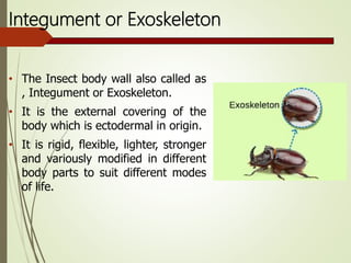
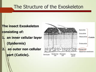
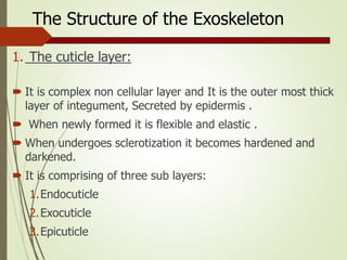
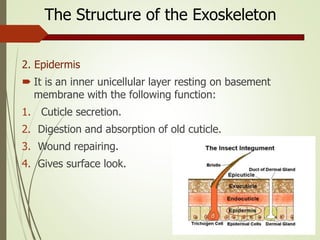
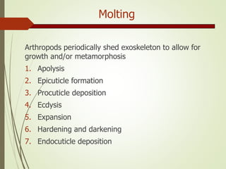
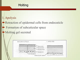
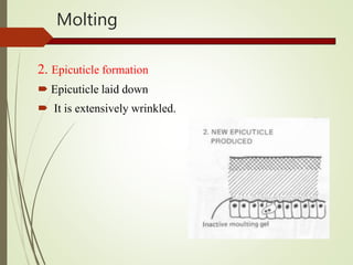
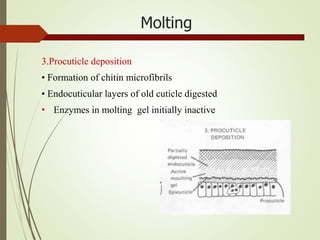
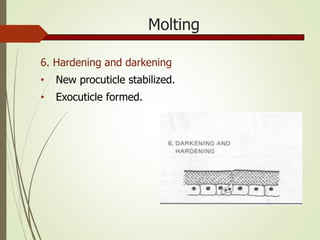
Kingdom of Saudi Arabia
Al Imam Mohammad Ibn Saud Islamic University
College of Science
Department of Biology
Entomology
353 Bio
T. Amani Alsharidah
1441 – 2019
Lecture 3
The Structure and Functions
of Insect Cuticle
(Integument) and Molting
Integument or Exoskeleton
• The Insect body wall also called as
, Integument or Exoskeleton.
• It is the external covering of the
body which is ectodermal in origin.
• It is rigid, flexible, lighter, stronger
and variously modified in different
body parts to suit different modes
of life.
The Structure of the Exoskeleton
The insect Exoskeleton
consisting of:
1. an inner cellular layer
(Epidermis)
2. an outer non cellular
part (Cuticle).
Cuticle
The Structure of the Exoskeleton
1. The cuticle layer:
 It is complex non cellular layer and It is the outer most thick
layer of integument, Secreted by epidermis .
 When newly formed it is flexible and elastic .
 When undergoes sclerotization it becomes hardened and
darkened.
 It is comprising of three sub layers:
1.Endocuticle
2.Exocuticle
3.Epicuticle
The Structure of the Exoskeleton
1. The cuticle layer:
1- Endocuticle
Compared to others it is the inner and thickest layer. This
layer is made up of Chitin and arthropodin. This layer is
colourless, soft and flexible.
1. The cuticle layer:
2. Exocuticle
Outer layer, much thicker with the composition of Chitin
and sclerotin. This layer is dark in colour and rigid.
The Structure of the Exoskeleton
1. The cuticle layer:
3. Epicuticle:
Outer most layer which is very thin. Pore canals present in the
exocuticle helps in the deposition of epiculticle. This layer is
differentiated into the following layers:
a. Inner epicuticle: It contains wax filaments.
b. Outer epicuticle: It makes the contact with cuticulin.
c. Cuticulin : Non chitinous polymerised lipoprotein layer.
d. Wax layer: It contains closely packed wax molecules which
prevents desiccation.
e. Cement layer: Outer most layer formed by lipid and tanned
protein. It protects wax layer. (see the picture slide 9)
The Structure of the Exoskeleton
a. Structure of Exoskeleton
b. Layer of epicuticle
The Structure of the Exoskeleton
2. Epidermis
 It is an inner unicellular layer resting on basement
membrane with the following function:
1. Cuticle secretion.
2. Digestion and absorption of old cuticle.
3. Wound repairing.
4. Gives surface look.
Composition of cuticle
1-Chitin:
It is the main constituent of cuticle. It is water insoluble
but soluble in dilute acids, alkalies and organic solvents.
2- Arthropodin:
An untanned cuticular protein, which is water soluble.
3- Sclerotin:
Tanned cuticular protein, which is water insoluble.
4- Resilin:
An elastic cuticular protein responsible for the flexibility
of sclerites, e.g., wing
Functions of exoskeleton
1. Acts as external armour and strengthen external organs
like jaws and ovipositor.
2. Protects the organs against physical aberation, injurious
chemicals, parasites, predators and pathogen.
3. Internally protects the vital organs, foregut, hindgut and
trachea.
4. Provides space for muscle attachment and gives shape
to the body.
5. Prevents water loss from the body.
6. Cuticular sensory organs helps in sensing the
environment.
7. Cuticular pigments give colour.
Molting
Arthropods periodically shed exoskeleton to allow for
growth and/or metamorphosis
1. Apolysis
2. Epicuticle formation
3. Procuticle deposition
4. Ecdysis
5. Expansion
6. Hardening and darkening
7. Endocuticle deposition
Molting
1. Apolysis
Retraction of epidermal cells from endocuticle
 Formation of subcuticular space
Molting gel secreted
Molting
2. Epicuticle formation
 Epicuticle laid down
 It is extensively wrinkled.
Molting
3.Procuticle deposition
• Formation of chitin microfibrils
• Endocuticular layers of old cuticle digested
• Enzymes in molting gel initially inactive
Molting
4. Ecdysis
• Old cuticle splits along middorsal suture.
• Cast skin = epicuticle and exocuticle.
• Endocuticle recovered and recycled into new
procuticle.
Molting
5-Expansion
• Insect swallows air.
• Insect swells, removes wrinkles. in epicuticle
Molting
6. Hardening and darkening
• New procuticle stabilized.
• Exocuticle formed.
Molting
7. Endocuticle deposition
• Depositing chitin and protein takes time
• Some insects deposit one lamina of endocuticle every 24
hours.
Cuticular Appendages
1. Non-cellular:
Non-cellular appendages have no epidermal association,
but rigidly attached. e.g. minute hairs and thorns.
2. Cellular:
Cellular appendages have eipdermal association.
Types Of Protuberance
 Four basic types of protuberance, all with sclerotized
cuticle, can be recognized on morphological, functional,
and developmental grounds:
1. Spines: are multicellular with undifferentiated epidermal
cells.
2. Setae: also called hairs, macrotrichia, or trichoid
sensilla, are multicellular with specialized cells.
3. Acanthae: are unicellular in origin
4. Microtrichia: are subcellular, with several to many
extensions per cell.
The four basic types of cuticular protuberance: (a) a multicellular
spine; (b) a seta, or trichoid sensillum; (c) acanthae; and (d)
microtrichia.
Color Production
The diverse colors of insects are produced by the inter-
action of light with cuticle and/or underlying cells or fluid by
two different mechanisms:
1- Physical (structural) colors result from light scattering,
interference, and diffraction. All physical colors derive from
the cuticle and its protuberances.
2- Interference colors, such as iridescence and ultraviolet,
are produced by refraction from varyingly spaced, close
reflective layers produced by microfibrillar orientation within
the exocuticle.
Insect pigment
 Insect pigments are produced in three ways:
1. Insect’s metabolism.
2. Sequestering from a plant source.
3. Rarely, by microbial endosymbionts.
 Pigments may be located in the cuticle, epidermis,
hemolymph, or fat body.
 Colors have an array of functions in addition to the obvious
roles of color patterns in sexual and defensive display.

The Structure and Function of Insect.

  • 1.
    Kingdom of SaudiArabia Al Imam Mohammad Ibn Saud Islamic University College of Science Department of Biology Entomology 353 Bio T. Amani Alsharidah 1441 – 2019
  • 2.
    Lecture 3 The Structureand Functions of Insect Cuticle (Integument) and Molting
  • 3.
    Integument or Exoskeleton •The Insect body wall also called as , Integument or Exoskeleton. • It is the external covering of the body which is ectodermal in origin. • It is rigid, flexible, lighter, stronger and variously modified in different body parts to suit different modes of life.
  • 4.
    The Structure ofthe Exoskeleton The insect Exoskeleton consisting of: 1. an inner cellular layer (Epidermis) 2. an outer non cellular part (Cuticle). Cuticle
  • 5.
    The Structure ofthe Exoskeleton 1. The cuticle layer:  It is complex non cellular layer and It is the outer most thick layer of integument, Secreted by epidermis .  When newly formed it is flexible and elastic .  When undergoes sclerotization it becomes hardened and darkened.  It is comprising of three sub layers: 1.Endocuticle 2.Exocuticle 3.Epicuticle
  • 6.
    The Structure ofthe Exoskeleton 1. The cuticle layer: 1- Endocuticle Compared to others it is the inner and thickest layer. This layer is made up of Chitin and arthropodin. This layer is colourless, soft and flexible.
  • 7.
    1. The cuticlelayer: 2. Exocuticle Outer layer, much thicker with the composition of Chitin and sclerotin. This layer is dark in colour and rigid. The Structure of the Exoskeleton
  • 8.
    1. The cuticlelayer: 3. Epicuticle: Outer most layer which is very thin. Pore canals present in the exocuticle helps in the deposition of epiculticle. This layer is differentiated into the following layers: a. Inner epicuticle: It contains wax filaments. b. Outer epicuticle: It makes the contact with cuticulin. c. Cuticulin : Non chitinous polymerised lipoprotein layer. d. Wax layer: It contains closely packed wax molecules which prevents desiccation. e. Cement layer: Outer most layer formed by lipid and tanned protein. It protects wax layer. (see the picture slide 9) The Structure of the Exoskeleton
  • 9.
    a. Structure ofExoskeleton b. Layer of epicuticle
  • 10.
    The Structure ofthe Exoskeleton 2. Epidermis  It is an inner unicellular layer resting on basement membrane with the following function: 1. Cuticle secretion. 2. Digestion and absorption of old cuticle. 3. Wound repairing. 4. Gives surface look.
  • 11.
    Composition of cuticle 1-Chitin: Itis the main constituent of cuticle. It is water insoluble but soluble in dilute acids, alkalies and organic solvents. 2- Arthropodin: An untanned cuticular protein, which is water soluble. 3- Sclerotin: Tanned cuticular protein, which is water insoluble. 4- Resilin: An elastic cuticular protein responsible for the flexibility of sclerites, e.g., wing
  • 12.
    Functions of exoskeleton 1.Acts as external armour and strengthen external organs like jaws and ovipositor. 2. Protects the organs against physical aberation, injurious chemicals, parasites, predators and pathogen. 3. Internally protects the vital organs, foregut, hindgut and trachea. 4. Provides space for muscle attachment and gives shape to the body. 5. Prevents water loss from the body. 6. Cuticular sensory organs helps in sensing the environment. 7. Cuticular pigments give colour.
  • 13.
    Molting Arthropods periodically shedexoskeleton to allow for growth and/or metamorphosis 1. Apolysis 2. Epicuticle formation 3. Procuticle deposition 4. Ecdysis 5. Expansion 6. Hardening and darkening 7. Endocuticle deposition
  • 14.
    Molting 1. Apolysis Retraction ofepidermal cells from endocuticle  Formation of subcuticular space Molting gel secreted
  • 15.
    Molting 2. Epicuticle formation Epicuticle laid down  It is extensively wrinkled.
  • 16.
    Molting 3.Procuticle deposition • Formationof chitin microfibrils • Endocuticular layers of old cuticle digested • Enzymes in molting gel initially inactive
  • 17.
    Molting 4. Ecdysis • Oldcuticle splits along middorsal suture. • Cast skin = epicuticle and exocuticle. • Endocuticle recovered and recycled into new procuticle.
  • 18.
    Molting 5-Expansion • Insect swallowsair. • Insect swells, removes wrinkles. in epicuticle
  • 19.
    Molting 6. Hardening anddarkening • New procuticle stabilized. • Exocuticle formed.
  • 20.
    Molting 7. Endocuticle deposition •Depositing chitin and protein takes time • Some insects deposit one lamina of endocuticle every 24 hours.
  • 21.
    Cuticular Appendages 1. Non-cellular: Non-cellularappendages have no epidermal association, but rigidly attached. e.g. minute hairs and thorns. 2. Cellular: Cellular appendages have eipdermal association.
  • 22.
    Types Of Protuberance Four basic types of protuberance, all with sclerotized cuticle, can be recognized on morphological, functional, and developmental grounds: 1. Spines: are multicellular with undifferentiated epidermal cells. 2. Setae: also called hairs, macrotrichia, or trichoid sensilla, are multicellular with specialized cells. 3. Acanthae: are unicellular in origin 4. Microtrichia: are subcellular, with several to many extensions per cell.
  • 23.
    The four basictypes of cuticular protuberance: (a) a multicellular spine; (b) a seta, or trichoid sensillum; (c) acanthae; and (d) microtrichia.
  • 24.
    Color Production The diversecolors of insects are produced by the inter- action of light with cuticle and/or underlying cells or fluid by two different mechanisms: 1- Physical (structural) colors result from light scattering, interference, and diffraction. All physical colors derive from the cuticle and its protuberances. 2- Interference colors, such as iridescence and ultraviolet, are produced by refraction from varyingly spaced, close reflective layers produced by microfibrillar orientation within the exocuticle.
  • 25.
    Insect pigment  Insectpigments are produced in three ways: 1. Insect’s metabolism. 2. Sequestering from a plant source. 3. Rarely, by microbial endosymbionts.  Pigments may be located in the cuticle, epidermis, hemolymph, or fat body.  Colors have an array of functions in addition to the obvious roles of color patterns in sexual and defensive display.

Editor's Notes

  • #4 Insects are part of a larger group of animals called the arthropods. All insects, arachnids, crustaceans, and centipedes and millipedes belong to the arthropod group. Animals in the group are characterized by a hard exoskeleton and jointed appendages. The hard exoskeleton is made of a substance called chitin and muscles connect to the interior of the shell, rather than to bones as they do in mammals. Along with jointed appendages, arthropods also have segmented bodies. Sometimes people think that slugs, snails, and earthworms are arthropods, but they are not as they lack the exoskeleton.
  • #10 للفهم
  • #15 من شريحة 14 الى 20 ليس من ضمن الفاينل