This document provides information about infective endocarditis:
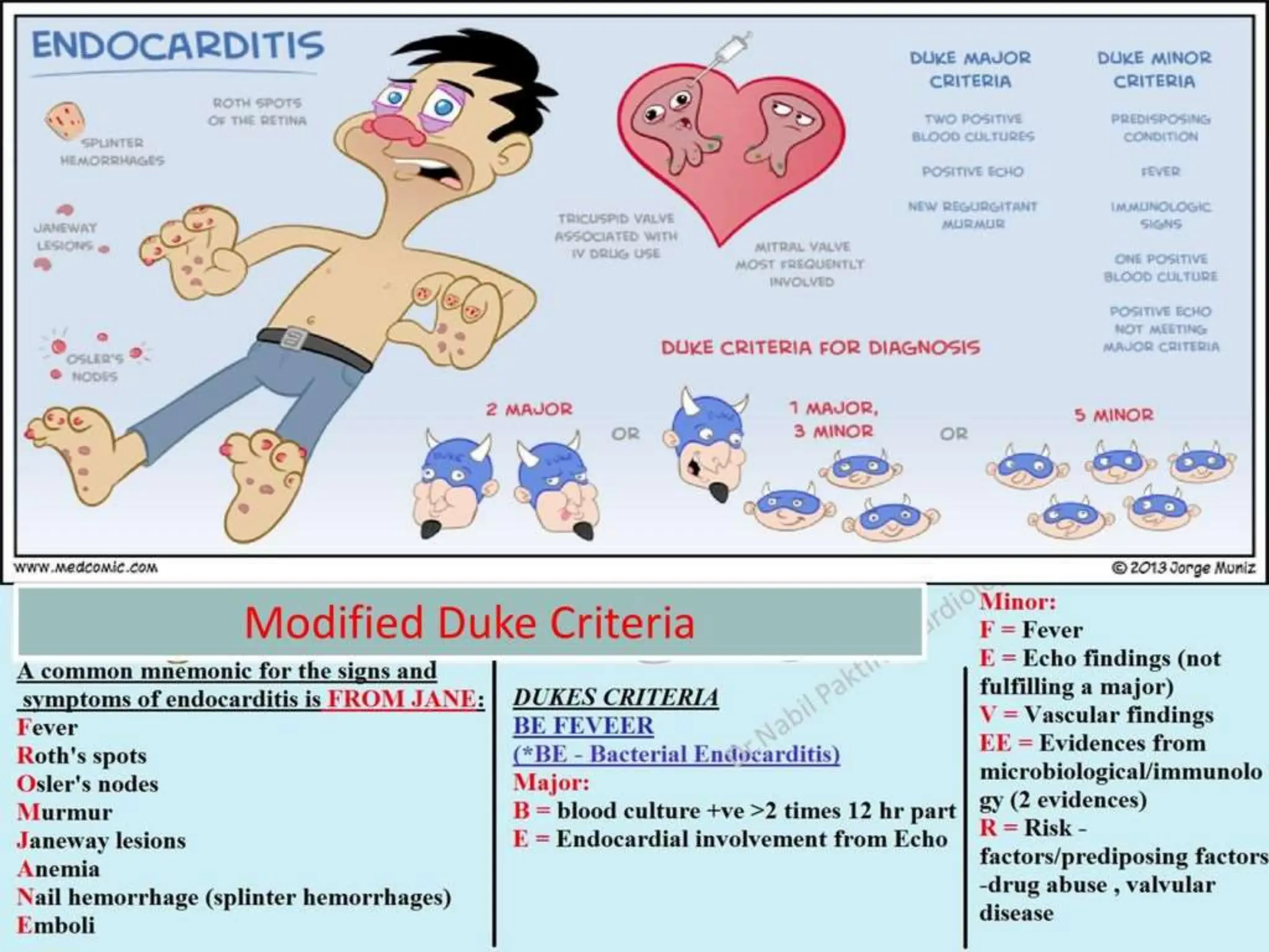
- Infective endocarditis involves infection of the heart valves and inner lining of the heart. Common causes are bacteria entering the bloodstream from dental, respiratory, or other procedures.
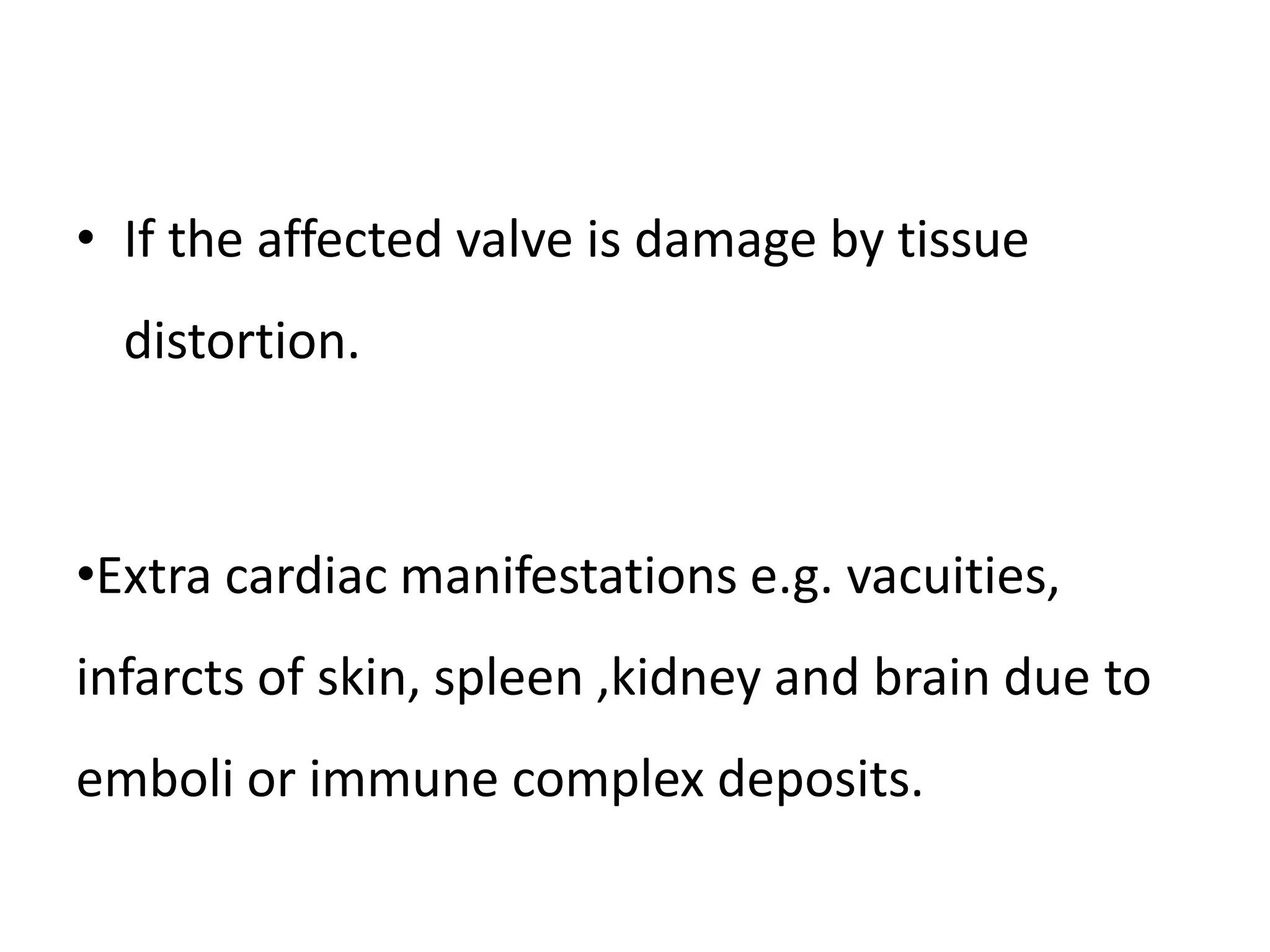
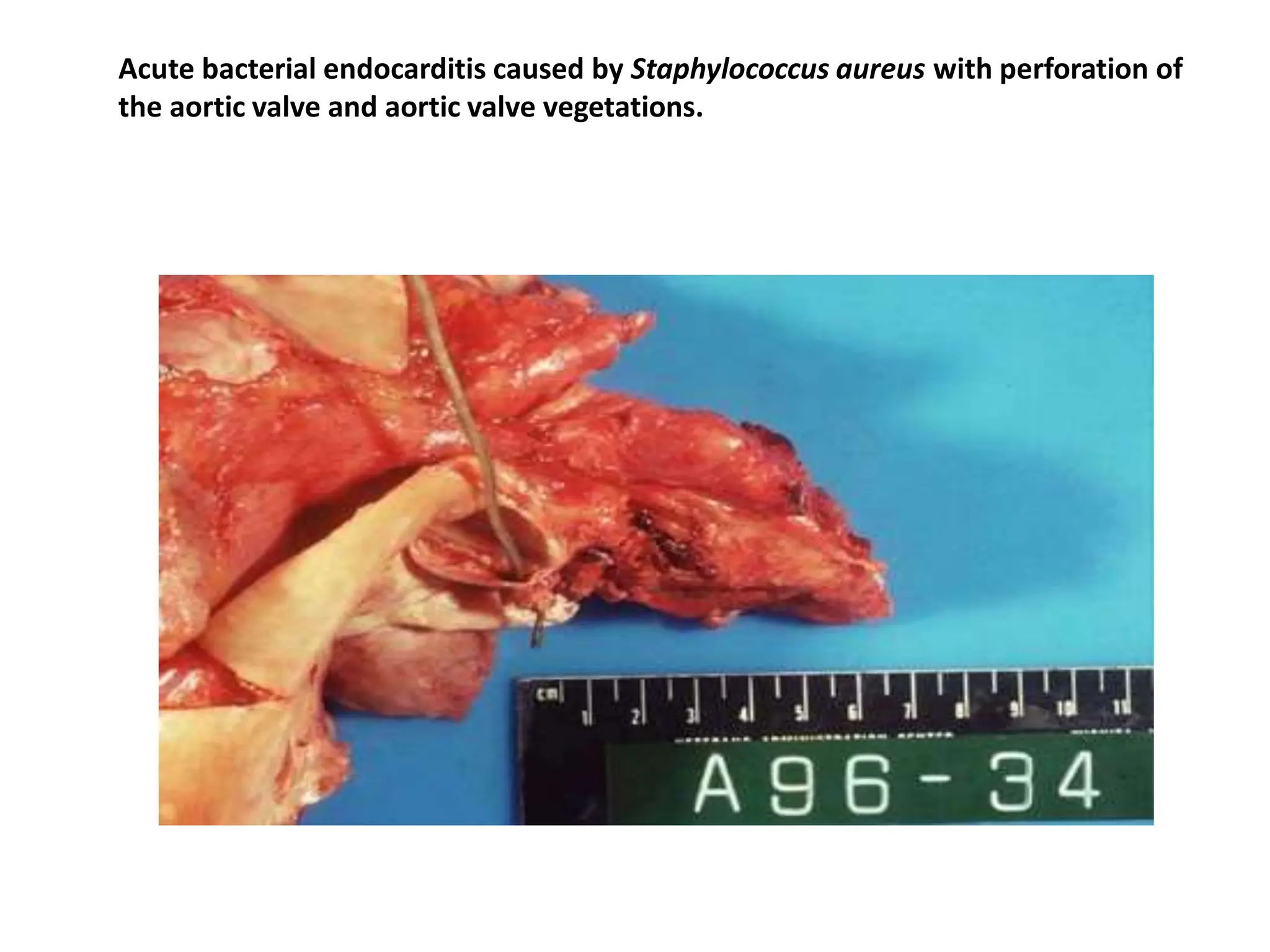
- The infection can cause growths (vegetations) on the heart valves that can break off and block blood vessels in the brain, lungs, kidneys or other organs.
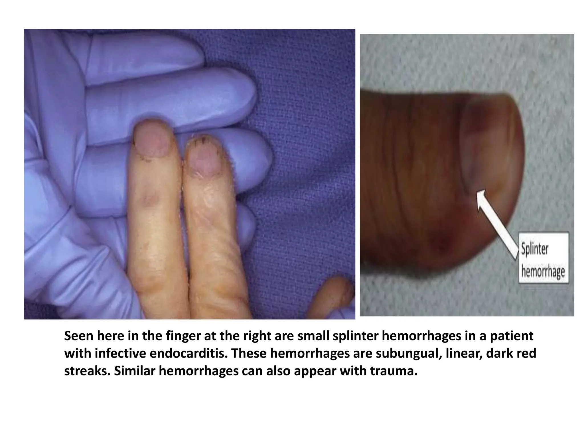
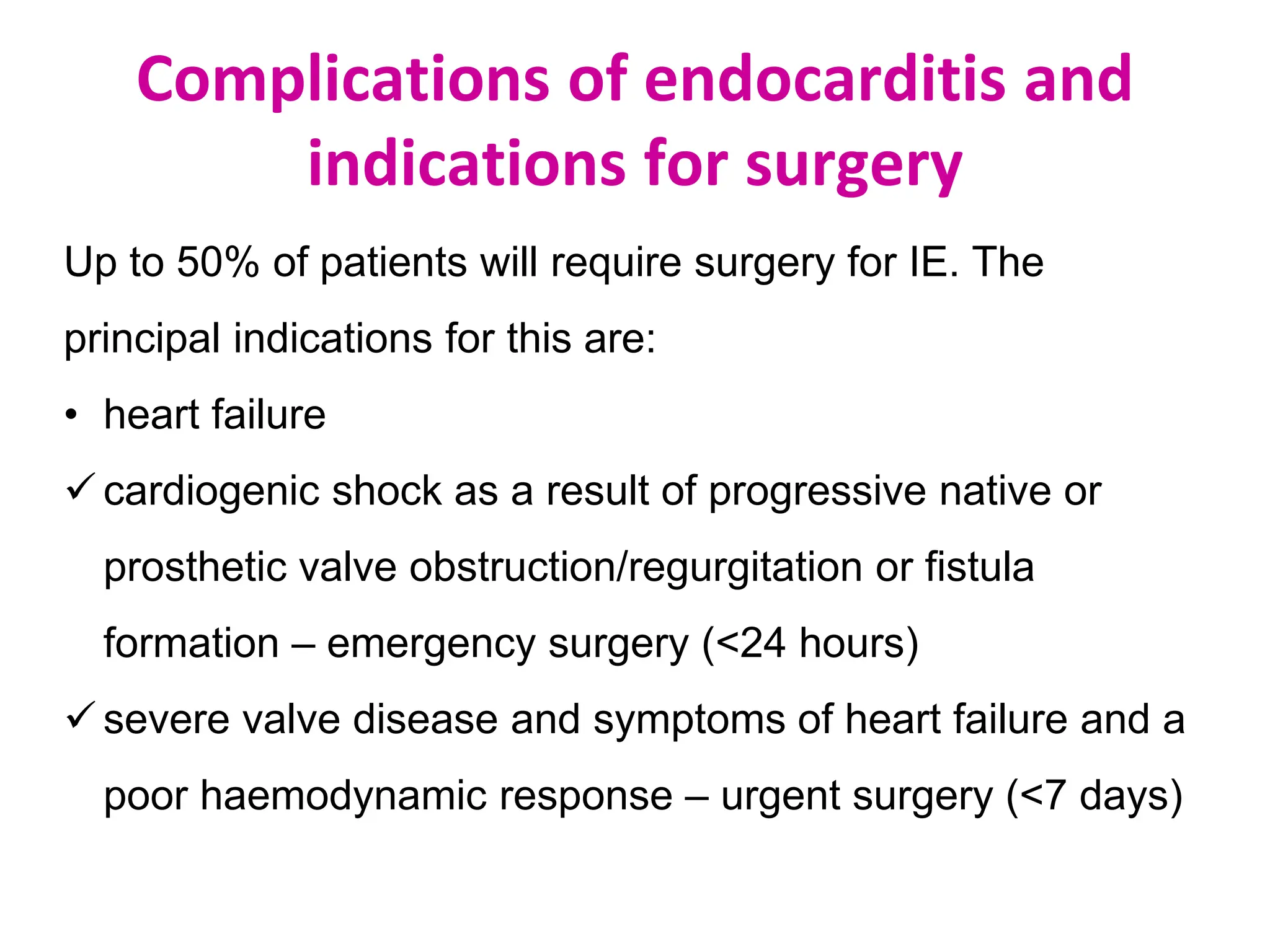
- Risk factors include previous heart damage, dental and surgical procedures, and some reproductive or congenital conditions. Investigations include blood tests, cultures, ECG and echocardiography. Complications may require surgery to repair or replace damaged valves.


![Cont.
Results: Among the 2761 case-patients with definite endocarditis enrolled in ICE-PCS,
49 (1.8%) had endocarditis (20 native valve, 29 prosthetic valve or device) due to non-
HACEK, gram-negative bacilli. Escherichia coli (14 patients [29%]) and Pseudomonas
aeruginosa (11 patients [22%]) were the most common pathogens. Most patients (57%)
with non-HACEK gram-negative bacillus endocarditis had health care-associated
infection, whereas injection drug use was rare (4%). Implanted endovascular devices
were frequently associated with non-HACEK gram-negative bacillus endocarditis
compared with other causes of endocarditis (29% vs. 11%; P < 0.001). The in-hospital
mortality rate of patients with endocarditis due to non-HACEK gram-negative bacilli was
high (24%) despite high rates of cardiac surgery (51%).](https://image.slidesharecdn.com/endocarditis-240212165151-ba740bb4/75/Infective-Endocarditis-IE-Lecture-pptx-37-2048.jpg)


![Infective Endocarditis Group [A7] Seminar](https://cdn.slidesharecdn.com/ss_thumbnails/infectiveendocarditisa7-250627114414-bb3356a5-thumbnail.jpg?width=640&height=640&fit=bounds)


