SlideShare a Scribd company logo
1 of 123
Download to read offline
Demonstration
on
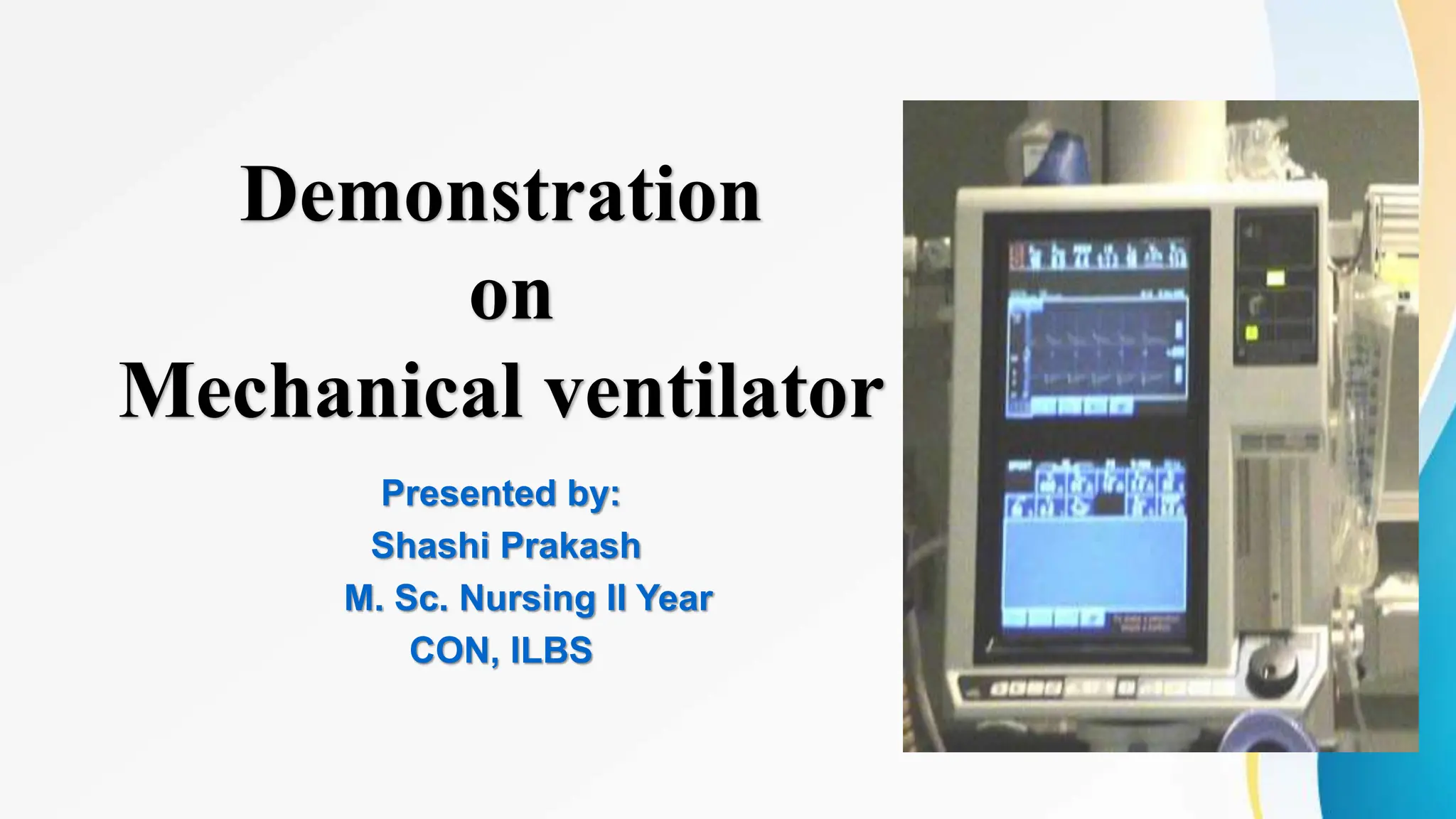
Mechanical ventilator
Presented by:
Shashi Prakash
M. Sc. Nursing II Year
CON, ILBS
Objectives
 Definition of mechanical ventilator
 Purpose of mechanical ventilator
 Indications of mechanical ventilations
 Normal cycle of Respiration
 Lung volumes
 Modes of ventilator Types of mechanical ventilators
 Describe the alarms of mechanical ventilator
 Contraindications of mechanical ventilation
 Complication of mechanical ventilator
 Role of nurses during weaning and care of patient with VAP
DEFINITION
• Mechanical ventilation is the medical term for artificial
ventilation where mechanical means is used to assist or
replace spontaneous breathing
• Mechanical ventilation is ventilation of the lungs by artificial
means usually by a ventilator
PURPOSE
INDICATIONS
1. Acute respiratory failure due to:
• Mechanical failure, includes neuromuscular diseases as Myasthenia
Gravis, Guillian-Barre Syndrome, and poliomyelitis (failure of the
normal respiratory neuromuscular system ).
• Musculoskeletal abnormalities, such as chest wall trauma (flial
chest)
• Infectious diseases, of the lungs such as Pneumonia,Tuberculosis.
CONT...
2. Abnormalities of Pulmonary gas exchage as in:
• Obstructive lung disease in the form of Asthma, Chronic
bronchitis emphysema
• Conditions such as Pulmonary edema, atelectasis
Pulmonary fibrosis.
• Patient who has received general anaesthesia as well as
post cardiac arrest patients often require ventilatory
support until they have recovered from the effects of the
anaesthesia or the insult of an arrest.
Two basic forms of respiratory failure
Hypoxic
• Sao2 < 90% at Fio2 > or 60%
• Examples are acute exacerbation of COPD, severe pneumonia,
and pulmonary edema.
Hypercapnic
• PaCO2 > 50 mm Hg & pH < 7.3
• Hypercapnic respiratory failure includes neuromuscular disease,
such as myasthenia gravis, and other diseases causing
respiratory muscle fatigue, such as asthma and COPD.
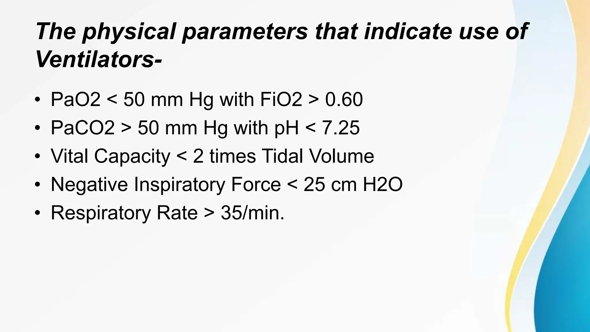
The physical parameters that indicate use of
Ventilators-
• PaO2 < 50 mm Hg with FiO2 > 0.60
• PaCO2 > 50 mm Hg with pH < 7.25
• Vital Capacity < 2 times Tidal Volume
• Negative Inspiratory Force < 25 cm H2O
• Respiratory Rate > 35/min.
NORMAL CYCLE OF RESPIRATION
LUNG CAPACITIES AND VOLUMES
• Tidal volume (TV) – air that moves into and out of the lungs with each breath
(approximately 500 ml)
• Inspiratory reserve volume (IRV) – air that can be inspired forcibly beyond
the tidal volume (2100–3200 ml)
• Expiratory reserve volume (ERV) – air that can be evacuated from the
lungs after a tidal expiration (1000–1200 ml)
• Residual volume (RV) – air left in the lungs after strenuous expiration (1200
ml)
CONT...
• Inspiratory capacity (IC) – total amount of air that can be
inspired after a tidal expiration (IRV + TV)
• Functional residual capacity (FRC) – amount of air remaining in
the lungs after a tidal expiration (RV + ERV)
• Vital capacity (VC) – the total amount of exchangeable air (TV +
IRV + ERV)
• Total lung capacity (TLC) – sum of all lung volumes
(approximately 6000 ml in males)
Types/ classification of ventilators
• According to the method by which they support
ventilation two general categories are-
I. Negative pressure ventilators
II. Positive pressure ventilators
Negative pressure ventilators
I. Negative pressure ventilators
– Exert a negative pressure
– Decrease the intrathoracic pressure
– No tracheostomy or endotracheal tube.
Disadvantages…
– Limits access to the patient by health care providers.
– It is cumbersome and leads to patient discomfort
– Cardiac output tends to decrease from pooling of venous
blood in the lower torso.
Types of negative – pressure ventilators
Iron Lung (drinker respirator tank)
• Body wrap ( pneumo- wrap)
• Chest Cuirass (tortoise shell)
A cuirass, or shell unit, allows
negative pressure to be applied
only to the patient's chest by using
a combination of a form-fitted shell
and a soft bladder.
Positive pressure ventilators
II Positive pressure ventilators
• The positive nature of the pressure causes the gas to
flow into the lungs until the ventilator breath is
terminated. As the airway pressure drops to zero,
elastic recoil of the chest accomplishes passive
exhalation by pushing the tidal volume out.
Classifications of Positive-Pressure
Ventilators
• They are classified by their method of cycling from the
inspiratory phase to the expiratory phase. That is, after
that parameter that signals the termination of the positive-
pressure inspiration cycle of the machine. Include-
• Time-cycled ventilators
• Pressure-cycled ventilators
• Volume-cycled ventilators
Time-cycled ventilators
• Time-cycled ventilation occurs as the Inspiratory phase begins
and gas flows through the ventilator circuit into the patient's lungs
until a timing mechanism in the ventilator reaches a preset
duration.
• Once the preset time is reached, the Inspiratory phase ends and
the patient passively exhale.
• During time-cycled ventilation, tidal volume is not controlled
directly.
Time-cycled ventilators
• tidal volume dependent on gas flow rate and lung
compliance.
• A higher flow rate results in a shorter Inspiratory time and
vice versa.
• As lung compliance decreases, the peak Inspiratory
pressure and potential for alveolar hyperinflation increase.
Pressure-cycled ventilators
• inspiratory phase begins and gas flows through the
ventilator circuit into the patient's lungs until a pressure-
sensing mechanism in the ventilator reaches a preset level.
• Once this level is reached, the inspiratory phase ends and
the patient passively exhale.
Delivery of adequate tidal volume during pressure-cycled
ventilation is determined by the initial cycling pressure
selected, the flow rate (L/min), and lung characteristics,
including both pulmonary compliance and airway
resistance.
Pressure-cycled ventilators
• In the clinical setting, reduced lung compliance and
increased airway resistance shorten the inspiratory
phase, thus lowering the tidal volume delivered.
• Pressure-cycled ventilators are used as portable
ventilators in health care facilities, mobile intensive care
units, and life-flight helicopters.
Volume-cycled ventilators
• In volume-cycled ventilation, the most common form of
ventilation cycling used for adult patients, the signal to
terminate the Inspiratory activity of the machine is a
preset volume.
– variable modes of ventilation
– improved patient-ventilator synchrony
– simplicity of tidal volume adjustment to better facilitate the
ventilation process.
Noninvasive Positive Pressure Ventilation
• Delivered by nasal or face mask, therefore eliminating the
need for endotracheal intubation.
• Includes-
– Bi-level positive airway pressure (BiPAP)
– Continuous Positive Airway Pressure
Bi-level positive airway pressure
Inspiratory positive airway pressure the pressure support
ventilation level
Expiratory positive airway pressure the CPAP
• It integrates inspiratory with expiratory positive airway pressure to
achieve adequate alveolar ventilation.
• Is the mode of choice for NIPPV because it provides continuous
high-flow positive airway pressures that cycle between preset high
and low pressure levels.
• Clinically indicated for patients with chronic stable or slowly
progressing respiratory failure, such as those with COPD plus
severe hypercapnia, acute pulmonary edema, asthma,
hypoxemia, or pneumonia, and those who refuse intubation.
• With BiPAP, two pressures (inspiratory and expiratory) are used.
Bi-level positive airway pressure
• As the patient is inclined to an angle of at least 45 degrees,
inspiratory positive airway pressure is usually started at 8–10 cm
H2O; a mask is placed over the patient's nose or mouth and fitted
for comfort.
• A back-up mandatory ventilator rate is typically set at 4–12
breaths/minute in case the patient experiences apnea.
• Expiratory positive airway pressure is started at 4–6 cm H2O,
which can be increased in increments of 2–3 cm H2O for patients
experiencing hypoxia.
• Oxygen may be titrated into the circuit at 0.5–6 L/minute to
alleviate hypoxemia.
Continuous Positive Airway Pressure
• Continuous positive airway pressure is used for patients with
obstructive sleep apnea and heart failure with respiratory muscle
weakness. It is usually started at 2.5–15 cm H2O, based on
ventilatory goals. Oxygen may be titrated into the circuit at 0.5–6
L/minute to alleviate hypoxemia.
• Traditional weaning mode
• Both CPAP and BiPAP devices have limitations, including poor
patient compliance due to discomfort of the device.
Diaphragm Pacing
• An electronic pacer stimulates the diaphragm to contract, thus
assisting breathing by "bellows" motion of the diaphragm.
• This method is used by patients who have high (C1-C2) spinal
cord injury, and with tracheostomy in some children who cannot
breathe spontaneously because of a problem with central control
of breathing.
Contra indications of Noninvasive Positive
Pressure Ventilation
• Patients who have experienced respiratory arrest
• Serious dysrhythmias
• Cognitive impairment
• Head or facial trauma
Articles
• Laryngoscope
• Endotracheal tube
• Stylet
• Syringe
• Suction catheter
• Carbon dioxide detector
• Oral airway
• Nasal airway
cont....
• Bag-valve-mask
• Nasal cannula
• Stethoscope
• Magills forcep
• Ambu bag
• with reserviour
• HME filter
• Catheter mount
• Ventilator circuit
Ventilator Modes
Traditional Modes of Invasive Mechanical Ventilation
• Traditional modes of invasive mechanical ventilation use volume, pressure,
and time to cycle from the inspiratory to the expiratory phase and can be
administered as full or partial ventilator support.
• Full support provides the patient with adequate ventilatory requirements to
meet metabolic demands without supplementation by the patient.
• Partial support provides partial ventilator assistance but requires patients to
actively participate in their own spontaneous ventilation.
Control Mode Ventilation
• Control mode ventilation (CMV) is a time-cycling process used primarily with
patients unable or not required to generate a voluntary respiratory effort.
• The timing mechanism generates the inspiratory tidal volume breath
independent of the patient's respiratory effort.
• the ventilator does not allow the patient to self-generate a tidal volume
breath.
• Patients waking from sedation or pharmacologic paralysis may experience
agitation and "air hunger" since they cannot interface with the ventilator.
Assist Control Ventilation
• During assist control ventilation (A/C) the patient receives a predetermined
mechanical respiratory rate and tidal volume, as with CMV, but is able to self-
generate additional tidal volume breaths.
• Negative pressure starts the inspiratory breath. By tradition, a pressure of -2
cm H2O is selected.
• Patients who have apnea or are unable to self-generate a tidal volume breath
are guaranteed the predetermined mechanical respiratory rate and tidal
volume.
• Control mode and A/C ventilation are differentiated only by the preset
triggering threshold for A/C ventilation.
• Assist control is highly successful as a primary ventilator mode but has
limitations.
– The ventilator delivers a preset tidal volume breath and respiratory rate, resulting in an
expired minute ventilation (e.g., tidal volume 500 ml x 14 breaths/min = 7 L/min).
– Patients can also self-generate additional tidal volume breaths (e.g., tidal volume 500 ml
x 10 breaths/min = 5 L/min).
– The result is hyperventilation and possible respiratory alkalosis due to excessive minute
ventilation (e.g., 500 ml x 24 breaths/min = 12 L/min).
Synchronized Intermittent Mandatory
Ventilation
• Synchronized intermittent mandatory ventilation is almost identical to IMV;
the difference is that the patient's own spontaneous breathing pattern is
synchronous with the SIMV rate.
• Machine breaths plus self-generated additional breaths equal the total
respiratory rate (e.g., a SIMV preset rate of 8 breaths/min and 4 self-
generated spontaneous breaths would be documented as 8/12).
• This prevents the "stacking"
• Synchronized intermittent mandatory ventilation, like A/C ventilation, is used
primarily as an initial ventilator mode.
Synchronized Intermittent Mandatory
Ventilation
Pressure Control Ventilation
• Pressure control ventilation is a pressure-limiting, time-cycled mode in which a constant
pressure is maintained throughout the preset inspiratory time.
• As gas flows through the ventilator circuit and into the patient's lungs, the airway pressure
rises, resulting in increased alveolar volume. The outcome is that delivered airway pressure
equals intrapulmonary pressure, ending the inspiratory phase.
• Clinicians often change the ventilation mode from volume controlled (CMV, A/C, or SIMV) to
pressure control ventilation if adequate ventilation or oxygenation goals are not achieved, or
if excessive peak airway pressures are required to optimize gas exchange.
• Patients with acute lung injury or acute respiratory distress syndrome often require pressure
control ventilation, primarily due to difficulties in optimizing ventilation and oxygenation goals
with conventional volume-controlled ventilation.
Pressure Support Ventilation
• Pressure support ventilation provides preset pressure assistance during each
spontaneous patient breath to overcome airway resistance of the
endotracheal tube and dead space of the ventilator circuit to decrease the
patient work of breathing.
• Pressure support ventilation is used as a tool for ventilator weaning since it
requires full spontaneous respiratory effort by the patient.
• Pressure support ventilation is widely used in the critical care setting in
combination with other modes, such as SIMV. This combination provides
mandated mechanical ventilator breaths from SIMV augmented with pressure
support ventilation during spontaneous breathing by the patient.
volume pressure curve for-
low lung volume and stiff non-compliant lung
Normal lung
pressure support ventilation
Continuous Positive Airway Pressure
• Continuous positive airway pressure (CPAP) allows for constant pressure
maintained above atmospheric pressure throughout the respiratory cycle.
• A CPAP of 5 cm H2O is traditionally started to maintain pressure above
atmospheric pressure.
• Continuous positive airway pressure can be provided alone or in
combination with pressure support ventilation.
• Continuous positive airway pressure by itself or in combination with pressure
support ventilation is the primary mode used to promote spontaneous
ventilation to assist with discontinuation of mechanical ventilation.
Inverse ratio ventilation ( IRV)
• proportion of inspiratory to expiratory time is greater than 1:2
• Can be initiated using pressure controlled breaths (PC- IRV) or volume
controlled breaths ( VC-IRV)
• IRV is used in clients with hypoxemia refractory to PEEP. the longer
inspiratory time increases functional residual capacity and improves
oxygenation by opening collapsed alveoli, and the shorter expiratory time
induces auto- PEEP that prevent alveoli from re-collapsing.
– Increase intra- thoracic pressure can result in excessive air trapping and
– decrease cardiac output.
High frequency ventilation (HFV)
• delivers a small volume of gas at rapid rate.
• HFPPV ( high frequency positive pressure ventilation) delivers 60-
100 breaths / minute.
• High frequency jet ventilation ( HFJV):
– delivers 100-600 cycles / minute.
• High frequency oscillation (HFO)
– delivers 900- 3000 cycles / minute
High frequency ventilation (HFV)
• Compromised hemodynamic stability
• Bronco pulmonary fistula
• During short term procedures
• Diseases that create risk of volutrauma.
– sedation and/ or pharmacological paralysis required.
– assessment of breath sounds is difficult.
Independent lung ventilation (ILV)
• each lung is ventilated separately.
• ILV s used in patients with unilateral lung disease,
broncho pleural fistulas, and B/L asymmetric lung
disease.
– Requires a double lumen endotracheal tube,
– Two ventilators,
– Sedation and/ or pharmacological paralysis.
Proportional assist ventilation (PAV)-
• It provides partial ventilatory support in which the ventilator generates
pressure in proportion to the patients ventilatory efforts.
• With every breath, the ventilator synchronizes with the patients ventilatory
efforts.
• The more inspiratory pressure the patient generates, the more pressure the
ventilator generates, amplifying the patients inspiratory effort without any
specific preselected target pressure or volume.
• It generally adds “additional muscle” to the patients effort: the depth and
frequency of breath are controlled by the patient.
Volume assured pressure support
• This mode has been called pressure augmentation according to various manufacturers.
• This mode can switch from pressure control to volume control within a single specific breath
cycle.
• After a breath is triggered, rapid and variable flow creates pressure to reach the set level of
pressure support.
• The tidal volume that is delivered from the machine is monitored. If the tidal volume equals
the minimum set tidal volume, the patient receives a typical pressure-supported breath,
which makes this mode essentially like volume support.
• However, if the tidal volume is less than the set tidal volume, the ventilator switches to a
volume-controlled breath with constant flow rate until the set tidal volume is reached.
Ventilator settings
• A variety of settings on the ventilator allows the
ventilator parameters to be individualized to patients
needs and the mode of ventilation selected.
Settings
• PEEP
– The purpose of peep is to expand collapsed small airways and atelectic
alveoli, thus improving alveolar ventilation.
• PEEP is generally administered at pressure not exceeding 7-10
cm H2O.
• PEEP up to 20 cm H2O may occasionally be given in some ARDS
patients with very low compliance and severe respiratory
insufficiency.
Optimal PEEP is characterized by-
• A decrease in the percentage of shunt
• An increase in FRC
• No significant increase in PCWP as monitored by swan
Ganz catheter.
• Maintenance of BP
• increase in Pao2
• increase in lung compliance
• an improvement in serial cardiac outputs.
Settings
• RATE
– Usually set around 10-14 / min.
• TIDAL VOLUME
– 10- 15 cc/kg
• PEAK PRESSURE
– Preferably not to exceed 35 cm H2o
• Sensitivity
– 2 cm H2O Inspiratory Force
Settings
• SIGH VOLUME
– Usually 1.5 times the tidal volume, 1-3 times / hour.
• CIRCUIT TEMPERATURE
– The inspiratory air temperature should not exceed 39 degree Celsius at
the point of entry into the patient.
• Fio2
– Titrated to the lowest required for normal pao2 ( 80-100 mm Hg )
• Minute volume
– Tidal volume x respiratory rate, usually 6-8 L/ min
Complications of Mechanical Ventilation
• Volutrauma
• Barotrauma
• Oxygen toxicity
• Ventilator-associated pneumonia
• Intrinsic PEEP or auto-PEEP
• Sinusitis
• Gastrointestinal Complications
• Cardiovascular Effects
Problems with mechanical ventilation
• Increase in peak airway pressure
It can be a result of coughing or plugged airway tube, patient
“bucking” ventilator, decreasing lung compliance, tubing kinked,
pneumothorax, atelectasis or bronchospasm.
• Decrease in pressure or loss of volume
It can result due to increase in compliance or leak in ventilator or
tubing; cuff on tube/ humidifier not tight.
TROUBLESHOOTING ALARMS
• HIGH CONTINOUS PRESSURE
• Possible Cause-
– airway pressure higher than set PEEP plus 15 cm H2O for
more than 15 seconds.
• Remedy-
– check client
– check circuit
– check ventilator settings and alarm limits
TROUBLESHOOTING ALARMS
• CHECK TUBING
• Possible Cause
– disconnected pressure transducer ( expiratory)
– blocked pressure transducer
– water in expiratory limb of ventilator
– wet bacterial filter
– clogged bacterial filter
Remedy-
– check ventilator internals on expiratory side
– check heated wires in humidifier if present
– refer to service
– replace filter
– remove water from tubing and check humidifier settings ( relative humidity)
TROUBLESHOOTING ALARMS
• AIRWAY PRESSURE TOO HIGH
• Possible Cause-
– kinked or blocked client tubing
– mucus or secretion plug in ET tube or in airway
– client coughing or fighting ventilator.
– inspiratory flow rate too high
– improper alarm setting
• Remedy-
– check client
– check ventilator settings and alarm limits.
TROUBLESHOOTING ALARMS
• LIMITED PRESSURE (alarm active only in PRVC and VS mode)
• Possible Cause-
– kinked or blocked client tubing
– mucus or secretion plug in ET tube or in airway
– client coughing or fighting ventilator.
– improper alarm setting
– client's lung/ thorax compliance decreasing
– client's airway resistance increasing
• Remedy-
– check client
– check ventilator settings and alarm limits.
TROUBLESHOOTING ALARMS
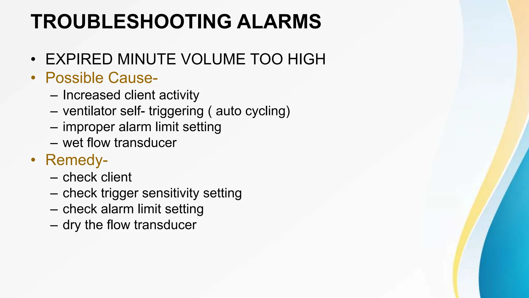
• EXPIRED MINUTE VOLUME TOO HIGH
• Possible Cause-
– Increased client activity
– ventilator self- triggering ( auto cycling)
– improper alarm limit setting
– wet flow transducer
• Remedy-
– check client
– check trigger sensitivity setting
– check alarm limit setting
– dry the flow transducer
TROUBLESHOOTING ALARMS
• EXPIRED MINUTE VOLUME TOO LOW
• Possible Cause-
– low spontaneous client breathing activity
– leakage in cuff
– leakage in client circuit
– improper alarm limit setting
• Remedy-
– check client
– check cuff pressure
– check client circuit ( perform leakage test if necessary)
– check pause time and graphics to verify
– consider more ventilatory support for client
TROUBLESHOOTING ALARMS
• EXPIRED MINUTE VOLUME DISPLAY READS ZERO
• Possible Cause-
– flow transducer faulty
– circuit disconnected from patient.
• Remedy-
– replace flow transducer
– connect Y- piece to client
TROUBLESHOOTING ALARMS
• APNEA ALARM
• Possible Cause-
– time between two consecutive inspiratoey efforts exceeds……
– adult: 20 seconds
– pediatric: 15 seconds
– Neonate : 10 seconds
• Remedy-
– check client
– check ventilator settings
TROUBLESHOOTING ALARMS
• PEEP/ CPAP AND/ OR PLATEAU PRESSURE FAILS TO BE MAINTAINED.
• Possible Cause-
– leakage in cuff
– leakage in client circuit
– improper alarm limit setting
• Remedy-
– check cuff pressure
– check client circuit ( perform leakage test if necessary)
– check pause time and graphics to verify
– consider more ventilatory support for client
Weaning
• Respiratory weaning, the process of withdrawing the patient from
dependence on the ventilator, takes place in three stages: the
patient is gradually removed from the ventilator, then from the
tune, and finally from oxygen. Weaning from mechanical
ventilation is performed at the earliest possible time consistent
with patient safety.
Weaning Criteria
• weaning can be done if client tolerates spontaneous
breathing without undue anxiety or inadequate
cardiopulmonary function.
• The following indicate readiness for weaning-
Weaning Criteria
• Fio2 < 50%
• PEEP of < 5 CM H2O
• Respiratory Rate < 30/ minute
• Minute Volume < 10 liter / minute
• Static compliance > 25 – 30 cm
H2O
• Max inspiratory pressure < -20 cm
H2O
• Vital Capacity > 10 -15 ml /kg
• Spontaneous Tidal Volume > 4 -5
ml / kg
• Max voluntary ventilation> twice
minute ventilation
• Alveolar arterial O2 tension
difference < 350 torr on an FiO2 of
100%
• Shunt < 15% - 20%
• PaCo2 within normal limit
• PaO2 > 60% on an FiO2 of < 50%
Weaning include….
• Weaning from ventilator
• Weaning from the tube
• Weaning from oxygen
Weaning from ventilator
• Assist-control ventilation
• SIMV
• PAV
• CPAP
• When the patient can breath spontaneously, weaning trials using
a T-Piece are conducted with the patient disconnected from the
ventilator, receiving humidified oxygen only and performing all
work of breathing
Weaning from ventilator
• During weaning, the patient is maintained on the same or a higher oxygen
concentration than when receiving mechanical ventilation.
• While the patient is using the T- piece, he or she is observed for signs and
symptoms of hypoxia, increasing respiratory muscle fatigue or systemic
fatigue.
• These include
– restlessness
– increased respiratory rate
– use of accessory muscles
– tachycardia with PVC and
– paradoxical chest movements.
Weaning from ventilator
• If clinically stable the patient usually can be extubated within 2 or
3 hours after weaning and allowed spontaneous ventilation by
means of a mask with humidified oxygen.
• Patients who have had prolonged ventilatory assistance usually
require more gradual weaning. It may take days or even weeks.
– They are weaned primarily during the day and placed back on
the ventilator at night to rest.
Weaning from ventilator
• Because, patients respond in different manners to the various weaning
methods there is no definite way to assess which method is best.
• ongoing assessment of respiratory status is essential to monitor patient
progress.
• These patients still have borderline pulmonary function and need vigorous
supportive therapy before their respiratory status returns to a level that
supports activities of daily living.
supplemented by intensive pulmonary care
• Oxygen therapy
• Arterial blood gas evaluation
• Pulse oxymetry
• Broncodilator therapy
• CPT
• Adequate nutrition, hydration and humidification
• Incentive spirometry
Weaning from the tube
• Weaning from the tube is considered when the patient can
breathe spontaneously, maintain an adequate airway by
effectively coughing up secretions, swallow and move the jaw.
• If frequent suctioning is needed to clear secretion, the tube
weaning may be unsuccessful.
• Secretions clearance and aspiration risk are assessed to
determine weather active pharyngeal and laryngeal reflexes are
intact.
Weaning from the tube
• Once the patient can clear secretions adequately, a trial period of
mouth breathing or nose breathing is conducted. This can be
accomplished by several methods.
• The first method requires changing to a smaller size tube to
increase the resistance to air flow or plugging the tracheostomy
tube. The smaller tube is sometimes replaced by a cuffless
tracheostomy tube, which allows the tube to be plugged at
lengthening intervals to monitor patient progress.
Fenestrated Tube
• A second method involves
changing to a fenestrated
tube. This permits air to flow
around and through the tube
to the upper airway and
enables talking.
Tracheostomy Button
• A third method involves
switching to a smaller
tracheostomy button. A
tracheostomy button is a plastic
tube approximately one inch long
that helps keep the wind pipe
open after the larger
tracheostomy has been removed.
Weaning from the tube
• Finally, when the patient demonstrates the ability to
maintain a patent airway, the tube can be removed.
• An occlusive dressing is placed over the stoma, which
heals in several days to weeks.
Weaning from oxygen
• The patient who has been successfully been weaned from the
ventilator, cuff and tube and has adequate respiratory function is
then weaned from oxygen.
• The Fio2 is gradually reduced until the Pao2 is in the range of 70
– 100 mm Hg.
• While the patient is breathing room air, if the Pao2 is less than 70
mm Hg on room air, supplemental oxygen Is recommended.
Complications of weaning
• Complications of weaning include-
– CO2 retention
– hypoxemia
– cardiovascular instability.
Do not attempt weaning in-
• A client who has too little strength to inspire.
• Hypovolemia
• Life threatening dysrhythmias
• A low cardiac index
Do not attempt weaning in
• An increased oxygen consumption
• Uncorrected acidosis or alkalosis
• Severe catabolism
• A Vd/ Vt ratio of 60% or greater.
– Normal is 30
• When patients with COPD are weaned their PaO2 and
PaCo2 should be as close to their normal levels as
possible.
Intolerance Indicators
• Increase or decrease of heart rate by 20 beats,
respiratory rate by 10 breaths or blood pressure by 20
mm Hg from baseline. ( or a difference of 10% from
baseline)
• Tidal volume of less than 300ml
• PaO2 less than 60 mm Hg.
• PaCo2 more than 55 mm of Hg.
Intolerance Indicators
• Ph less than 7.35
• Spo2 less than 90%
• Increase in minute ventilation more than 5 liter/ minute.
Failure to wean a patient from ventilator is usually
caused by
• An increase in the alveolar ventilation requirement in the presence
of an increase in Vd/ Vt ratio or an increase in the Paco2 level.
• A decrease in muscle strength secondary to metabolic wasting,
inadequate nutrition, or respiratory disco-ordination.
• An increase in the work of breathing after a decrease in lung
compliance or an increase in airway resistance due to
bronchospasm or mucous obstruction or narrowing of the air
conduits with edema.
Nursing Management of a Client on Ventilator
• Nursing care of the mechanically ventilated patient
requires expert technical and interpersonal skills.
• Two general nursing interventions important are-
– Pulmonary auscultation and
– Interpretation of ABG measurements.
Impaired gas exchange and ineffective breathing pattern R/T
underlying disease process and artificial airway and ventilator
system.
• The alteration in gas exchange may be caused by the underlying illness or by mechanical factors related to
adjustment of the machine to the patient.
• Assess the patient for adequate gas exchange, signs and symptoms of hypoxia and response to treatment.
• Nursing interventions to promote optimal gas exchange include judicious administration of analgesic
agents to relive pain without suppressing the respiratory drive and frequent repositioning to diminish the
pulmonary effects of immobility.
• The nurse also monitor s for adequate fluid balance by assessing for presence of peripheral edema,
calculating daily initial and output and monitoring daily weight.
• The nurse administers medications prescribed to control the primary disease and monitors for their side-
effect
Enhancing gas exchange
Ineffective airway clearance R/T inability to cough and
stimulation of increased secretion formation in the
lower tracheobronchial tree from the ET tube.
• Continuous positive pressure ventilation increases the production of secretions regardless of the
patients underlying condition.
• The nurse assess for the presence of secretions by lung auscultation at least every 2-4 hours.
Measures to clear the airway of secretions include suctioning, chest physiotherapy, frequent position
changes and increased mobility as soon as possible. Frequency of suctioning should be determined by
patient assessment.
• Humidification of the airway via the ventilator is maintained to help liquefy secretions so that they are
more easily removed.
Promoting effective airway clearance
• Bronco dilatators administered to dilate the bronchioles and are classified as adrenergic or
anti- cholinergic. Adrenergic are mostly inhaled and work while stimulating the beta receptor
sites, mimicking the effects of epinephrine in the body. The desired effect is smooth muscle
relaxation, which dilates the constricted bronchial tube. Medications include albuterol,
isoproterenol, metaproterenol, salmeterol and terbutalin.
• Tachycardia, heart palpitations and tremors have been reported with use of these
medications. Anti- cholinergic bronco dilatators such as ipratropium and ipratropium with
albuterol produce airway relaxation by blocking cholinergic induced bronco-constriction.
Promoting effective airway clearance
• Patients receiving bronco-dilators therapy of either type should be monitored
for adverse effects, including dizziness, nausea, decreased oxygen
saturation, hypokalemia, increased heart rate and urine retention.
• Mucolytic agents’ acetylcysteine (mucomyst) are administered to liquefy
secretions so that they are more easily mobilized.
• Nursing management of patients receiving mucolytic therapy includes
assessment of adequate cough reflex, sputum characteristic and
improvement in incentive spirometry.
• Side – effect include nausea, vomiting, bronchospasm, stomatitis, urticaria
and rhinorrhea.
Risk for infection R/T impaired defenses in respiratory
tract
• Position the ventilator tubing so that there is minimal pulling or distortion of the tube in the
trachea, reducing the risk of trauma to the trachea. Cuff pressure is monitored every 6-8
hours to maintain pressure at less than 25 mmHg.
• Tracheostomy care is performed every 8 hours.
• Ventilator circuit tubing and inline suction tubing to be replaced periodically.
• Administer oral hygiene frequently as it is the primary source of contamination of lungs in the
intubated client
• Position the patient with head elevated above the stomach as much as possible to decrease
incidence of aspiration pneumonia.
Preventing infection
Risk for infection R/T impaired defenses in
respiratory tract
• Evaluate color, amount, consistency, and odor of sputum with
each suctioning.
• Collect specimens for culture and sensitivity, as indicated.
• Maintain sterile technique when suctioning.
• Change ventilator tubing q24-72h.
• Perform oral hygiene every shift.
• Monitor vital signs for indications of infection.
• Drain condensed water in ventillator tubing externally away
from airway & humidifier reservoir.
• Wash hands frequently.
• Follow universal precautions.
HIGH RISK TO DEVELOP INFECTION
Due to
• CENTRAL LINE
• ET Tubing , NG tubing.
• Catheters
• Drainages
• Wound sites
SOURCES OF INFECTION IN ICU
• Change every 72 hours (tubing, exhalation valve, and
humidifiers.
• Periodically drain and discard any condensate that
collects in the ventilator tubing's.
• Sterilize reusable breathing circuit between use on
different patients.
• Use sterile water to fill humidifiers.
Endotracheal suctioning
• Aseptic technique should be adhered to during suctioning.
• Use single use sterile suction catheters separately for oral
and ET suction.
• While disconnecting ET tube from circuit , keep the open
end of angle connection on a sterile pad.
• Change angle connection and corrugated tubing every 24
hrs. or when visibly soiled.
CENTRAL VENOUS CANNULATION
• Asepsis should be maintained during infusion and flushing.
• Do not keep the cap cover unsterile area and care should be
taken not to touch the middle part of the cap.
• Replace the gauze dressing after every 24 hrs.
• Fluids and drugs should not be given through the TPN line.
• Try to keep the IV set and the cannula as a closed system.
Repeat connections and disconnections should be avoided.
URINARY CATHETERIZATION
• Avoid raising the level of bag to avoid backflow.
• Hang the urine bag to the hook attached.
• Do not let the bag touch the floor.
• Unnecessary uncorking or disconnecting the bag
should not be done.
• Do not change the catheters routinely unless there is
definitive evidence of infection.
PERIPHERAL IV CANNULATION /BLOOD/ SAMPLING
• Do not rub the vein in the hope of facilitating the flow.
• Replace the tubings to administer blood, fat emulsion
within 24 hrs. of use.
• Replace the IV canulla every 72-96 hrs. or earlier if there
is any evidence of phlebitis.
High risk for alterations in tissue
perfusion: R/T PPV,PEEP, hypotension.
• Monitor vital signs, LOC, and input and output.
• Hyperoxygenate/hyperventilate prior to suctioning .
• Assess for impaired peripheral circulation: color,
temperature, capillary refill time, numbness.
Promote optimal nutrition
Imbalance nutrition: Less than body requirement R/T lack of ability to eat
while on a ventilator and increased metabolic needs.
• provide adequate nutrition (high calorie intake, protein, vitamins, minerals)
• begin tube feeding a ssoon as it is evident that client will remain on CMV for a length time (
usually 2-3 days).
• avoid excessive CHO load.
• monitor intake and output.
• if the client cannot tolerate enteral feeding, consider total parenteral nutrition.
• monitor bowel sounds.
• Maintain high caloric intake by tube feedings, total
parenteral nutrition, and intralipids.
• Avoid high carbohydrate loads which can elevate PaCO2
levels during weaning.
• Monitor intake & output.
• Monitor the serum electrolyte levels.
• Provide oral hygiene every 2 hrly.
• Observe for evidence of gastrointestinal bleeding.
• Supplemental vitamins and minerals should be
administered as prescribed.
• If tube feedings cannot be tolerated, parenteral hvperalimentation
is considered. Observe strict aseptic technique to minimize the
risk of infection.
• Basic caloric requirements are usually increased by 25% for
hospital activity and stress associated with treatment.
• High amino acid infusion also increases oxygen consumption. All
ventilator patients who require long-term ventilation need 2,000 to
2,500 calories per day.
High risk for injury: R/T mechanical
ventilation, ET tube, anxiety, stress.
• Monitor ventilator for sharp increases on pressure gauge.
• Monitor cuff pressures q2-4h; maintain cuff pressures 20 mm Hg.
• Restrain patient to prevent selfextubation.
• Remove NG ASAP or replace with small feeding tube.
• Position ventilator tubing to prevent traction on endotracheal tube
.
• Administer antacids and H2 gastric blockers, as ordered.
• Sedate patient, as needed.
• Monitor patient for abdominal distension, pH of NG
aspirate, Hgb and Hct, auscultate bowel sounds, and
check stool for occult blood.
High risk for alteration in fluid volume excess: R/T
positive water balance during mechanical ventilation
• Monitor ventilator temperature and humidifier q2-4h.
• Monitor input and output.
• Weigh patient daily.
• Calculate lung compliance q2-4h.
• Monitor serum sodium.
• Monitor CXR for signs of water retention.
• Check skin turgor and edema.
• Auscultate lungs for rales and wheezes q2h .
• Assess for edema, oliguria & weight gain.
Pain: R/T mechanical ventillation, ET placement
• Maintain tubing position to. prevent pulling or jarring of the
endotracheal tube.
• Adjust ventilator flow rates for comfort.
• Adjust ventilator sensitivity to decrease patient's effort to initiate
breathing.
• Position patient with head of bed up unless contraindicated.
Change position q2h.
• Administer analgesic medications as ordered.
Impaired verbal communication R/T mute
state when the ET tube is in place
• Offer several appropriate communication approaches like lip
reading, pad and pencil or magic slate, communication board,
gesturing, sign language or electric larynx.
Promoting optimal communication
Promoting coping ability
Anxiety R/T dependence on MV for breathing
• Encourage the family to verbalize their feelings about the
ventilator, the patient’s condition and the environment in general.
• Encourage the patient to participate in decision about care,
schedules and treatment when possible.
• Inform the patient about when appropriate. It is important to
provide diversions such as watching T.V, playing music or taking a
walk.
High risk for complications of MV and PPV
• Alteration in cardiac function
• Positive intrathoracic pressure during inspiration compresses the heart and great vessels,
thereby reducing venous return and cardiac output. This is usually corrected during
exhalation when the positive pressure is off.
• Observe for signs and symptoms of hypoxia (restlessness, apprehension, tachycardia,
tachypnea, labored breathing, pallor progressing to cyanosis, diaphoresis, transient
hypertension and decreased urine output)
Monitoring and managing potential
complications
Monitoring and managing potential
complications
• If a pulmonary artery catheter is in place cardiac output, cardiac index and
other hemodynamic values can be used to assess the patient status.
• Barotrauma and pneumothorax
• Consider any sudden change in oxygen saturation or the onset of respiratory
distress to be a life threatening emergency requiring immediate action.
• Pulmonary infection
• Report fever or a change in the color or odor of sputum to the physician for
follow- up.
Promoting optimal level of mobility
• Help patient to get out of bed and move to a chair as soon
as possible. Mobility and muscle activity are beneficial as
they stimulate respiration and improve morale.
• Encourage performance of active range of motion
exercise every 6-8 hours.
Promoting optimal level of mobility
The main components of nursing care include:
• 1. Performing frequent assessments including level of consciousness and vital signs.
• 2. Verifying prescribed ventilator settings and appropriate alarm limits. Nurses should also
properly secure the endotracheal tube and respond to and troubleshoot ventilator alarms,
adhere to infection control guidelines, and identify complications or mechanical problems
associated with MV, such as an air leak or kink in the ventilator circuit.
• 3. Ensuring emergency equipment, such as manual resuscitation bags and oropharyngeal
and nasopharyngeal airways, are immediately available.
• 4. Assessing the adequacy of cardiac output. MV compromises hemodynamic status and
predisposes patients to hypotension and renal dysfunction. Maintaining adequate perfusion
is paramount.
The main components of nursing care include:
• 5. Evaluating the adequacy of oxygenation. Oxygen saturation and partial
pressure of arterial oxygen (PaO2) are key indicators of oxygenation.
• 6. Assessing the adequacy of ventilation. It is essential to monitor the
patient's PaO2, PaCO2, and acid-base balance.
• 7. Monitoring the patient-ventilator interaction. Using accessory muscles for
breathing suggests increasing work of breathing. Common causes include
increasing airway resistance as a result of bronchospasm, excessive sputum,
or small ETT size.
• 8. Educating patients and their families (with the patient's consent) about the
patient's illness, the need for respiratory support and the application of MV.
VIDEOS
• https://youtu.be/NveO8rmpQcs
• https://youtu.be/6JFHiiEkjlk

More Related Content

Similar to Demonstration on Mechanical Ventilator.pptx

Mechanical Ventilation.pptx
Mechanical Ventilation.pptxMechanical Ventilation.pptx
Mechanical Ventilation.pptxSapana Shrestha
 
modesofventilationppt.pptx
modesofventilationppt.pptxmodesofventilationppt.pptx
modesofventilationppt.pptxJitenLad2
 
Mechanical ventilator basic setting and modes
Mechanical ventilator  basic  setting and modesMechanical ventilator  basic  setting and modes
Mechanical ventilator basic setting and modesDr.Ashutosh Kumar Singh
 
ventilator mode.pdf
ventilator mode.pdfventilator mode.pdf
ventilator mode.pdfsyedumair76
 
Mechanical ventilation and physiotherapy management
Mechanical ventilation and physiotherapy managementMechanical ventilation and physiotherapy management
Mechanical ventilation and physiotherapy managementMuskan Rastogi
 
Mechanical Ventilation.pptx
Mechanical Ventilation.pptxMechanical Ventilation.pptx
Mechanical Ventilation.pptxPunitTiwari40
 
Mechanical Ventilation
Mechanical VentilationMechanical Ventilation
Mechanical Ventilationsonalikoiri1
 
Mechanical ventilation
Mechanical ventilationMechanical ventilation
Mechanical ventilationRoy Shilanjan
 
New عرض تقديمي من Microsoft PowerPoint.pptx
New عرض تقديمي من Microsoft PowerPoint.pptxNew عرض تقديمي من Microsoft PowerPoint.pptx
New عرض تقديمي من Microsoft PowerPoint.pptxMustafaALShlash1
 
Mechanical ventilation ppt
Mechanical ventilation pptMechanical ventilation ppt
Mechanical ventilation pptBibini Bab
 
overview of mechanical ventilation and nursing care
overview of mechanical ventilation and nursing careoverview of mechanical ventilation and nursing care
overview of mechanical ventilation and nursing carePallavi Rai
 
MECHANICAL VENTILATION.ppt
MECHANICAL VENTILATION.pptMECHANICAL VENTILATION.ppt
MECHANICAL VENTILATION.pptMustafaALShlash1
 
VENTILATOR ppt basics.ppt
VENTILATOR ppt basics.pptVENTILATOR ppt basics.ppt
VENTILATOR ppt basics.pptDrSangitaEram
 
MECHANICAL VENTILATION.ppt..............
MECHANICAL VENTILATION.ppt..............MECHANICAL VENTILATION.ppt..............
MECHANICAL VENTILATION.ppt..............MuhammadKhalil858111
 

Similar to Demonstration on Mechanical Ventilator.pptx (20)

Mechanical Ventilation.pptx
Mechanical Ventilation.pptxMechanical Ventilation.pptx
Mechanical Ventilation.pptx
 
modesofventilationppt.pptx
modesofventilationppt.pptxmodesofventilationppt.pptx
modesofventilationppt.pptx
 
Mechanical ventilator basic setting and modes
Mechanical ventilator  basic  setting and modesMechanical ventilator  basic  setting and modes
Mechanical ventilator basic setting and modes
 
ventilator mode.pdf
ventilator mode.pdfventilator mode.pdf
ventilator mode.pdf
 
Ventilator mode
Ventilator modeVentilator mode
Ventilator mode
 
Mechanical ventilation and physiotherapy management
Mechanical ventilation and physiotherapy managementMechanical ventilation and physiotherapy management
Mechanical ventilation and physiotherapy management
 
Mechanical Ventilation.pptx
Mechanical Ventilation.pptxMechanical Ventilation.pptx
Mechanical Ventilation.pptx
 
mechanical ventilation
mechanical ventilationmechanical ventilation
mechanical ventilation
 
Mechanical Ventilation
Mechanical VentilationMechanical Ventilation
Mechanical Ventilation
 
Mechanical ventilation
Mechanical ventilationMechanical ventilation
Mechanical ventilation
 
New عرض تقديمي من Microsoft PowerPoint.pptx
New عرض تقديمي من Microsoft PowerPoint.pptxNew عرض تقديمي من Microsoft PowerPoint.pptx
New عرض تقديمي من Microsoft PowerPoint.pptx
 
Mechanical ventilation ppt
Mechanical ventilation pptMechanical ventilation ppt
Mechanical ventilation ppt
 
Spirometry
SpirometrySpirometry
Spirometry
 
overview of mechanical ventilation and nursing care
overview of mechanical ventilation and nursing careoverview of mechanical ventilation and nursing care
overview of mechanical ventilation and nursing care
 
Ventilator in Critical Care
Ventilator in Critical CareVentilator in Critical Care
Ventilator in Critical Care
 
MECHANICAL VENTILATION.ppt
MECHANICAL VENTILATION.pptMECHANICAL VENTILATION.ppt
MECHANICAL VENTILATION.ppt
 
VENTILATOR ppt basics.ppt
VENTILATOR ppt basics.pptVENTILATOR ppt basics.ppt
VENTILATOR ppt basics.ppt
 
MECHANICAL VENTILATION (1).ppt
MECHANICAL VENTILATION (1).pptMECHANICAL VENTILATION (1).ppt
MECHANICAL VENTILATION (1).ppt
 
MECHANICAL VENTILATION.ppt
MECHANICAL VENTILATION.pptMECHANICAL VENTILATION.ppt
MECHANICAL VENTILATION.ppt
 
MECHANICAL VENTILATION.ppt..............
MECHANICAL VENTILATION.ppt..............MECHANICAL VENTILATION.ppt..............
MECHANICAL VENTILATION.ppt..............
 

More from Shashi Prakash

Gastrointestinal (GI) examination. Seminar ppt.
Gastrointestinal (GI) examination. Seminar  ppt.Gastrointestinal (GI) examination. Seminar  ppt.
Gastrointestinal (GI) examination. Seminar ppt.Shashi Prakash
 
Renal System Examination. Seminar pptx..
Renal System Examination. Seminar pptx..Renal System Examination. Seminar pptx..
Renal System Examination. Seminar pptx..Shashi Prakash
 
HISTORY OF DEVELOPMENT OF NSG. PROF..pptx
HISTORY OF DEVELOPMENT OF NSG. PROF..pptxHISTORY OF DEVELOPMENT OF NSG. PROF..pptx
HISTORY OF DEVELOPMENT OF NSG. PROF..pptxShashi Prakash
 
Viral hepatitis & Related Real Life Experience.pptx
Viral hepatitis & Related Real Life Experience.pptxViral hepatitis & Related Real Life Experience.pptx
Viral hepatitis & Related Real Life Experience.pptxShashi Prakash
 
Food Poisoning Lecture cum Discussion ppt
Food Poisoning Lecture cum Discussion pptFood Poisoning Lecture cum Discussion ppt
Food Poisoning Lecture cum Discussion pptShashi Prakash
 
Gannt Chart Class Presentation pptx
Gannt  Chart  Class  Presentation   pptxGannt  Chart  Class  Presentation   pptx
Gannt Chart Class Presentation pptxShashi Prakash
 
BETTY NEUMANN’S SYSTEM MODEL THEORY .pptx
BETTY NEUMANN’S SYSTEM MODEL THEORY .pptxBETTY NEUMANN’S SYSTEM MODEL THEORY .pptx
BETTY NEUMANN’S SYSTEM MODEL THEORY .pptxShashi Prakash
 
Topic: Gastritis Nursing Lecture ppt.ppt
Topic: Gastritis Nursing Lecture ppt.pptTopic: Gastritis Nursing Lecture ppt.ppt
Topic: Gastritis Nursing Lecture ppt.pptShashi Prakash
 
Upper GI bleeding (UGIB) Lecture Ppt.pptx
Upper GI bleeding (UGIB) Lecture Ppt.pptxUpper GI bleeding (UGIB) Lecture Ppt.pptx
Upper GI bleeding (UGIB) Lecture Ppt.pptxShashi Prakash
 
MANAGING CONFLICTS (Nursing Management)pptx
MANAGING CONFLICTS (Nursing Management)pptxMANAGING CONFLICTS (Nursing Management)pptx
MANAGING CONFLICTS (Nursing Management)pptxShashi Prakash
 
RUMINATION SYNDROME, BELCHING, AEROPHAGIA.pptx
RUMINATION SYNDROME, BELCHING, AEROPHAGIA.pptxRUMINATION SYNDROME, BELCHING, AEROPHAGIA.pptx
RUMINATION SYNDROME, BELCHING, AEROPHAGIA.pptxShashi Prakash
 
Anorectal Disorders - Gastroenterology pptx
Anorectal Disorders - Gastroenterology pptxAnorectal Disorders - Gastroenterology pptx
Anorectal Disorders - Gastroenterology pptxShashi Prakash
 
Injuries to bowel and mesentery. Lecture pptx
Injuries to bowel and mesentery.  Lecture pptxInjuries to bowel and mesentery.  Lecture pptx
Injuries to bowel and mesentery. Lecture pptxShashi Prakash
 
Hirschprung's Disease (HD) Lecture.pptx
Hirschprung's  Disease (HD) Lecture.pptxHirschprung's  Disease (HD) Lecture.pptx
Hirschprung's Disease (HD) Lecture.pptxShashi Prakash
 
Infective Endocarditis (IE) Lecture pptx
Infective Endocarditis (IE) Lecture pptxInfective Endocarditis (IE) Lecture pptx
Infective Endocarditis (IE) Lecture pptxShashi Prakash
 
Pott's Spine. (Tuberculosis Spine) pptx
Pott's Spine.  (Tuberculosis Spine) pptxPott's Spine.  (Tuberculosis Spine) pptx
Pott's Spine. (Tuberculosis Spine) pptxShashi Prakash
 

More from Shashi Prakash (16)

Gastrointestinal (GI) examination. Seminar ppt.
Gastrointestinal (GI) examination. Seminar  ppt.Gastrointestinal (GI) examination. Seminar  ppt.
Gastrointestinal (GI) examination. Seminar ppt.
 
Renal System Examination. Seminar pptx..
Renal System Examination. Seminar pptx..Renal System Examination. Seminar pptx..
Renal System Examination. Seminar pptx..
 
HISTORY OF DEVELOPMENT OF NSG. PROF..pptx
HISTORY OF DEVELOPMENT OF NSG. PROF..pptxHISTORY OF DEVELOPMENT OF NSG. PROF..pptx
HISTORY OF DEVELOPMENT OF NSG. PROF..pptx
 
Viral hepatitis & Related Real Life Experience.pptx
Viral hepatitis & Related Real Life Experience.pptxViral hepatitis & Related Real Life Experience.pptx
Viral hepatitis & Related Real Life Experience.pptx
 
Food Poisoning Lecture cum Discussion ppt
Food Poisoning Lecture cum Discussion pptFood Poisoning Lecture cum Discussion ppt
Food Poisoning Lecture cum Discussion ppt
 
Gannt Chart Class Presentation pptx
Gannt  Chart  Class  Presentation   pptxGannt  Chart  Class  Presentation   pptx
Gannt Chart Class Presentation pptx
 
BETTY NEUMANN’S SYSTEM MODEL THEORY .pptx
BETTY NEUMANN’S SYSTEM MODEL THEORY .pptxBETTY NEUMANN’S SYSTEM MODEL THEORY .pptx
BETTY NEUMANN’S SYSTEM MODEL THEORY .pptx
 
Topic: Gastritis Nursing Lecture ppt.ppt
Topic: Gastritis Nursing Lecture ppt.pptTopic: Gastritis Nursing Lecture ppt.ppt
Topic: Gastritis Nursing Lecture ppt.ppt
 
Upper GI bleeding (UGIB) Lecture Ppt.pptx
Upper GI bleeding (UGIB) Lecture Ppt.pptxUpper GI bleeding (UGIB) Lecture Ppt.pptx
Upper GI bleeding (UGIB) Lecture Ppt.pptx
 
MANAGING CONFLICTS (Nursing Management)pptx
MANAGING CONFLICTS (Nursing Management)pptxMANAGING CONFLICTS (Nursing Management)pptx
MANAGING CONFLICTS (Nursing Management)pptx
 
RUMINATION SYNDROME, BELCHING, AEROPHAGIA.pptx
RUMINATION SYNDROME, BELCHING, AEROPHAGIA.pptxRUMINATION SYNDROME, BELCHING, AEROPHAGIA.pptx
RUMINATION SYNDROME, BELCHING, AEROPHAGIA.pptx
 
Anorectal Disorders - Gastroenterology pptx
Anorectal Disorders - Gastroenterology pptxAnorectal Disorders - Gastroenterology pptx
Anorectal Disorders - Gastroenterology pptx
 
Injuries to bowel and mesentery. Lecture pptx
Injuries to bowel and mesentery.  Lecture pptxInjuries to bowel and mesentery.  Lecture pptx
Injuries to bowel and mesentery. Lecture pptx
 
Hirschprung's Disease (HD) Lecture.pptx
Hirschprung's  Disease (HD) Lecture.pptxHirschprung's  Disease (HD) Lecture.pptx
Hirschprung's Disease (HD) Lecture.pptx
 
Infective Endocarditis (IE) Lecture pptx
Infective Endocarditis (IE) Lecture pptxInfective Endocarditis (IE) Lecture pptx
Infective Endocarditis (IE) Lecture pptx
 
Pott's Spine. (Tuberculosis Spine) pptx
Pott's Spine.  (Tuberculosis Spine) pptxPott's Spine.  (Tuberculosis Spine) pptx
Pott's Spine. (Tuberculosis Spine) pptx
 

Recently uploaded

Call Girls Service Navi Mumbai Samaira 8617697112 Independent Escort Service ...
Call Girls Service Navi Mumbai Samaira 8617697112 Independent Escort Service ...Call Girls Service Navi Mumbai Samaira 8617697112 Independent Escort Service ...
Call Girls Service Navi Mumbai Samaira 8617697112 Independent Escort Service ...Call girls in Ahmedabad High profile
 
Best Rate (Hyderabad) Call Girls Jahanuma ⟟ 8250192130 ⟟ High Class Call Girl...
Best Rate (Hyderabad) Call Girls Jahanuma ⟟ 8250192130 ⟟ High Class Call Girl...Best Rate (Hyderabad) Call Girls Jahanuma ⟟ 8250192130 ⟟ High Class Call Girl...
Best Rate (Hyderabad) Call Girls Jahanuma ⟟ 8250192130 ⟟ High Class Call Girl...astropune
 
Call Girl Number in Vashi Mumbai📲 9833363713 💞 Full Night Enjoy
Call Girl Number in Vashi Mumbai📲 9833363713 💞 Full Night EnjoyCall Girl Number in Vashi Mumbai📲 9833363713 💞 Full Night Enjoy
Call Girl Number in Vashi Mumbai📲 9833363713 💞 Full Night Enjoybabeytanya
 
High Profile Call Girls Coimbatore Saanvi☎️ 8250192130 Independent Escort Se...
High Profile Call Girls Coimbatore Saanvi☎️  8250192130 Independent Escort Se...High Profile Call Girls Coimbatore Saanvi☎️  8250192130 Independent Escort Se...
High Profile Call Girls Coimbatore Saanvi☎️ 8250192130 Independent Escort Se...narwatsonia7
 
Russian Escorts Girls Nehru Place ZINATHI 🔝9711199012 ☪ 24/7 Call Girls Delhi
Russian Escorts Girls  Nehru Place ZINATHI 🔝9711199012 ☪ 24/7 Call Girls DelhiRussian Escorts Girls  Nehru Place ZINATHI 🔝9711199012 ☪ 24/7 Call Girls Delhi
Russian Escorts Girls Nehru Place ZINATHI 🔝9711199012 ☪ 24/7 Call Girls DelhiAlinaDevecerski
 
VIP Call Girls Tirunelveli Aaradhya 8250192130 Independent Escort Service Tir...
VIP Call Girls Tirunelveli Aaradhya 8250192130 Independent Escort Service Tir...VIP Call Girls Tirunelveli Aaradhya 8250192130 Independent Escort Service Tir...
VIP Call Girls Tirunelveli Aaradhya 8250192130 Independent Escort Service Tir...narwatsonia7
 
Russian Call Girls in Pune Riya 9907093804 Short 1500 Night 6000 Best call gi...
Russian Call Girls in Pune Riya 9907093804 Short 1500 Night 6000 Best call gi...Russian Call Girls in Pune Riya 9907093804 Short 1500 Night 6000 Best call gi...
Russian Call Girls in Pune Riya 9907093804 Short 1500 Night 6000 Best call gi...Miss joya
 
💎VVIP Kolkata Call Girls Parganas🩱7001035870🩱Independent Girl ( Ac Rooms Avai...
💎VVIP Kolkata Call Girls Parganas🩱7001035870🩱Independent Girl ( Ac Rooms Avai...💎VVIP Kolkata Call Girls Parganas🩱7001035870🩱Independent Girl ( Ac Rooms Avai...
💎VVIP Kolkata Call Girls Parganas🩱7001035870🩱Independent Girl ( Ac Rooms Avai...Taniya Sharma
 
♛VVIP Hyderabad Call Girls Chintalkunta🖕7001035870🖕Riya Kappor Top Call Girl ...
♛VVIP Hyderabad Call Girls Chintalkunta🖕7001035870🖕Riya Kappor Top Call Girl ...♛VVIP Hyderabad Call Girls Chintalkunta🖕7001035870🖕Riya Kappor Top Call Girl ...
♛VVIP Hyderabad Call Girls Chintalkunta🖕7001035870🖕Riya Kappor Top Call Girl ...astropune
 
VIP Mumbai Call Girls Hiranandani Gardens Just Call 9920874524 with A/C Room ...
VIP Mumbai Call Girls Hiranandani Gardens Just Call 9920874524 with A/C Room ...VIP Mumbai Call Girls Hiranandani Gardens Just Call 9920874524 with A/C Room ...
VIP Mumbai Call Girls Hiranandani Gardens Just Call 9920874524 with A/C Room ...Garima Khatri
 
Call Girls Service Jaipur Grishma WhatsApp ❤8445551418 VIP Call Girls Jaipur
Call Girls Service Jaipur Grishma WhatsApp ❤8445551418 VIP Call Girls JaipurCall Girls Service Jaipur Grishma WhatsApp ❤8445551418 VIP Call Girls Jaipur
Call Girls Service Jaipur Grishma WhatsApp ❤8445551418 VIP Call Girls Jaipurparulsinha
 
Aspirin presentation slides by Dr. Rewas Ali
Aspirin presentation slides by Dr. Rewas AliAspirin presentation slides by Dr. Rewas Ali
Aspirin presentation slides by Dr. Rewas AliRewAs ALI
 
Vip Call Girls Anna Salai Chennai 👉 8250192130 ❣️💯 Top Class Girls Available
Vip Call Girls Anna Salai Chennai 👉 8250192130 ❣️💯 Top Class Girls AvailableVip Call Girls Anna Salai Chennai 👉 8250192130 ❣️💯 Top Class Girls Available
Vip Call Girls Anna Salai Chennai 👉 8250192130 ❣️💯 Top Class Girls AvailableNehru place Escorts
 
Call Girls Horamavu WhatsApp Number 7001035870 Meeting With Bangalore Escorts
Call Girls Horamavu WhatsApp Number 7001035870 Meeting With Bangalore EscortsCall Girls Horamavu WhatsApp Number 7001035870 Meeting With Bangalore Escorts
Call Girls Horamavu WhatsApp Number 7001035870 Meeting With Bangalore Escortsvidya singh
 
VIP Call Girls Pune Sanjana 9907093804 Short 1500 Night 6000 Best call girls ...
VIP Call Girls Pune Sanjana 9907093804 Short 1500 Night 6000 Best call girls ...VIP Call Girls Pune Sanjana 9907093804 Short 1500 Night 6000 Best call girls ...
VIP Call Girls Pune Sanjana 9907093804 Short 1500 Night 6000 Best call girls ...Miss joya
 
Call Girls Service Surat Samaira ❤️🍑 8250192130 👄 Independent Escort Service ...
Call Girls Service Surat Samaira ❤️🍑 8250192130 👄 Independent Escort Service ...Call Girls Service Surat Samaira ❤️🍑 8250192130 👄 Independent Escort Service ...
Call Girls Service Surat Samaira ❤️🍑 8250192130 👄 Independent Escort Service ...CALL GIRLS
 
Premium Call Girls Cottonpet Whatsapp 7001035870 Independent Escort Service
Premium Call Girls Cottonpet Whatsapp 7001035870 Independent Escort ServicePremium Call Girls Cottonpet Whatsapp 7001035870 Independent Escort Service
Premium Call Girls Cottonpet Whatsapp 7001035870 Independent Escort Servicevidya singh
 

Recently uploaded (20)

Call Girls Service Navi Mumbai Samaira 8617697112 Independent Escort Service ...
Call Girls Service Navi Mumbai Samaira 8617697112 Independent Escort Service ...Call Girls Service Navi Mumbai Samaira 8617697112 Independent Escort Service ...
Call Girls Service Navi Mumbai Samaira 8617697112 Independent Escort Service ...
 
Best Rate (Hyderabad) Call Girls Jahanuma ⟟ 8250192130 ⟟ High Class Call Girl...
Best Rate (Hyderabad) Call Girls Jahanuma ⟟ 8250192130 ⟟ High Class Call Girl...Best Rate (Hyderabad) Call Girls Jahanuma ⟟ 8250192130 ⟟ High Class Call Girl...
Best Rate (Hyderabad) Call Girls Jahanuma ⟟ 8250192130 ⟟ High Class Call Girl...
 
Call Girl Number in Vashi Mumbai📲 9833363713 💞 Full Night Enjoy
Call Girl Number in Vashi Mumbai📲 9833363713 💞 Full Night EnjoyCall Girl Number in Vashi Mumbai📲 9833363713 💞 Full Night Enjoy
Call Girl Number in Vashi Mumbai📲 9833363713 💞 Full Night Enjoy
 
High Profile Call Girls Coimbatore Saanvi☎️ 8250192130 Independent Escort Se...
High Profile Call Girls Coimbatore Saanvi☎️  8250192130 Independent Escort Se...High Profile Call Girls Coimbatore Saanvi☎️  8250192130 Independent Escort Se...
High Profile Call Girls Coimbatore Saanvi☎️ 8250192130 Independent Escort Se...
 
Russian Escorts Girls Nehru Place ZINATHI 🔝9711199012 ☪ 24/7 Call Girls Delhi
Russian Escorts Girls  Nehru Place ZINATHI 🔝9711199012 ☪ 24/7 Call Girls DelhiRussian Escorts Girls  Nehru Place ZINATHI 🔝9711199012 ☪ 24/7 Call Girls Delhi
Russian Escorts Girls Nehru Place ZINATHI 🔝9711199012 ☪ 24/7 Call Girls Delhi
 
Russian Call Girls in Delhi Tanvi ➡️ 9711199012 💋📞 Independent Escort Service...
Russian Call Girls in Delhi Tanvi ➡️ 9711199012 💋📞 Independent Escort Service...Russian Call Girls in Delhi Tanvi ➡️ 9711199012 💋📞 Independent Escort Service...
Russian Call Girls in Delhi Tanvi ➡️ 9711199012 💋📞 Independent Escort Service...
 
Escort Service Call Girls In Sarita Vihar,, 99530°56974 Delhi NCR
Escort Service Call Girls In Sarita Vihar,, 99530°56974 Delhi NCREscort Service Call Girls In Sarita Vihar,, 99530°56974 Delhi NCR
Escort Service Call Girls In Sarita Vihar,, 99530°56974 Delhi NCR
 
VIP Call Girls Tirunelveli Aaradhya 8250192130 Independent Escort Service Tir...
VIP Call Girls Tirunelveli Aaradhya 8250192130 Independent Escort Service Tir...VIP Call Girls Tirunelveli Aaradhya 8250192130 Independent Escort Service Tir...
VIP Call Girls Tirunelveli Aaradhya 8250192130 Independent Escort Service Tir...
 
Russian Call Girls in Pune Riya 9907093804 Short 1500 Night 6000 Best call gi...
Russian Call Girls in Pune Riya 9907093804 Short 1500 Night 6000 Best call gi...Russian Call Girls in Pune Riya 9907093804 Short 1500 Night 6000 Best call gi...
Russian Call Girls in Pune Riya 9907093804 Short 1500 Night 6000 Best call gi...
 
💎VVIP Kolkata Call Girls Parganas🩱7001035870🩱Independent Girl ( Ac Rooms Avai...
💎VVIP Kolkata Call Girls Parganas🩱7001035870🩱Independent Girl ( Ac Rooms Avai...💎VVIP Kolkata Call Girls Parganas🩱7001035870🩱Independent Girl ( Ac Rooms Avai...
💎VVIP Kolkata Call Girls Parganas🩱7001035870🩱Independent Girl ( Ac Rooms Avai...
 
♛VVIP Hyderabad Call Girls Chintalkunta🖕7001035870🖕Riya Kappor Top Call Girl ...
♛VVIP Hyderabad Call Girls Chintalkunta🖕7001035870🖕Riya Kappor Top Call Girl ...♛VVIP Hyderabad Call Girls Chintalkunta🖕7001035870🖕Riya Kappor Top Call Girl ...
♛VVIP Hyderabad Call Girls Chintalkunta🖕7001035870🖕Riya Kappor Top Call Girl ...
 
VIP Mumbai Call Girls Hiranandani Gardens Just Call 9920874524 with A/C Room ...
VIP Mumbai Call Girls Hiranandani Gardens Just Call 9920874524 with A/C Room ...VIP Mumbai Call Girls Hiranandani Gardens Just Call 9920874524 with A/C Room ...
VIP Mumbai Call Girls Hiranandani Gardens Just Call 9920874524 with A/C Room ...
 
sauth delhi call girls in Bhajanpura 🔝 9953056974 🔝 escort Service
sauth delhi call girls in Bhajanpura 🔝 9953056974 🔝 escort Servicesauth delhi call girls in Bhajanpura 🔝 9953056974 🔝 escort Service
sauth delhi call girls in Bhajanpura 🔝 9953056974 🔝 escort Service
 
Call Girls Service Jaipur Grishma WhatsApp ❤8445551418 VIP Call Girls Jaipur
Call Girls Service Jaipur Grishma WhatsApp ❤8445551418 VIP Call Girls JaipurCall Girls Service Jaipur Grishma WhatsApp ❤8445551418 VIP Call Girls Jaipur
Call Girls Service Jaipur Grishma WhatsApp ❤8445551418 VIP Call Girls Jaipur
 
Aspirin presentation slides by Dr. Rewas Ali
Aspirin presentation slides by Dr. Rewas AliAspirin presentation slides by Dr. Rewas Ali
Aspirin presentation slides by Dr. Rewas Ali
 
Vip Call Girls Anna Salai Chennai 👉 8250192130 ❣️💯 Top Class Girls Available
Vip Call Girls Anna Salai Chennai 👉 8250192130 ❣️💯 Top Class Girls AvailableVip Call Girls Anna Salai Chennai 👉 8250192130 ❣️💯 Top Class Girls Available
Vip Call Girls Anna Salai Chennai 👉 8250192130 ❣️💯 Top Class Girls Available
 
Call Girls Horamavu WhatsApp Number 7001035870 Meeting With Bangalore Escorts
Call Girls Horamavu WhatsApp Number 7001035870 Meeting With Bangalore EscortsCall Girls Horamavu WhatsApp Number 7001035870 Meeting With Bangalore Escorts
Call Girls Horamavu WhatsApp Number 7001035870 Meeting With Bangalore Escorts
 
VIP Call Girls Pune Sanjana 9907093804 Short 1500 Night 6000 Best call girls ...
VIP Call Girls Pune Sanjana 9907093804 Short 1500 Night 6000 Best call girls ...VIP Call Girls Pune Sanjana 9907093804 Short 1500 Night 6000 Best call girls ...
VIP Call Girls Pune Sanjana 9907093804 Short 1500 Night 6000 Best call girls ...
 
Call Girls Service Surat Samaira ❤️🍑 8250192130 👄 Independent Escort Service ...
Call Girls Service Surat Samaira ❤️🍑 8250192130 👄 Independent Escort Service ...Call Girls Service Surat Samaira ❤️🍑 8250192130 👄 Independent Escort Service ...
Call Girls Service Surat Samaira ❤️🍑 8250192130 👄 Independent Escort Service ...
 
Premium Call Girls Cottonpet Whatsapp 7001035870 Independent Escort Service
Premium Call Girls Cottonpet Whatsapp 7001035870 Independent Escort ServicePremium Call Girls Cottonpet Whatsapp 7001035870 Independent Escort Service
Premium Call Girls Cottonpet Whatsapp 7001035870 Independent Escort Service
 

Demonstration on Mechanical Ventilator.pptx

  • 1. Demonstration on Mechanical ventilator Presented by: Shashi Prakash M. Sc. Nursing II Year CON, ILBS
  • 2. Objectives  Definition of mechanical ventilator  Purpose of mechanical ventilator  Indications of mechanical ventilations  Normal cycle of Respiration  Lung volumes  Modes of ventilator Types of mechanical ventilators  Describe the alarms of mechanical ventilator  Contraindications of mechanical ventilation  Complication of mechanical ventilator  Role of nurses during weaning and care of patient with VAP
  • 3. DEFINITION • Mechanical ventilation is the medical term for artificial ventilation where mechanical means is used to assist or replace spontaneous breathing • Mechanical ventilation is ventilation of the lungs by artificial means usually by a ventilator
  • 5. INDICATIONS 1. Acute respiratory failure due to: • Mechanical failure, includes neuromuscular diseases as Myasthenia Gravis, Guillian-Barre Syndrome, and poliomyelitis (failure of the normal respiratory neuromuscular system ). • Musculoskeletal abnormalities, such as chest wall trauma (flial chest) • Infectious diseases, of the lungs such as Pneumonia,Tuberculosis.
  • 6. CONT... 2. Abnormalities of Pulmonary gas exchage as in: • Obstructive lung disease in the form of Asthma, Chronic bronchitis emphysema • Conditions such as Pulmonary edema, atelectasis Pulmonary fibrosis. • Patient who has received general anaesthesia as well as post cardiac arrest patients often require ventilatory support until they have recovered from the effects of the anaesthesia or the insult of an arrest.
  • 7. Two basic forms of respiratory failure Hypoxic • Sao2 < 90% at Fio2 > or 60% • Examples are acute exacerbation of COPD, severe pneumonia, and pulmonary edema. Hypercapnic • PaCO2 > 50 mm Hg & pH < 7.3 • Hypercapnic respiratory failure includes neuromuscular disease, such as myasthenia gravis, and other diseases causing respiratory muscle fatigue, such as asthma and COPD.
  • 8. The physical parameters that indicate use of Ventilators- • PaO2 < 50 mm Hg with FiO2 > 0.60 • PaCO2 > 50 mm Hg with pH < 7.25 • Vital Capacity < 2 times Tidal Volume • Negative Inspiratory Force < 25 cm H2O • Respiratory Rate > 35/min.
  • 9. NORMAL CYCLE OF RESPIRATION
  • 10.
  • 11. LUNG CAPACITIES AND VOLUMES • Tidal volume (TV) – air that moves into and out of the lungs with each breath (approximately 500 ml) • Inspiratory reserve volume (IRV) – air that can be inspired forcibly beyond the tidal volume (2100–3200 ml) • Expiratory reserve volume (ERV) – air that can be evacuated from the lungs after a tidal expiration (1000–1200 ml) • Residual volume (RV) – air left in the lungs after strenuous expiration (1200 ml)
  • 12. CONT... • Inspiratory capacity (IC) – total amount of air that can be inspired after a tidal expiration (IRV + TV) • Functional residual capacity (FRC) – amount of air remaining in the lungs after a tidal expiration (RV + ERV) • Vital capacity (VC) – the total amount of exchangeable air (TV + IRV + ERV) • Total lung capacity (TLC) – sum of all lung volumes (approximately 6000 ml in males)
  • 13.
  • 14. Types/ classification of ventilators • According to the method by which they support ventilation two general categories are- I. Negative pressure ventilators II. Positive pressure ventilators
  • 15. Negative pressure ventilators I. Negative pressure ventilators – Exert a negative pressure – Decrease the intrathoracic pressure – No tracheostomy or endotracheal tube.
  • 16. Disadvantages… – Limits access to the patient by health care providers. – It is cumbersome and leads to patient discomfort – Cardiac output tends to decrease from pooling of venous blood in the lower torso.
  • 17. Types of negative – pressure ventilators Iron Lung (drinker respirator tank)
  • 18.
  • 19. • Body wrap ( pneumo- wrap) • Chest Cuirass (tortoise shell) A cuirass, or shell unit, allows negative pressure to be applied only to the patient's chest by using a combination of a form-fitted shell and a soft bladder.
  • 20. Positive pressure ventilators II Positive pressure ventilators • The positive nature of the pressure causes the gas to flow into the lungs until the ventilator breath is terminated. As the airway pressure drops to zero, elastic recoil of the chest accomplishes passive exhalation by pushing the tidal volume out.
  • 21. Classifications of Positive-Pressure Ventilators • They are classified by their method of cycling from the inspiratory phase to the expiratory phase. That is, after that parameter that signals the termination of the positive- pressure inspiration cycle of the machine. Include- • Time-cycled ventilators • Pressure-cycled ventilators • Volume-cycled ventilators
  • 22. Time-cycled ventilators • Time-cycled ventilation occurs as the Inspiratory phase begins and gas flows through the ventilator circuit into the patient's lungs until a timing mechanism in the ventilator reaches a preset duration. • Once the preset time is reached, the Inspiratory phase ends and the patient passively exhale. • During time-cycled ventilation, tidal volume is not controlled directly.
  • 23. Time-cycled ventilators • tidal volume dependent on gas flow rate and lung compliance. • A higher flow rate results in a shorter Inspiratory time and vice versa. • As lung compliance decreases, the peak Inspiratory pressure and potential for alveolar hyperinflation increase.
  • 24. Pressure-cycled ventilators • inspiratory phase begins and gas flows through the ventilator circuit into the patient's lungs until a pressure- sensing mechanism in the ventilator reaches a preset level. • Once this level is reached, the inspiratory phase ends and the patient passively exhale. Delivery of adequate tidal volume during pressure-cycled ventilation is determined by the initial cycling pressure selected, the flow rate (L/min), and lung characteristics, including both pulmonary compliance and airway resistance.
  • 25. Pressure-cycled ventilators • In the clinical setting, reduced lung compliance and increased airway resistance shorten the inspiratory phase, thus lowering the tidal volume delivered. • Pressure-cycled ventilators are used as portable ventilators in health care facilities, mobile intensive care units, and life-flight helicopters.
  • 26. Volume-cycled ventilators • In volume-cycled ventilation, the most common form of ventilation cycling used for adult patients, the signal to terminate the Inspiratory activity of the machine is a preset volume. – variable modes of ventilation – improved patient-ventilator synchrony – simplicity of tidal volume adjustment to better facilitate the ventilation process.
  • 27. Noninvasive Positive Pressure Ventilation • Delivered by nasal or face mask, therefore eliminating the need for endotracheal intubation. • Includes- – Bi-level positive airway pressure (BiPAP) – Continuous Positive Airway Pressure
  • 28. Bi-level positive airway pressure Inspiratory positive airway pressure the pressure support ventilation level Expiratory positive airway pressure the CPAP • It integrates inspiratory with expiratory positive airway pressure to achieve adequate alveolar ventilation. • Is the mode of choice for NIPPV because it provides continuous high-flow positive airway pressures that cycle between preset high and low pressure levels. • Clinically indicated for patients with chronic stable or slowly progressing respiratory failure, such as those with COPD plus severe hypercapnia, acute pulmonary edema, asthma, hypoxemia, or pneumonia, and those who refuse intubation. • With BiPAP, two pressures (inspiratory and expiratory) are used.
  • 29. Bi-level positive airway pressure • As the patient is inclined to an angle of at least 45 degrees, inspiratory positive airway pressure is usually started at 8–10 cm H2O; a mask is placed over the patient's nose or mouth and fitted for comfort. • A back-up mandatory ventilator rate is typically set at 4–12 breaths/minute in case the patient experiences apnea. • Expiratory positive airway pressure is started at 4–6 cm H2O, which can be increased in increments of 2–3 cm H2O for patients experiencing hypoxia. • Oxygen may be titrated into the circuit at 0.5–6 L/minute to alleviate hypoxemia.
  • 30. Continuous Positive Airway Pressure • Continuous positive airway pressure is used for patients with obstructive sleep apnea and heart failure with respiratory muscle weakness. It is usually started at 2.5–15 cm H2O, based on ventilatory goals. Oxygen may be titrated into the circuit at 0.5–6 L/minute to alleviate hypoxemia. • Traditional weaning mode • Both CPAP and BiPAP devices have limitations, including poor patient compliance due to discomfort of the device.
  • 31. Diaphragm Pacing • An electronic pacer stimulates the diaphragm to contract, thus assisting breathing by "bellows" motion of the diaphragm. • This method is used by patients who have high (C1-C2) spinal cord injury, and with tracheostomy in some children who cannot breathe spontaneously because of a problem with central control of breathing.
  • 32. Contra indications of Noninvasive Positive Pressure Ventilation • Patients who have experienced respiratory arrest • Serious dysrhythmias • Cognitive impairment • Head or facial trauma
  • 33. Articles • Laryngoscope • Endotracheal tube • Stylet • Syringe • Suction catheter • Carbon dioxide detector • Oral airway • Nasal airway
  • 34. cont.... • Bag-valve-mask • Nasal cannula • Stethoscope • Magills forcep • Ambu bag • with reserviour • HME filter • Catheter mount • Ventilator circuit
  • 35.
  • 36. Ventilator Modes Traditional Modes of Invasive Mechanical Ventilation • Traditional modes of invasive mechanical ventilation use volume, pressure, and time to cycle from the inspiratory to the expiratory phase and can be administered as full or partial ventilator support. • Full support provides the patient with adequate ventilatory requirements to meet metabolic demands without supplementation by the patient. • Partial support provides partial ventilator assistance but requires patients to actively participate in their own spontaneous ventilation.
  • 37. Control Mode Ventilation • Control mode ventilation (CMV) is a time-cycling process used primarily with patients unable or not required to generate a voluntary respiratory effort. • The timing mechanism generates the inspiratory tidal volume breath independent of the patient's respiratory effort. • the ventilator does not allow the patient to self-generate a tidal volume breath. • Patients waking from sedation or pharmacologic paralysis may experience agitation and "air hunger" since they cannot interface with the ventilator.
  • 38. Assist Control Ventilation • During assist control ventilation (A/C) the patient receives a predetermined mechanical respiratory rate and tidal volume, as with CMV, but is able to self- generate additional tidal volume breaths. • Negative pressure starts the inspiratory breath. By tradition, a pressure of -2 cm H2O is selected. • Patients who have apnea or are unable to self-generate a tidal volume breath are guaranteed the predetermined mechanical respiratory rate and tidal volume.
  • 39. • Control mode and A/C ventilation are differentiated only by the preset triggering threshold for A/C ventilation. • Assist control is highly successful as a primary ventilator mode but has limitations. – The ventilator delivers a preset tidal volume breath and respiratory rate, resulting in an expired minute ventilation (e.g., tidal volume 500 ml x 14 breaths/min = 7 L/min). – Patients can also self-generate additional tidal volume breaths (e.g., tidal volume 500 ml x 10 breaths/min = 5 L/min). – The result is hyperventilation and possible respiratory alkalosis due to excessive minute ventilation (e.g., 500 ml x 24 breaths/min = 12 L/min).
  • 40. Synchronized Intermittent Mandatory Ventilation • Synchronized intermittent mandatory ventilation is almost identical to IMV; the difference is that the patient's own spontaneous breathing pattern is synchronous with the SIMV rate. • Machine breaths plus self-generated additional breaths equal the total respiratory rate (e.g., a SIMV preset rate of 8 breaths/min and 4 self- generated spontaneous breaths would be documented as 8/12). • This prevents the "stacking" • Synchronized intermittent mandatory ventilation, like A/C ventilation, is used primarily as an initial ventilator mode.
  • 42. Pressure Control Ventilation • Pressure control ventilation is a pressure-limiting, time-cycled mode in which a constant pressure is maintained throughout the preset inspiratory time. • As gas flows through the ventilator circuit and into the patient's lungs, the airway pressure rises, resulting in increased alveolar volume. The outcome is that delivered airway pressure equals intrapulmonary pressure, ending the inspiratory phase. • Clinicians often change the ventilation mode from volume controlled (CMV, A/C, or SIMV) to pressure control ventilation if adequate ventilation or oxygenation goals are not achieved, or if excessive peak airway pressures are required to optimize gas exchange. • Patients with acute lung injury or acute respiratory distress syndrome often require pressure control ventilation, primarily due to difficulties in optimizing ventilation and oxygenation goals with conventional volume-controlled ventilation.
  • 43. Pressure Support Ventilation • Pressure support ventilation provides preset pressure assistance during each spontaneous patient breath to overcome airway resistance of the endotracheal tube and dead space of the ventilator circuit to decrease the patient work of breathing. • Pressure support ventilation is used as a tool for ventilator weaning since it requires full spontaneous respiratory effort by the patient. • Pressure support ventilation is widely used in the critical care setting in combination with other modes, such as SIMV. This combination provides mandated mechanical ventilator breaths from SIMV augmented with pressure support ventilation during spontaneous breathing by the patient.
  • 44. volume pressure curve for- low lung volume and stiff non-compliant lung Normal lung
  • 46. Continuous Positive Airway Pressure • Continuous positive airway pressure (CPAP) allows for constant pressure maintained above atmospheric pressure throughout the respiratory cycle. • A CPAP of 5 cm H2O is traditionally started to maintain pressure above atmospheric pressure. • Continuous positive airway pressure can be provided alone or in combination with pressure support ventilation. • Continuous positive airway pressure by itself or in combination with pressure support ventilation is the primary mode used to promote spontaneous ventilation to assist with discontinuation of mechanical ventilation.
  • 47. Inverse ratio ventilation ( IRV) • proportion of inspiratory to expiratory time is greater than 1:2 • Can be initiated using pressure controlled breaths (PC- IRV) or volume controlled breaths ( VC-IRV) • IRV is used in clients with hypoxemia refractory to PEEP. the longer inspiratory time increases functional residual capacity and improves oxygenation by opening collapsed alveoli, and the shorter expiratory time induces auto- PEEP that prevent alveoli from re-collapsing. – Increase intra- thoracic pressure can result in excessive air trapping and – decrease cardiac output.
  • 48. High frequency ventilation (HFV) • delivers a small volume of gas at rapid rate. • HFPPV ( high frequency positive pressure ventilation) delivers 60- 100 breaths / minute. • High frequency jet ventilation ( HFJV): – delivers 100-600 cycles / minute. • High frequency oscillation (HFO) – delivers 900- 3000 cycles / minute
  • 49. High frequency ventilation (HFV) • Compromised hemodynamic stability • Bronco pulmonary fistula • During short term procedures • Diseases that create risk of volutrauma. – sedation and/ or pharmacological paralysis required. – assessment of breath sounds is difficult.
  • 50. Independent lung ventilation (ILV) • each lung is ventilated separately. • ILV s used in patients with unilateral lung disease, broncho pleural fistulas, and B/L asymmetric lung disease. – Requires a double lumen endotracheal tube, – Two ventilators, – Sedation and/ or pharmacological paralysis.
  • 51. Proportional assist ventilation (PAV)- • It provides partial ventilatory support in which the ventilator generates pressure in proportion to the patients ventilatory efforts. • With every breath, the ventilator synchronizes with the patients ventilatory efforts. • The more inspiratory pressure the patient generates, the more pressure the ventilator generates, amplifying the patients inspiratory effort without any specific preselected target pressure or volume. • It generally adds “additional muscle” to the patients effort: the depth and frequency of breath are controlled by the patient.
  • 52. Volume assured pressure support • This mode has been called pressure augmentation according to various manufacturers. • This mode can switch from pressure control to volume control within a single specific breath cycle. • After a breath is triggered, rapid and variable flow creates pressure to reach the set level of pressure support. • The tidal volume that is delivered from the machine is monitored. If the tidal volume equals the minimum set tidal volume, the patient receives a typical pressure-supported breath, which makes this mode essentially like volume support. • However, if the tidal volume is less than the set tidal volume, the ventilator switches to a volume-controlled breath with constant flow rate until the set tidal volume is reached.
  • 53. Ventilator settings • A variety of settings on the ventilator allows the ventilator parameters to be individualized to patients needs and the mode of ventilation selected.
  • 54. Settings • PEEP – The purpose of peep is to expand collapsed small airways and atelectic alveoli, thus improving alveolar ventilation. • PEEP is generally administered at pressure not exceeding 7-10 cm H2O. • PEEP up to 20 cm H2O may occasionally be given in some ARDS patients with very low compliance and severe respiratory insufficiency.
  • 55. Optimal PEEP is characterized by- • A decrease in the percentage of shunt • An increase in FRC • No significant increase in PCWP as monitored by swan Ganz catheter. • Maintenance of BP • increase in Pao2 • increase in lung compliance • an improvement in serial cardiac outputs.
  • 56. Settings • RATE – Usually set around 10-14 / min. • TIDAL VOLUME – 10- 15 cc/kg • PEAK PRESSURE – Preferably not to exceed 35 cm H2o • Sensitivity – 2 cm H2O Inspiratory Force
  • 57. Settings • SIGH VOLUME – Usually 1.5 times the tidal volume, 1-3 times / hour. • CIRCUIT TEMPERATURE – The inspiratory air temperature should not exceed 39 degree Celsius at the point of entry into the patient. • Fio2 – Titrated to the lowest required for normal pao2 ( 80-100 mm Hg ) • Minute volume – Tidal volume x respiratory rate, usually 6-8 L/ min
  • 58. Complications of Mechanical Ventilation • Volutrauma • Barotrauma • Oxygen toxicity • Ventilator-associated pneumonia • Intrinsic PEEP or auto-PEEP • Sinusitis • Gastrointestinal Complications • Cardiovascular Effects
  • 59. Problems with mechanical ventilation • Increase in peak airway pressure It can be a result of coughing or plugged airway tube, patient “bucking” ventilator, decreasing lung compliance, tubing kinked, pneumothorax, atelectasis or bronchospasm. • Decrease in pressure or loss of volume It can result due to increase in compliance or leak in ventilator or tubing; cuff on tube/ humidifier not tight.
  • 60. TROUBLESHOOTING ALARMS • HIGH CONTINOUS PRESSURE • Possible Cause- – airway pressure higher than set PEEP plus 15 cm H2O for more than 15 seconds. • Remedy- – check client – check circuit – check ventilator settings and alarm limits
  • 61. TROUBLESHOOTING ALARMS • CHECK TUBING • Possible Cause – disconnected pressure transducer ( expiratory) – blocked pressure transducer – water in expiratory limb of ventilator – wet bacterial filter – clogged bacterial filter Remedy- – check ventilator internals on expiratory side – check heated wires in humidifier if present – refer to service – replace filter – remove water from tubing and check humidifier settings ( relative humidity)
  • 62. TROUBLESHOOTING ALARMS • AIRWAY PRESSURE TOO HIGH • Possible Cause- – kinked or blocked client tubing – mucus or secretion plug in ET tube or in airway – client coughing or fighting ventilator. – inspiratory flow rate too high – improper alarm setting • Remedy- – check client – check ventilator settings and alarm limits.
  • 63. TROUBLESHOOTING ALARMS • LIMITED PRESSURE (alarm active only in PRVC and VS mode) • Possible Cause- – kinked or blocked client tubing – mucus or secretion plug in ET tube or in airway – client coughing or fighting ventilator. – improper alarm setting – client's lung/ thorax compliance decreasing – client's airway resistance increasing • Remedy- – check client – check ventilator settings and alarm limits.
  • 64. TROUBLESHOOTING ALARMS • EXPIRED MINUTE VOLUME TOO HIGH • Possible Cause- – Increased client activity – ventilator self- triggering ( auto cycling) – improper alarm limit setting – wet flow transducer • Remedy- – check client – check trigger sensitivity setting – check alarm limit setting – dry the flow transducer
  • 65. TROUBLESHOOTING ALARMS • EXPIRED MINUTE VOLUME TOO LOW • Possible Cause- – low spontaneous client breathing activity – leakage in cuff – leakage in client circuit – improper alarm limit setting • Remedy- – check client – check cuff pressure – check client circuit ( perform leakage test if necessary) – check pause time and graphics to verify – consider more ventilatory support for client
  • 66. TROUBLESHOOTING ALARMS • EXPIRED MINUTE VOLUME DISPLAY READS ZERO • Possible Cause- – flow transducer faulty – circuit disconnected from patient. • Remedy- – replace flow transducer – connect Y- piece to client
  • 67. TROUBLESHOOTING ALARMS • APNEA ALARM • Possible Cause- – time between two consecutive inspiratoey efforts exceeds…… – adult: 20 seconds – pediatric: 15 seconds – Neonate : 10 seconds • Remedy- – check client – check ventilator settings
  • 68. TROUBLESHOOTING ALARMS • PEEP/ CPAP AND/ OR PLATEAU PRESSURE FAILS TO BE MAINTAINED. • Possible Cause- – leakage in cuff – leakage in client circuit – improper alarm limit setting • Remedy- – check cuff pressure – check client circuit ( perform leakage test if necessary) – check pause time and graphics to verify – consider more ventilatory support for client
  • 69. Weaning • Respiratory weaning, the process of withdrawing the patient from dependence on the ventilator, takes place in three stages: the patient is gradually removed from the ventilator, then from the tune, and finally from oxygen. Weaning from mechanical ventilation is performed at the earliest possible time consistent with patient safety.
  • 70. Weaning Criteria • weaning can be done if client tolerates spontaneous breathing without undue anxiety or inadequate cardiopulmonary function. • The following indicate readiness for weaning-
  • 71. Weaning Criteria • Fio2 < 50% • PEEP of < 5 CM H2O • Respiratory Rate < 30/ minute • Minute Volume < 10 liter / minute • Static compliance > 25 – 30 cm H2O • Max inspiratory pressure < -20 cm H2O • Vital Capacity > 10 -15 ml /kg • Spontaneous Tidal Volume > 4 -5 ml / kg • Max voluntary ventilation> twice minute ventilation • Alveolar arterial O2 tension difference < 350 torr on an FiO2 of 100% • Shunt < 15% - 20% • PaCo2 within normal limit • PaO2 > 60% on an FiO2 of < 50%
  • 72. Weaning include…. • Weaning from ventilator • Weaning from the tube • Weaning from oxygen
  • 73. Weaning from ventilator • Assist-control ventilation • SIMV • PAV • CPAP • When the patient can breath spontaneously, weaning trials using a T-Piece are conducted with the patient disconnected from the ventilator, receiving humidified oxygen only and performing all work of breathing
  • 74. Weaning from ventilator • During weaning, the patient is maintained on the same or a higher oxygen concentration than when receiving mechanical ventilation. • While the patient is using the T- piece, he or she is observed for signs and symptoms of hypoxia, increasing respiratory muscle fatigue or systemic fatigue. • These include – restlessness – increased respiratory rate – use of accessory muscles – tachycardia with PVC and – paradoxical chest movements.
  • 75. Weaning from ventilator • If clinically stable the patient usually can be extubated within 2 or 3 hours after weaning and allowed spontaneous ventilation by means of a mask with humidified oxygen. • Patients who have had prolonged ventilatory assistance usually require more gradual weaning. It may take days or even weeks. – They are weaned primarily during the day and placed back on the ventilator at night to rest.
  • 76. Weaning from ventilator • Because, patients respond in different manners to the various weaning methods there is no definite way to assess which method is best. • ongoing assessment of respiratory status is essential to monitor patient progress. • These patients still have borderline pulmonary function and need vigorous supportive therapy before their respiratory status returns to a level that supports activities of daily living.
  • 77. supplemented by intensive pulmonary care • Oxygen therapy • Arterial blood gas evaluation • Pulse oxymetry • Broncodilator therapy • CPT • Adequate nutrition, hydration and humidification • Incentive spirometry
  • 78. Weaning from the tube • Weaning from the tube is considered when the patient can breathe spontaneously, maintain an adequate airway by effectively coughing up secretions, swallow and move the jaw. • If frequent suctioning is needed to clear secretion, the tube weaning may be unsuccessful. • Secretions clearance and aspiration risk are assessed to determine weather active pharyngeal and laryngeal reflexes are intact.
  • 79. Weaning from the tube • Once the patient can clear secretions adequately, a trial period of mouth breathing or nose breathing is conducted. This can be accomplished by several methods. • The first method requires changing to a smaller size tube to increase the resistance to air flow or plugging the tracheostomy tube. The smaller tube is sometimes replaced by a cuffless tracheostomy tube, which allows the tube to be plugged at lengthening intervals to monitor patient progress.
  • 80. Fenestrated Tube • A second method involves changing to a fenestrated tube. This permits air to flow around and through the tube to the upper airway and enables talking.
  • 81. Tracheostomy Button • A third method involves switching to a smaller tracheostomy button. A tracheostomy button is a plastic tube approximately one inch long that helps keep the wind pipe open after the larger tracheostomy has been removed.
  • 82. Weaning from the tube • Finally, when the patient demonstrates the ability to maintain a patent airway, the tube can be removed. • An occlusive dressing is placed over the stoma, which heals in several days to weeks.
  • 83. Weaning from oxygen • The patient who has been successfully been weaned from the ventilator, cuff and tube and has adequate respiratory function is then weaned from oxygen. • The Fio2 is gradually reduced until the Pao2 is in the range of 70 – 100 mm Hg. • While the patient is breathing room air, if the Pao2 is less than 70 mm Hg on room air, supplemental oxygen Is recommended.
  • 84. Complications of weaning • Complications of weaning include- – CO2 retention – hypoxemia – cardiovascular instability. Do not attempt weaning in- • A client who has too little strength to inspire. • Hypovolemia • Life threatening dysrhythmias • A low cardiac index
  • 85. Do not attempt weaning in • An increased oxygen consumption • Uncorrected acidosis or alkalosis • Severe catabolism • A Vd/ Vt ratio of 60% or greater. – Normal is 30 • When patients with COPD are weaned their PaO2 and PaCo2 should be as close to their normal levels as possible.
  • 86. Intolerance Indicators • Increase or decrease of heart rate by 20 beats, respiratory rate by 10 breaths or blood pressure by 20 mm Hg from baseline. ( or a difference of 10% from baseline) • Tidal volume of less than 300ml • PaO2 less than 60 mm Hg. • PaCo2 more than 55 mm of Hg.
  • 87. Intolerance Indicators • Ph less than 7.35 • Spo2 less than 90% • Increase in minute ventilation more than 5 liter/ minute.
  • 88. Failure to wean a patient from ventilator is usually caused by • An increase in the alveolar ventilation requirement in the presence of an increase in Vd/ Vt ratio or an increase in the Paco2 level. • A decrease in muscle strength secondary to metabolic wasting, inadequate nutrition, or respiratory disco-ordination. • An increase in the work of breathing after a decrease in lung compliance or an increase in airway resistance due to bronchospasm or mucous obstruction or narrowing of the air conduits with edema.
  • 89. Nursing Management of a Client on Ventilator • Nursing care of the mechanically ventilated patient requires expert technical and interpersonal skills. • Two general nursing interventions important are- – Pulmonary auscultation and – Interpretation of ABG measurements.
  • 90. Impaired gas exchange and ineffective breathing pattern R/T underlying disease process and artificial airway and ventilator system. • The alteration in gas exchange may be caused by the underlying illness or by mechanical factors related to adjustment of the machine to the patient. • Assess the patient for adequate gas exchange, signs and symptoms of hypoxia and response to treatment. • Nursing interventions to promote optimal gas exchange include judicious administration of analgesic agents to relive pain without suppressing the respiratory drive and frequent repositioning to diminish the pulmonary effects of immobility. • The nurse also monitor s for adequate fluid balance by assessing for presence of peripheral edema, calculating daily initial and output and monitoring daily weight. • The nurse administers medications prescribed to control the primary disease and monitors for their side- effect Enhancing gas exchange
  • 91. Ineffective airway clearance R/T inability to cough and stimulation of increased secretion formation in the lower tracheobronchial tree from the ET tube. • Continuous positive pressure ventilation increases the production of secretions regardless of the patients underlying condition. • The nurse assess for the presence of secretions by lung auscultation at least every 2-4 hours. Measures to clear the airway of secretions include suctioning, chest physiotherapy, frequent position changes and increased mobility as soon as possible. Frequency of suctioning should be determined by patient assessment. • Humidification of the airway via the ventilator is maintained to help liquefy secretions so that they are more easily removed. Promoting effective airway clearance
  • 92. • Bronco dilatators administered to dilate the bronchioles and are classified as adrenergic or anti- cholinergic. Adrenergic are mostly inhaled and work while stimulating the beta receptor sites, mimicking the effects of epinephrine in the body. The desired effect is smooth muscle relaxation, which dilates the constricted bronchial tube. Medications include albuterol, isoproterenol, metaproterenol, salmeterol and terbutalin. • Tachycardia, heart palpitations and tremors have been reported with use of these medications. Anti- cholinergic bronco dilatators such as ipratropium and ipratropium with albuterol produce airway relaxation by blocking cholinergic induced bronco-constriction. Promoting effective airway clearance
  • 93. Promoting effective airway clearance • Patients receiving bronco-dilators therapy of either type should be monitored for adverse effects, including dizziness, nausea, decreased oxygen saturation, hypokalemia, increased heart rate and urine retention. • Mucolytic agents’ acetylcysteine (mucomyst) are administered to liquefy secretions so that they are more easily mobilized. • Nursing management of patients receiving mucolytic therapy includes assessment of adequate cough reflex, sputum characteristic and improvement in incentive spirometry. • Side – effect include nausea, vomiting, bronchospasm, stomatitis, urticaria and rhinorrhea.
  • 94. Risk for infection R/T impaired defenses in respiratory tract • Position the ventilator tubing so that there is minimal pulling or distortion of the tube in the trachea, reducing the risk of trauma to the trachea. Cuff pressure is monitored every 6-8 hours to maintain pressure at less than 25 mmHg. • Tracheostomy care is performed every 8 hours. • Ventilator circuit tubing and inline suction tubing to be replaced periodically. • Administer oral hygiene frequently as it is the primary source of contamination of lungs in the intubated client • Position the patient with head elevated above the stomach as much as possible to decrease incidence of aspiration pneumonia. Preventing infection
  • 95. Risk for infection R/T impaired defenses in respiratory tract • Evaluate color, amount, consistency, and odor of sputum with each suctioning. • Collect specimens for culture and sensitivity, as indicated. • Maintain sterile technique when suctioning. • Change ventilator tubing q24-72h. • Perform oral hygiene every shift. • Monitor vital signs for indications of infection. • Drain condensed water in ventillator tubing externally away from airway & humidifier reservoir. • Wash hands frequently. • Follow universal precautions.
  • 96. HIGH RISK TO DEVELOP INFECTION Due to • CENTRAL LINE • ET Tubing , NG tubing. • Catheters • Drainages • Wound sites
  • 98. • Change every 72 hours (tubing, exhalation valve, and humidifiers. • Periodically drain and discard any condensate that collects in the ventilator tubing's. • Sterilize reusable breathing circuit between use on different patients. • Use sterile water to fill humidifiers.
  • 99. Endotracheal suctioning • Aseptic technique should be adhered to during suctioning. • Use single use sterile suction catheters separately for oral and ET suction. • While disconnecting ET tube from circuit , keep the open end of angle connection on a sterile pad. • Change angle connection and corrugated tubing every 24 hrs. or when visibly soiled.
  • 100. CENTRAL VENOUS CANNULATION • Asepsis should be maintained during infusion and flushing. • Do not keep the cap cover unsterile area and care should be taken not to touch the middle part of the cap. • Replace the gauze dressing after every 24 hrs. • Fluids and drugs should not be given through the TPN line. • Try to keep the IV set and the cannula as a closed system. Repeat connections and disconnections should be avoided.
  • 101. URINARY CATHETERIZATION • Avoid raising the level of bag to avoid backflow. • Hang the urine bag to the hook attached. • Do not let the bag touch the floor. • Unnecessary uncorking or disconnecting the bag should not be done. • Do not change the catheters routinely unless there is definitive evidence of infection.
  • 102. PERIPHERAL IV CANNULATION /BLOOD/ SAMPLING • Do not rub the vein in the hope of facilitating the flow. • Replace the tubings to administer blood, fat emulsion within 24 hrs. of use. • Replace the IV canulla every 72-96 hrs. or earlier if there is any evidence of phlebitis.
  • 103. High risk for alterations in tissue perfusion: R/T PPV,PEEP, hypotension. • Monitor vital signs, LOC, and input and output. • Hyperoxygenate/hyperventilate prior to suctioning . • Assess for impaired peripheral circulation: color, temperature, capillary refill time, numbness.
  • 104. Promote optimal nutrition Imbalance nutrition: Less than body requirement R/T lack of ability to eat while on a ventilator and increased metabolic needs. • provide adequate nutrition (high calorie intake, protein, vitamins, minerals) • begin tube feeding a ssoon as it is evident that client will remain on CMV for a length time ( usually 2-3 days). • avoid excessive CHO load. • monitor intake and output. • if the client cannot tolerate enteral feeding, consider total parenteral nutrition. • monitor bowel sounds.
  • 105. • Maintain high caloric intake by tube feedings, total parenteral nutrition, and intralipids. • Avoid high carbohydrate loads which can elevate PaCO2 levels during weaning. • Monitor intake & output.
  • 106. • Monitor the serum electrolyte levels. • Provide oral hygiene every 2 hrly. • Observe for evidence of gastrointestinal bleeding. • Supplemental vitamins and minerals should be administered as prescribed.
  • 107. • If tube feedings cannot be tolerated, parenteral hvperalimentation is considered. Observe strict aseptic technique to minimize the risk of infection. • Basic caloric requirements are usually increased by 25% for hospital activity and stress associated with treatment. • High amino acid infusion also increases oxygen consumption. All ventilator patients who require long-term ventilation need 2,000 to 2,500 calories per day.
  • 108. High risk for injury: R/T mechanical ventilation, ET tube, anxiety, stress. • Monitor ventilator for sharp increases on pressure gauge. • Monitor cuff pressures q2-4h; maintain cuff pressures 20 mm Hg. • Restrain patient to prevent selfextubation. • Remove NG ASAP or replace with small feeding tube. • Position ventilator tubing to prevent traction on endotracheal tube .
  • 109. • Administer antacids and H2 gastric blockers, as ordered. • Sedate patient, as needed. • Monitor patient for abdominal distension, pH of NG aspirate, Hgb and Hct, auscultate bowel sounds, and check stool for occult blood.
  • 110. High risk for alteration in fluid volume excess: R/T positive water balance during mechanical ventilation • Monitor ventilator temperature and humidifier q2-4h. • Monitor input and output. • Weigh patient daily. • Calculate lung compliance q2-4h. • Monitor serum sodium. • Monitor CXR for signs of water retention. • Check skin turgor and edema. • Auscultate lungs for rales and wheezes q2h . • Assess for edema, oliguria & weight gain.
  • 111. Pain: R/T mechanical ventillation, ET placement • Maintain tubing position to. prevent pulling or jarring of the endotracheal tube. • Adjust ventilator flow rates for comfort. • Adjust ventilator sensitivity to decrease patient's effort to initiate breathing. • Position patient with head of bed up unless contraindicated. Change position q2h. • Administer analgesic medications as ordered.
  • 112. Impaired verbal communication R/T mute state when the ET tube is in place • Offer several appropriate communication approaches like lip reading, pad and pencil or magic slate, communication board, gesturing, sign language or electric larynx. Promoting optimal communication
  • 113. Promoting coping ability Anxiety R/T dependence on MV for breathing • Encourage the family to verbalize their feelings about the ventilator, the patient’s condition and the environment in general. • Encourage the patient to participate in decision about care, schedules and treatment when possible. • Inform the patient about when appropriate. It is important to provide diversions such as watching T.V, playing music or taking a walk.
  • 114. High risk for complications of MV and PPV • Alteration in cardiac function • Positive intrathoracic pressure during inspiration compresses the heart and great vessels, thereby reducing venous return and cardiac output. This is usually corrected during exhalation when the positive pressure is off. • Observe for signs and symptoms of hypoxia (restlessness, apprehension, tachycardia, tachypnea, labored breathing, pallor progressing to cyanosis, diaphoresis, transient hypertension and decreased urine output) Monitoring and managing potential complications
  • 115. Monitoring and managing potential complications • If a pulmonary artery catheter is in place cardiac output, cardiac index and other hemodynamic values can be used to assess the patient status. • Barotrauma and pneumothorax • Consider any sudden change in oxygen saturation or the onset of respiratory distress to be a life threatening emergency requiring immediate action. • Pulmonary infection • Report fever or a change in the color or odor of sputum to the physician for follow- up.
  • 116. Promoting optimal level of mobility • Help patient to get out of bed and move to a chair as soon as possible. Mobility and muscle activity are beneficial as they stimulate respiration and improve morale. • Encourage performance of active range of motion exercise every 6-8 hours. Promoting optimal level of mobility
  • 117. The main components of nursing care include: • 1. Performing frequent assessments including level of consciousness and vital signs. • 2. Verifying prescribed ventilator settings and appropriate alarm limits. Nurses should also properly secure the endotracheal tube and respond to and troubleshoot ventilator alarms, adhere to infection control guidelines, and identify complications or mechanical problems associated with MV, such as an air leak or kink in the ventilator circuit. • 3. Ensuring emergency equipment, such as manual resuscitation bags and oropharyngeal and nasopharyngeal airways, are immediately available. • 4. Assessing the adequacy of cardiac output. MV compromises hemodynamic status and predisposes patients to hypotension and renal dysfunction. Maintaining adequate perfusion is paramount.
  • 118. The main components of nursing care include: • 5. Evaluating the adequacy of oxygenation. Oxygen saturation and partial pressure of arterial oxygen (PaO2) are key indicators of oxygenation. • 6. Assessing the adequacy of ventilation. It is essential to monitor the patient's PaO2, PaCO2, and acid-base balance. • 7. Monitoring the patient-ventilator interaction. Using accessory muscles for breathing suggests increasing work of breathing. Common causes include increasing airway resistance as a result of bronchospasm, excessive sputum, or small ETT size. • 8. Educating patients and their families (with the patient's consent) about the patient's illness, the need for respiratory support and the application of MV.
  • 119.
  • 120.
  • 121.
  • 122.