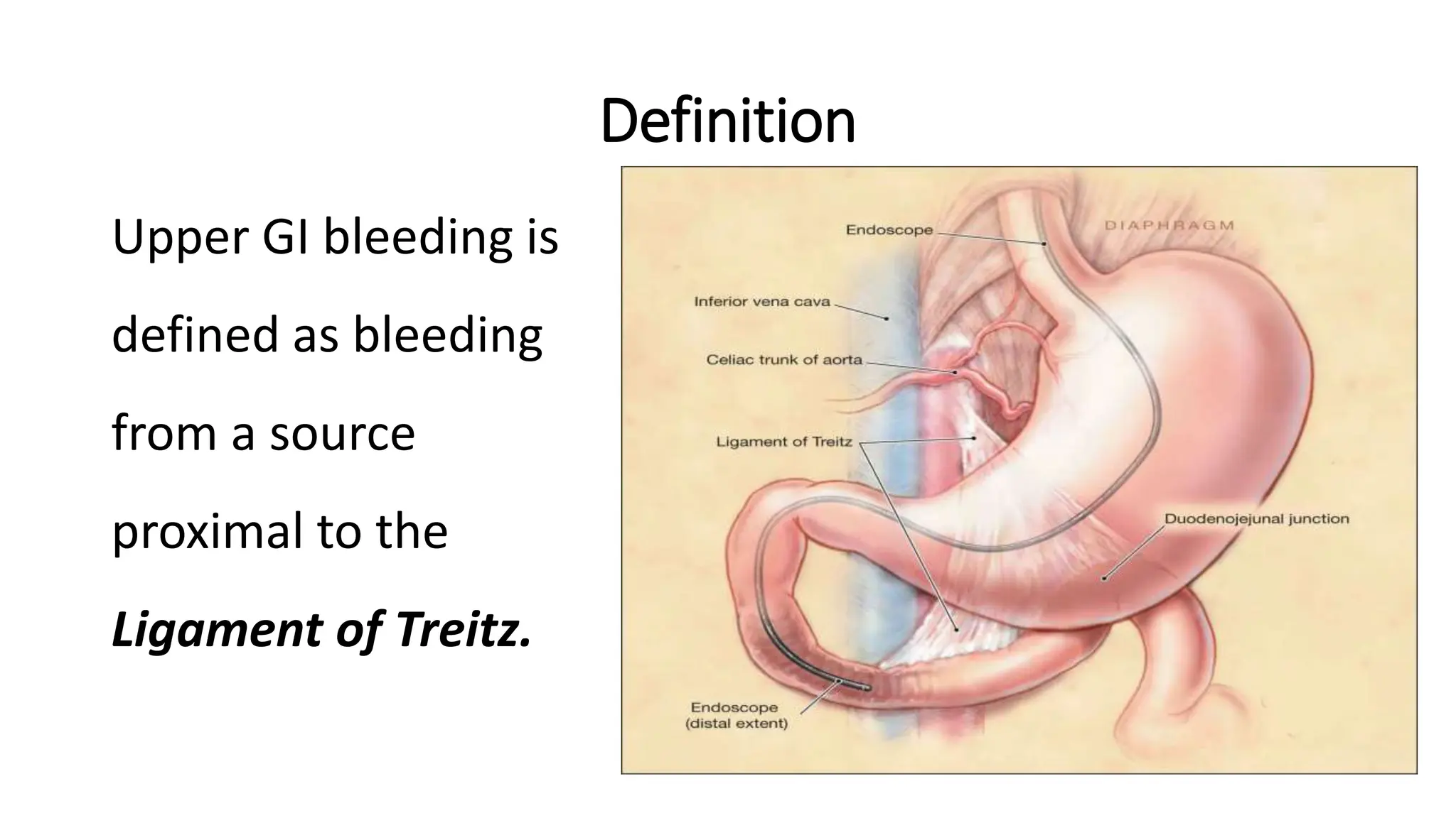
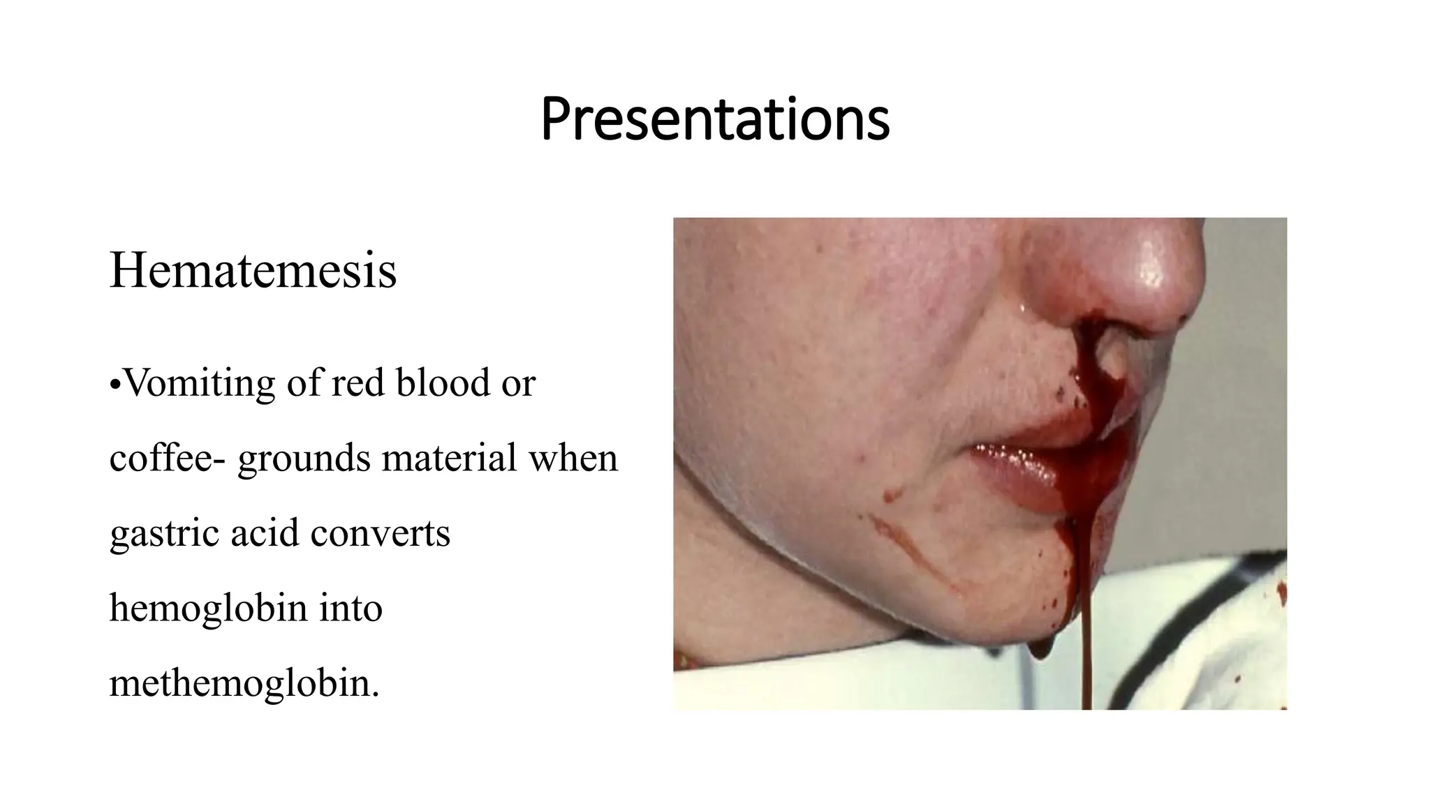
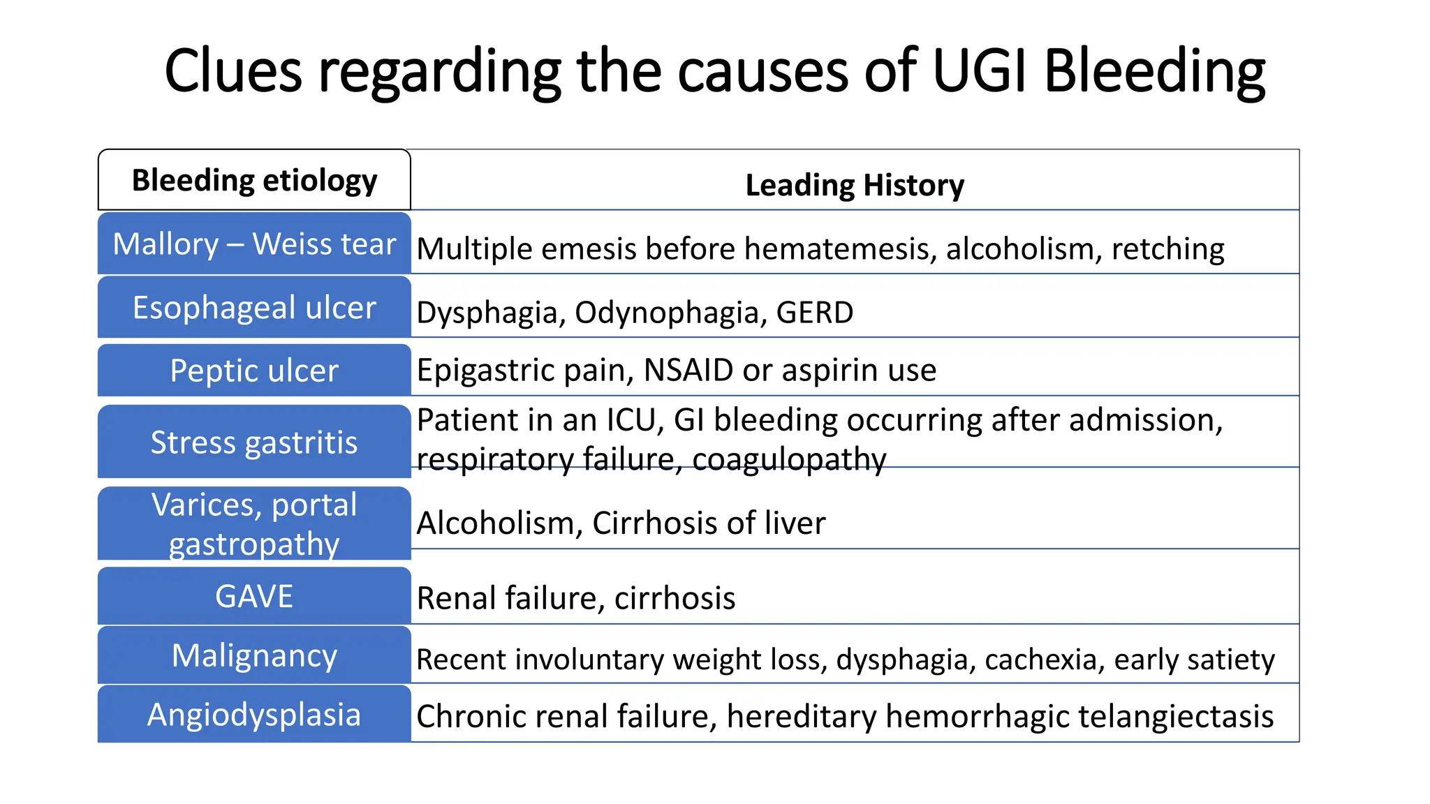
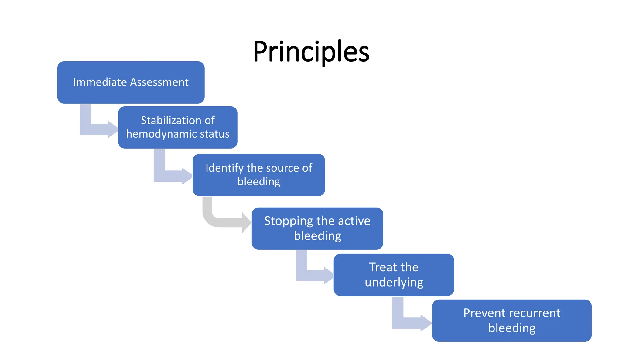
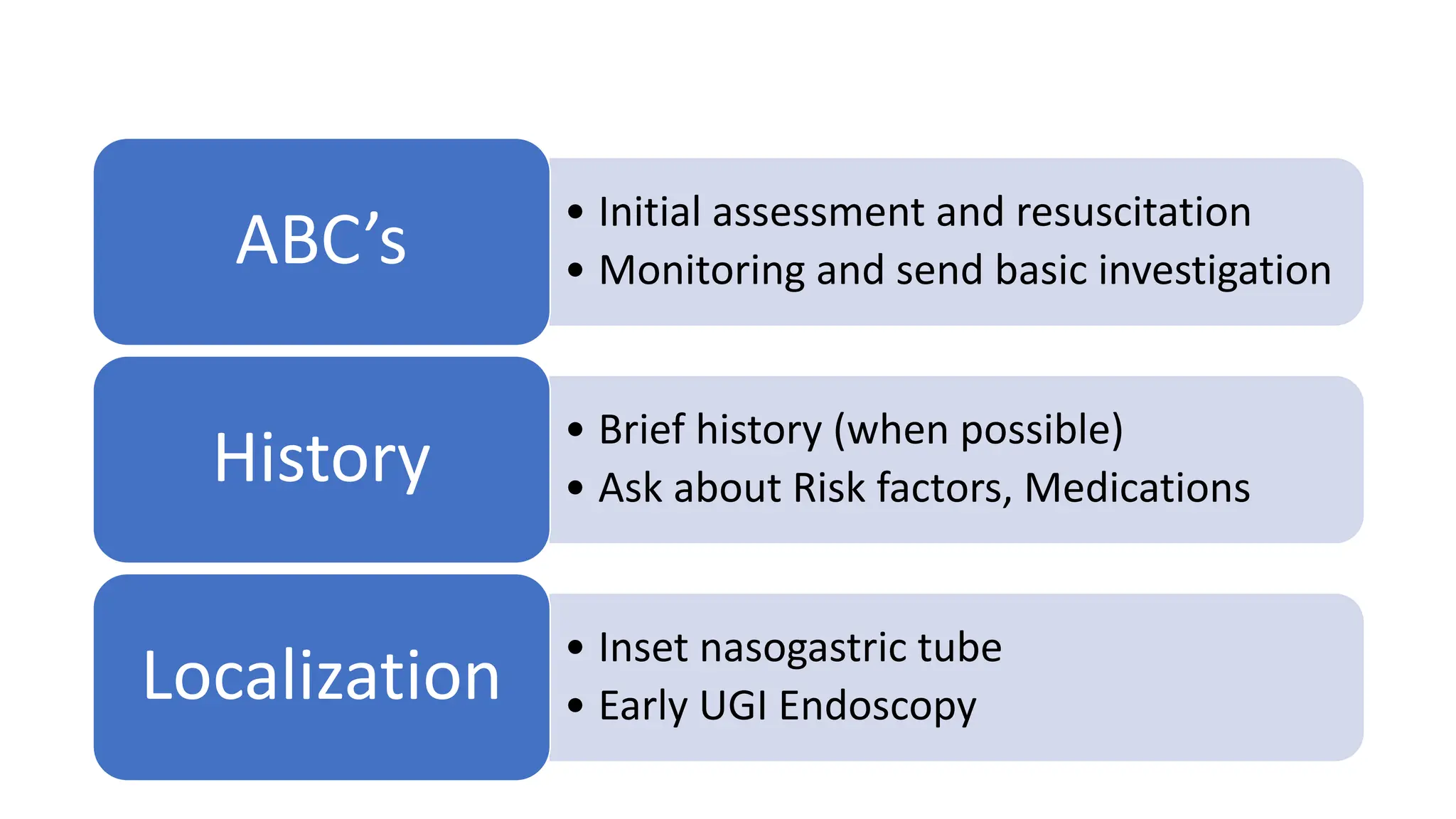
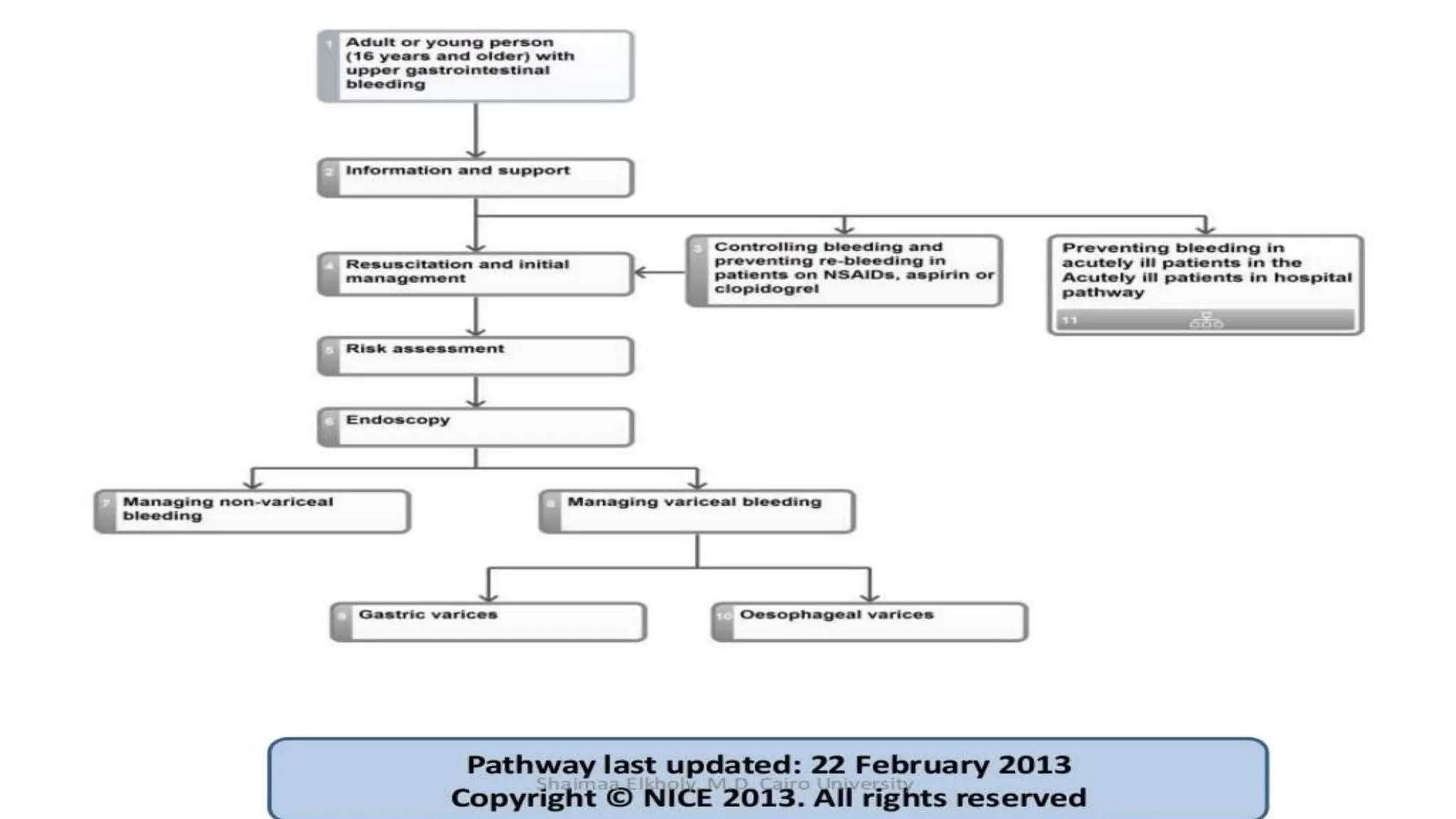
6. Melena
• Passage of black tarry stools.
• Blood loss between 50-100 ml /day will produce melena.
• The black color of melenic stools is caused by Hematin, the
product of oxidation of Heme by intestinal and bacterial enzymes.
• Non GI bleed – swallowed blood from epistaxis
• Blood for 14 hrs in the GI tract
• Oral iron and bismuth mimics melena.
7. Hematochezia
• It is defined as passage of bright-red or maroon blood from the
rectum.
• Common in bleeding from colon, rectum and anus.
• In case of brisk bleeding in the upper GIT, bright red blood may
come out unchanged in the stool.
• 10% of UGI bleed
8. Occult GI bleeding
• Absence of overt bleeding
• Fecal occult blood test - +ve
• Suspected in patients with iron deficiency anemia
• Normally 2.5 ml of blood is lost per day.
• OBT detects amount between 10-50 ml/d.
Symptoms of blood loss or anemia
• Light headedness, syncope, angina, or dyspnea
10. Causes
Esophageal causes
• Esophageal varices
• Esophagitis
• Esophageal cancer
• Esophageal ulcers
• Mallory-Weiss tear
Duodenal causes
• Duodenal ulcer
• Vascular malformation
including aorto- enteric
fistulae
• Hematobilia, or bleeding
from the biliary tree
• Hemosuccus pancreaticus,
or bleeding from the
pancreatic duct
• Severe superior
mesenteric artery syndrome
Esophageal causes
• Gastric ulcer
• Gastric cancer
• Gastritis
• Gastric varices
• Dieulafoy's lesions
• Gastric Antral Vascular
Ectasia
• Portal Hypertensive
Gastropathy
11. Causes of upper GI bleeding in chronic liver disease
patients
Causes of upper GI bleeding in chronic liver disease classified in two
classes:
• Portal hypertensive bleeding - the most common causes are
gastroesophageal varices and portal hypertensive gastropathy and
• Nonvariceal hemorrhage – gastric antral vascular ectasia (GAVE),
peptic ulcer and other lesions.
17. Assessment
Severity of bleeding can be determined:
• Level of consciousness
• Pulse rate >100bpm
• Postural hypotension.
• Severe blood loss—Vagal slowing of the heart
19. • Initial assessment and resuscitation
• Monitoring and send basic investigation
ABC’s
• Brief history (when possible)
• Ask about Risk factors, Medications
History
• Inset nasogastric tube
• Early UGI Endoscopy
Localization
20.
21. Primary Assessment:
• Firstly assess the patency of airway
• Assess the breathing, pulse, BP, temperature.
• Identifies emergency signs and symptoms i.e.
hypotension, bradycardia, tachycardia, dyspnea, weak and thready
pulse, shock, altered level of consciousness.
• Assess Glasgow coma scale.
• If the patient is having altered level of consciousness, give left lateral
position.
22. Initial evaluation
The initial evaluation of a patient with a suspected clinically significant acute
upper GI bleed includes a history, physical examination, and laboratory tests.
The goal of the evaluation is to assess the severity of the bleed, identify
potential sources of the bleed, and determine if there are conditions present
that may affect subsequent management.
The information gathered as part of the initial evaluation is used to guide
decisions regarding triage, resuscitation, empiric medical therapy, and
diagnostic testing.
25. Mode of enquiry
Onset ,episodes
True or spurious
Bleeding from oral cavity/nasopharynx
H/O retching with non bloody vomitus followed by hematemesis
H/O anorexia, dysphagia, rapid weight loss
26. Cont..
H/O Malena
H/O drug intake NSAIDs, aspirin or anti coagulants
H/O alcohol intake
Any skin telangiectasias?
Any pigmentation?
Perioral
Diffuse
27. Past history
H/O Chronic Liver Disease
H/O Peptic ulcer
H/O Bleeding disorders
Comorbidities -Pre existing CVS/Renal/CNS d/s may be
worsened by a/c bleeding
H/O medical illness/surgical intervention
Family history - hematemesis
28. Cont..
• Asks for if patient has previous hospitalization and past major illness.
• Gathers appropriate patient history: Alcohol intake, Complementary
alternative medicine, Antitubercular drugs, Mental confusion, Pain
abdomen. Gall bladder stones
• Any known medical history like, cirrhosis, Hepatitis, CLD, peptic ulcer
disease, Ascites, Malena, hematochezia, bleeding coagulopathies.
29. Cont..
• Assess vomitus for:
Bright red or coffee color ground granules
Amount of vomitus
• Assess skin for icterus/cyanosis
• Assess for presence of ecchymosis/petechiae
• Assess bowel sound
• Assess abdominal mass
33. Cont..
SEVERE BLEED (SR)
Tachycardia
Systolic blood pressure of less than 90 mm Hg
Cool extremities
Syncope
Ongoing brisk hematemesis or the occurrence of maroon or bright-
red stools, which requires rapid blood transfusion.
34.
35. Also Look For
• Any source of bleeding from oral cavity
• Telengiectasias in skin, conjunctiva, oral cavity
• Perioral/diffuse pigmentation
• Paraneoplastic syndromes
• STIGMATA OF CLD
36.
37.
38.
39.
40. Skin, nails and Hands
• Spider naevi - small telangiectatic superficial blood vessels with a central feeding vessel
• Clubbing
• Leukonychia - expansion of the paler half-moon at the base of the nail
• Palmar erythema - seen on the thenar and hypothenar eminences, often with a blotchy
appearance
• Bruising
• Dupuytren's contracture - can occur in the absence of liver disease
• Scratch marks - particularly in cholestatic liver disease
• Flapping Tremor
41. Cont..
Endocrine - due to excess oestrogens
• Gynaecomastia
• Testicular atrophy
• Loss of axillary and pubic hair
• Telengiectasias
Others
• Hepatic fetor - characteristic sweet-smelling breath
• Parotid swelling - particularly in alcohol-related liver disease
46. Clues From Initial Investigation
Parameter
Hb /HCT
MCV
Platelets
PT/INR
Disproportionate rise of BUN
Implications
24-72hrs
Need of transfusuion
Less than 80 Less than 80 with negative SOB
CLD
Hematological disorder
Coagulopathy
49. General management:
• Triage: “QUICK”
ICU admission
Hemodynamic instability (shock, orthostatic hypotension).
Active bleeding (manifested by hematemesis, bright red blood
per nasogastric tube, or hematochezia).
50. Cont..
• Support :
oxygen by nasal cannula.
NPO.
Two large caliber (16-18 gauge) peripheral I.V. Catheters.
Central venous line if possible.
Pulmonary artery catheter should be considered in patients with
hemodynamic instability or who need close monitoring during
resuscitation.
Elective endotracheal intubation in patients with ongoing hematemesis
with altered respiratory or mental status.
51. Cont..
• Fluid resuscitation:
resuscitation and stabilization is essential prior to endoscopy
Patients with active bleeding should receive intravenous fluids
(crystalloids or colloids)
while being typed and cross-matched for blood transfusion.
Patients at risk of fluid overload may require intensive
monitoring with a pulmonary artery catheter.
52. Cont..
• Indications of blood transfusion:
Hb below 7mg/dl (low risk).
High risk patients (old or comorbid) 10mg/dl.
Active (fresh) bleeding & Hypovolemia even with normal HB.
53. Cont..
• Indications of platelet & FFP transfusion:
low platelet count (<50,000/microL) OR INR > 1.5.
life-threatening bleeding receiving antiplatelet or anti coagulation.
Patients receiving massive blood transfusion due to dilutional
coagulopathy.
Over-transfuse patients with suspected variceal bleeding can
precipitate worsening of bleeding (10 mg/dl).
54. Cont..
• Nasogastric lavage:
• Its use before endoscopy in the ER remains controversial.
• Benefits:
To confirm an UGI source of bleeding (can still miss up to 15%)
Prognostic index for identifying high-risk lesions as presence fresh red blood
in the NGT aspirate.
May exclude false hematemesis.
To facilitate lavage of the upper GI tract to improve mucosal views at
subsequent endoscopy.
55. Balloon tamponade
• If intubated with persistent bleeding and no immediate definitive
treatment available, can try balloon tamponade (videos for placement
of Blakemore and Minnesota tubes)
56.
57.
58. Medications (pre- endoscopy):
• Acid suppression.
• Prokinetics.
• Somatostatin and its analogs in VUGIB.
• Antibiotics for patients with cirrhosis.
59. Cont..
• Acid suppression:
• start empirically on an I.V. PPI & continued until confirmation of
the cause of bleeding.
• I.V. of a PPI significantly reduces the rate of rebleeding
compared& hospital stay in comparison to H2 blockers.
• 80 mg bolus followed by 8 mg/hr infusion for 72 days then
switched to oral.
60.
61. Cont..
• Prokinetics: erythromycin & metchlopromide.
• Somatostatin, or its analog Octreotide
splanchnic vasoconstriction and decreased portal inflow
50 mcg bolus followed by a continuous infusion of 50 mcg per
hour and is continued for 3-5 days.
62.
63.
64. Cont..
• Antibiotics for patients with cirrhosis:
• The AASLD guidelines : (max.7 days)
Oral norfloxacin (400 mg twice daily) or intravenous
ciprofloxacin
In patients with advanced cirrhosis, I.V. ceftriaxone (1 g/day) &
with a high prevalence of quinolone-resistant organisms.
65.
66. Timing of endoscopy
• Patients with UGIB should generally undergo endoscopy within 24 h of
admission, following resuscitative efforts to optimize hemodynamic parameters
and other medical problems
• Patients who are hemodynamically stable and without serious comorbidities:
Endoscopy as soon as possible in a non-emergent setting to identify the
substantial proportion of patients with low-risk endoscopic findings who can be
safely discharged
• Patients with higher risk clinical features endoscopy within 12 h may be
considered to potentially improve clinical outcomes
67. Upper GI bleed: High risk factors
• Advanced AGE >65 years
• SHOCK on admission (pulse rate >100 beats/min; systolic blood pressure <
100mmHg)
• COMORBIDITY (particularly hepatic or renal failure, DM, HTN and
disseminated malignancy)
• Diagnosis (worst PROGNOSIS for advanced upper gastrointestinal
malignancy)
• ENDOSCOPIC FINDINGS (active, spurting hemorrhage from peptic ulcer;
non-bleeding visible vessel)
• RECURRENT BLEEDING (increases mortality 10 times)
• Where there is simultaneous upper and lower GI bleeding.
• On steroids or NSAIDs
• Alcoholic or tobacco smoker
69. Risk scoring
Rockall’s risk score
• Score that predicts poor prognosis, i.e. rebleeding and mortality
from upper GI haemorrhage
• It uses clinical criteria (increasing age, co-morbidity, shock) as
well as endoscopic finding (diagnosis, stigmata of spontaneous
haemorrhage -SSH)
70. Cont..
• A commonly used mnemonic is ABCDE - (Age, Blood pressure fall
(shock), Co-morbidity, Diagnosis and Evidence of bleeding).
71. Key independent risk factors
•Age. There is a close relationship between increasing age and
mortality. Patients 80 years of age or greater are at the highest
risk of death.
•Shock. Defined as a pulse rate of more than 100 beats/min and
systolic blood pressure less than 100 mm Hg.
72. Cont..
• Comorbidity. Mortality rates vary greatly depending on the number
and type of comorbidities. Heart failure, ischemic heart disease, or
any major comorbidity can have a moderate to high impact on
mortality rates. Renal failure, liver failure, or disseminated
malignancy carry the highest risk of death from gastrointestinal
bleeding.
73. Cont..
Endoscopic findings.
• Low risk patients:
• Normal upper gastrointestinal endoscopy
• Mallory-Weiss tear
• Finding of an ulcer with a clean base
74. Cont..
High risk:
• Active bleeding from a peptic ulcer in a shocked patient carried an
80% risk of continuing bleeding or of death (grade A).
• A non-bleeding visible vessel is associated with a 50% risk of
rebleeding in hospital.
75. Variable Score
0 1 2 3
Age (yrs) < 60 60 - 79 > 80
Hemodynamic
status
No shock P <
100 Syst BP >
100
P > 100 plus
Syst BP > 100
Hypotension
Comorbidity No or mild
coexisting
Moderate
coexisting
(e.g.,
hypertension)
Severe
coexisting
(e.g., CHF)
Life
threatenin
g (e.g., RF)
Diagnosis Mallory-Weiss
tear, normal
All other
diagnosis
Malignancy
of UGI tract
Major stigmata
of recent
None or dark
spot
Blood in UGI
tract