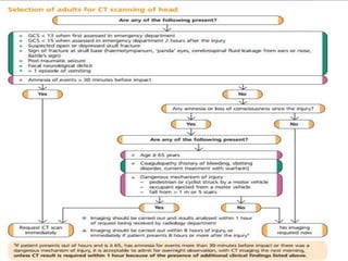
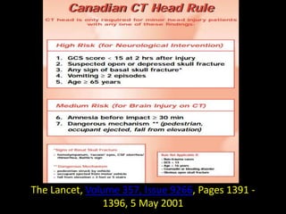
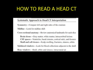
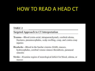
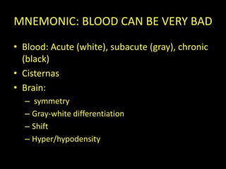
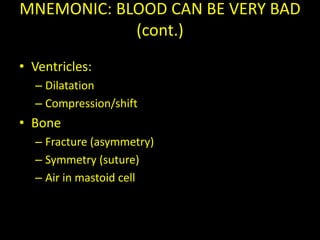
INTRODUCTION TO CRANIAL CT

       Hospital Eva Peron
      DR.LAPAL MA, DORIAN
INTRODUCTION
CT is the most useful neuroimaging study in
  emergency medicine it readily detects acute
  blood collections and intracranial lesions
  causing mass effect.
MRI is superior for imaging parenchymal
  abnormalities and has replaced CT in most
  nonemergency neurodiagnosis
GUIDELINES: (NICE)
• The national institute for health and clinical
  excellence (NICE) is a special health authority
  of the english nationa health service (NHS),
  serving, both English NHS and the Welsh NHS.
  It was set up as the National Institute for
  Clinical Excellence in 1999
The Lancet, Volume 357, Issue 9266, Pages 1391 -
                1396, 5 May 2001
BASIC PRINCIPALES OF CT
ATTENUATION COEFFICIENT
  The ability to block x rays as they pasoo
  through a substance (attenuation).For a given
  body tissue, the amount of attenuation is
  relatively constant an is known as that tissue’s
  attenuation coefficient.
  Sir Jeffery Hounsfield: mapping of attenuation
  coefficient
THE CT HOUNSFIELD SCALE
WINDOWING
• It allows the CT scan reader to focus on
  certain tissues within a set parameters. Most
  CT imaging includes windows that are
  optimized for brain, blood and bone.
• BRAIN : W 155 L 40
• STROKE : W 30 L 30
• SUBDURAL: W 150 L 5
• BONE: W 3000 L 570
WWINDOWING


BRAIN (W 155 L 40)   STROKE (W 30 L 30)
WINDOWING


BONE (W3000 L 570)   SUBDURAL (W150 L 5)
HEAD CT ARTIFACTS
Effects tha can potentially inhibit the ability to
   accuerately interpret the images
   Motion
   Metal artifacts
   Beam Hardening: when small amount of hypodense
   brain tissue is immediately adjacent to dense bone
   Partial volume: arises when the imaged area contains
   different types of tissue. Ej: brain and bone, an
   intermediate density will be represented that may have
   the appearace of blood
HEAD CT ARTIFACTS
HEAD CT ARTIFACTS
HEAD CT ARTIFACTS

•Oriented obliquely.
•Reduces the number
of slices that are
degrade by artifacts.
•CT: the frontal lobes
are anterior, but
posteriorly, the
cerebelum is seen,
rather than the
occipital lobes
HOW TO READ A HEAD CT
MNEMONIC: BLOOD CAN BE VERY BAD
• Blood: Acute (white), subacute (gray), chronic
  (black)
• Cisternas
• Brain:
  – symmetry
  – Gray-white differentiation
  – Shift
  – Hyper/hypodensity
MNEMONIC: BLOOD CAN BE VERY BAD
            (cont.)
• Ventricles:
  – Dilatation
  – Compression/shift
• Bone
  – Fracture (asymmetry)
  – Symmetry (suture)
  – Air in mastoid cell

Introduction to cranial ct

  • 1.
    INTRODUCTION TO CRANIALCT Hospital Eva Peron DR.LAPAL MA, DORIAN
  • 2.
    INTRODUCTION CT is themost useful neuroimaging study in emergency medicine it readily detects acute blood collections and intracranial lesions causing mass effect. MRI is superior for imaging parenchymal abnormalities and has replaced CT in most nonemergency neurodiagnosis
  • 3.
    GUIDELINES: (NICE) • Thenational institute for health and clinical excellence (NICE) is a special health authority of the english nationa health service (NHS), serving, both English NHS and the Welsh NHS. It was set up as the National Institute for Clinical Excellence in 1999
  • 6.
    The Lancet, Volume357, Issue 9266, Pages 1391 - 1396, 5 May 2001
  • 7.
    BASIC PRINCIPALES OFCT ATTENUATION COEFFICIENT The ability to block x rays as they pasoo through a substance (attenuation).For a given body tissue, the amount of attenuation is relatively constant an is known as that tissue’s attenuation coefficient. Sir Jeffery Hounsfield: mapping of attenuation coefficient
  • 8.
  • 9.
    WINDOWING • It allowsthe CT scan reader to focus on certain tissues within a set parameters. Most CT imaging includes windows that are optimized for brain, blood and bone. • BRAIN : W 155 L 40 • STROKE : W 30 L 30 • SUBDURAL: W 150 L 5 • BONE: W 3000 L 570
  • 10.
    WWINDOWING BRAIN (W 155L 40) STROKE (W 30 L 30)
  • 11.
    WINDOWING BONE (W3000 L570) SUBDURAL (W150 L 5)
  • 12.
    HEAD CT ARTIFACTS Effectstha can potentially inhibit the ability to accuerately interpret the images Motion Metal artifacts Beam Hardening: when small amount of hypodense brain tissue is immediately adjacent to dense bone Partial volume: arises when the imaged area contains different types of tissue. Ej: brain and bone, an intermediate density will be represented that may have the appearace of blood
  • 13.
  • 14.
  • 15.
    HEAD CT ARTIFACTS •Orientedobliquely. •Reduces the number of slices that are degrade by artifacts. •CT: the frontal lobes are anterior, but posteriorly, the cerebelum is seen, rather than the occipital lobes
  • 16.
    HOW TO READA HEAD CT
  • 17.
    HOW TO READA HEAD CT
  • 18.
    MNEMONIC: BLOOD CANBE VERY BAD • Blood: Acute (white), subacute (gray), chronic (black) • Cisternas • Brain: – symmetry – Gray-white differentiation – Shift – Hyper/hypodensity
  • 19.
    MNEMONIC: BLOOD CANBE VERY BAD (cont.) • Ventricles: – Dilatation – Compression/shift • Bone – Fracture (asymmetry) – Symmetry (suture) – Air in mastoid cell