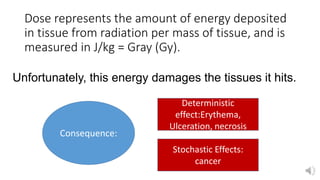
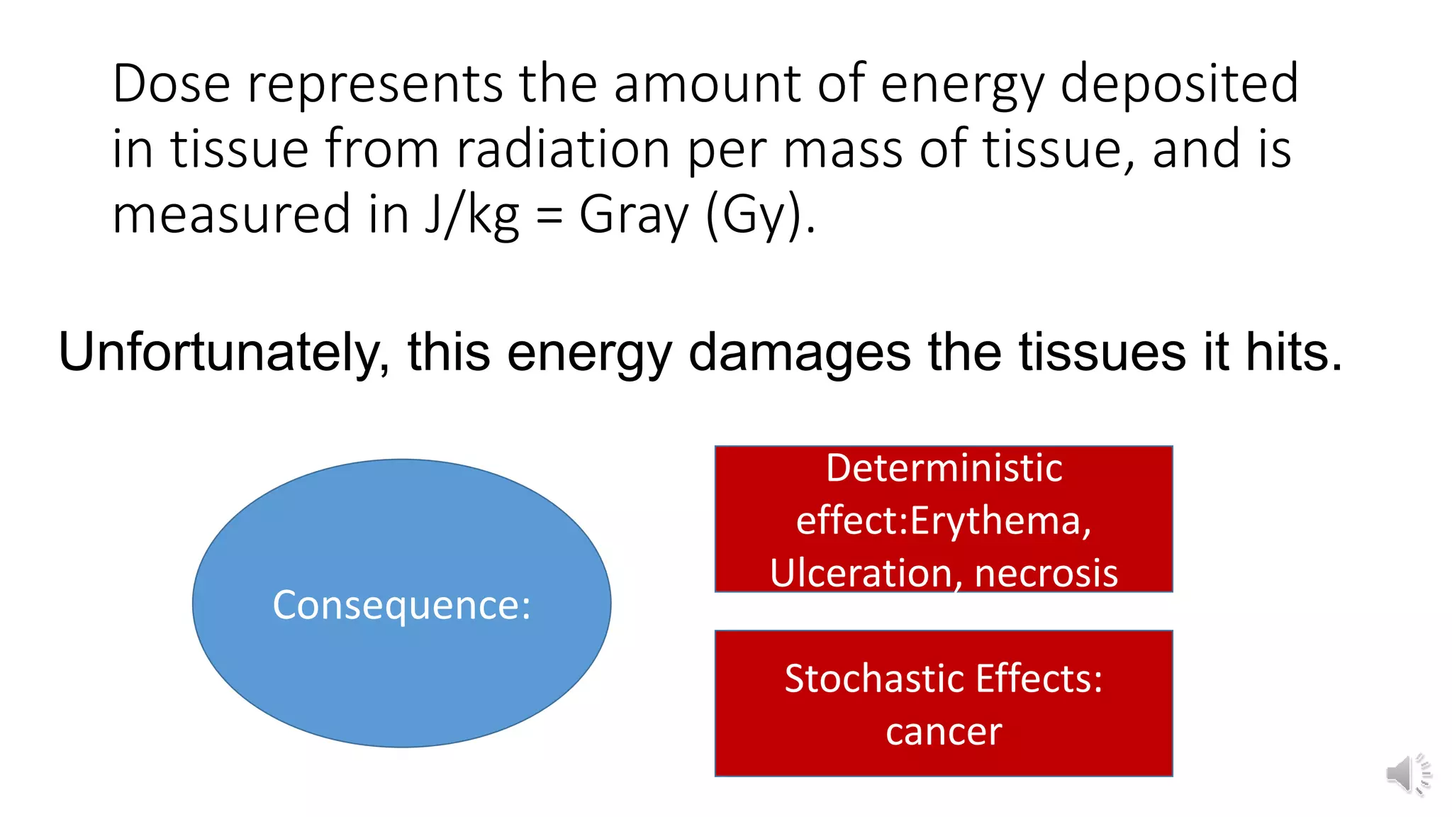
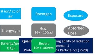
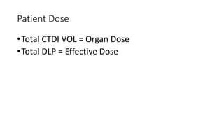
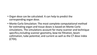
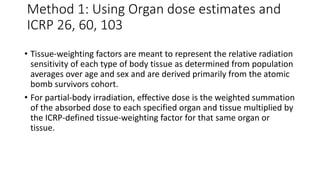
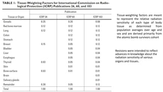
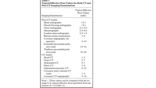
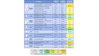
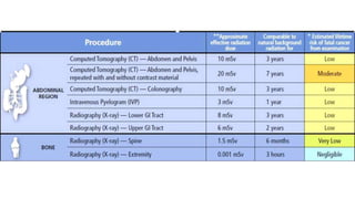
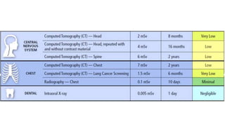
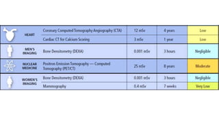
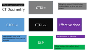
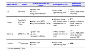
Dose represents the amount of energy deposited
in tissue from radiation per mass of tissue, and is
measured in J/kg = Gray (Gy).
Unfortunately, this energy damages the tissues it hits.
Consequence:
Deterministic
effect:Erythema,
Ulceration, necrosis
Stochastic Effects:
cancer
• Deterministic effects relate directly to the amount of radiation a
single cell receives; these require a large dose to become apparent.
• Stochastic effects can occur (randomly) with very small doses, and
even one cell can turn cancerous.
Generally speaking, in CT we are mostly
interested in the stochastic effects.
Exposure
Absorbed
Dose
Equivalent
Dose
Energy/gm
(Energy/gm)
X Q.F
# ion/ cc of
air
Roentgen
rem
rads
Quality factor :Cell killing ability of radiation
X ray , Gamma : 1
Proton, Neutron, Alpha Particle: >1 ( 2-20)
Gray:
1Gy = 100rad
Sievert
1Sv = 100rem
How is radiation dose measured in CT?
• CT dose is not measured directly on patient.
• CT dose is measured using standard phantoms.
• Measurements are then used to estimate patient dose.
Dose Gradient: Radiograph vs CT
How is Radiation dose measured?
CTDI 100 CTDI W CTDI VOL DLP Effective
dose
Dose
Distribution
Pitch
Scan
Length
Computed Tomography Dose Index (CTDI)
Measurement done at the centre of phantom of a single slice
CTDI
• Obtained by making
measurement in acryllic
cylinder phantom.
• Holes in centre and periphery
• Placement of pensil shaped
ionization chamber.
CTDI100 – Dose Distribution
In CT: the highest dose delivered is at the
periphery
Dose is uniform on the surface but
decreases towards centre.
• CTDI VOL :15mGy
• Independent of scan
length
Computed Tomography Dose Index (CTDI):
Measurement done at the centre of phantom
of a single slice:
cannot accommodate the anatomy covered
because we do just confine to single slice.
•Volume CTDI in mGy (CTDI VOL):
•X ray tube voltage (KV)
•X ray tube current (mA)
•X ray tube rotation time (s)
•CT Pitch (P)
•Phantom size (S or L)
• CTDI is the rate at which
you the energy is put on
the patient.
• DLP is total amount of the
energy that you are
putting the patient.
CTDI = Radiation Intensity
DLP = Total radiation used to perform a CT Scan.
CTDI Phantoms
• 16cm (S) (Head Phantom)
• 32cm (L) (Body Phantom)
Large CTDI is =
2 X Small CTDI
58.1/15.7
= 3.7
Abdomen CTDI VOL = 15.7 X 2 = 31.4 mGy (S)
58.1/31.4
= 1.85
Axial CT
•CTDI VOL is directly proportional to :
•Tube current (mA)
•Tube rotation time (s)
•mAs
Helical CT
• CTDI VOL is directly proportional to:
• mA,s (mAs)
• Inversely proportional to pitch:1/Pitch
• CTDI VOL is directly proportional to :
• Effective mAs: mAs/Pitch
• CTDI VOL = kV (2.6)
• 80 140 kV
• CTDI = > 400%
CTDI VOL “Universal Parameter”
• CTDI VOL for Head = 60mGy on Small Phantom
• KV
• X ray filter: Head, Body or pediatric
• Model: Type of CT Scanner
• Vendor: Philips/ Toshiba/ GE
• CTDI vol >>> mAs !
CTDI VOL/ DLP & Patient Dose
• CTDI Vol is the radiation incident on patient, not the patient dose.
• CTDI VOL = 20mGy:
CTDI Vol is not the
indicator of any kind of
patient dose.
Patient Dose
•Total CTDI VOL = Organ Dose
•Total DLP = Effective Dose
• Organ dose can be calculated. It can help to predict the
corresponding organ dose.
• Monte Carlo Simulation: The most complete computational method
for estimating organ and tissue doses is based on Monte Carlo
simulations. The simulations account for many scanner and technique
specifics,including scanner geometry, bow-tie filtration, beam
collimation, tube potential, and current as well as the CT dose index
(CTDI).
• Jones and Shrimpton used a simulated hermaphroditic patient (MIRD-
5 phantom) having mathematically modeled organs and tissues .
• The mathematic phantom was divided from head to mid thigh into
208 axial slabs of 5 mm thickness. Then, accounting for tube voltage
and using CT scanner–specific data for geometry and beam shaping,
they simulated a
• CT scan and calculated absorbed doses to all organs of the body for
the irradiation of each axial slab. Summing contributions from all
slabs exposed during a particular CT examination yielded the total
organ doses.
Effective Dose
• Effective dose is a parameter meant to reflect the relative risk from
exposure to ionizing radiation.
• The effective dose (E) is a measure of the risk of cancer induction in
the patient from the effects of the radiation.
• It takes into account the total amount of absorbed dose received and
averages it to give a whole body effective dose.
Method 1: Using Organ dose estimates and
ICRP 26, 60, 103
• Tissue-weighting factors are meant to represent the relative radiation
sensitivity of each type of body tissue as determined from population
averages over age and sex and are derived primarily from the atomic
bomb survivors cohort.
• For partial-body irradiation, effective dose is the weighted summation
of the absorbed dose to each specified organ and tissue multiplied by
the ICRP-defined tissue-weighting factor for that same organ or
tissue.
Revisions were intended to reflect
advances in knowledge about the
radiation sensitivity of various
organs and tissues.
Tissue-weighting factors are meant
to represent the relative radiation
sensitivity of each type of body
tissue as determined from
population averages over age and
sex and are derived primarily from
the atomic bomb survivors cohort
• E = Effective Dose
• T = all ICRP specified tissue and organ
• W t = ICRP specified tissue weighting factor
• H t = Dose to particular organ or tissue
• E T = overall tisue
• Ez = Overall irradiated Slabs
Using DLP and K Coefficients from the
European Guideline
• E = k × DLP,
• where k
coefficient
is specific
only to the
anatomic
region
scanned.
Measuring Effective Dose
Effective Dose in NCCT Head: (1100X 0.0021 ) + 4.4 X 0.0021 = 2.31
msv
Effective Dose in CT IVU: ((771.7 X 3) + 4.7 ) X 0.015 = 34.79 msv
Body
Region
KvP Effective
mAs
Scan
Length(cm)
CTDI
VOL(mGy)
DLP
(mGy-cm)
K factor Effective
Dose(mSv)
Head 120 390 17 60 (S) 1000 0.002 2
Chest 120 110 30 7.4 (L) 220 0.018 4.5
Abdomen 120 210 25 14 (L) 350 0.016 5.7
Pelvis 120 210 30 14 420 0.014 6
CT Dosimetry
CTDI 100 Effective dose
DLP
CTDIVOL
CTDIW
Measurement done on a
standard phantom using 100 cm
chamber.
Taking into account the distribution
variation based on large and small
patient.
When the scan involves pitch.
Taking account of the whole length
exposure
Risk estimation to the body
Automatic Tube Current Modulation
• mA is varied: Around the patient, Along the patient(scan length), &
between patient
Quality Reference mAs
• Quality Reference mAs = Effective mAs
• Effective mAs = mAs/pitch
• Once set in protocol and is different for different body region.
• Can convert Effective mAs in CTDI Vol i.e the Universal parameter.
• Modulation: Five Strength
Thankyou
Refrences
• Estimating Effective Dose forCT Using Dose–Length Product Compared With Using Organ Doses:
Consequences of Adopting International Commission on Radiological Protection Publication 103
or Dual-Energy Scanning, Jodi A Christner, Mayo Clinic, Available from www.ajronline.org,
DOI:10.2214/AJR.09.3462
• Videos from Walter Huda, available on Youtube.

Computed Tomography Dose Index

  • 1.
    Dose represents theamount of energy deposited in tissue from radiation per mass of tissue, and is measured in J/kg = Gray (Gy). Unfortunately, this energy damages the tissues it hits. Consequence: Deterministic effect:Erythema, Ulceration, necrosis Stochastic Effects: cancer
  • 2.
    • Deterministic effectsrelate directly to the amount of radiation a single cell receives; these require a large dose to become apparent. • Stochastic effects can occur (randomly) with very small doses, and even one cell can turn cancerous. Generally speaking, in CT we are mostly interested in the stochastic effects.
  • 3.
    Exposure Absorbed Dose Equivalent Dose Energy/gm (Energy/gm) X Q.F # ion/cc of air Roentgen rem rads Quality factor :Cell killing ability of radiation X ray , Gamma : 1 Proton, Neutron, Alpha Particle: >1 ( 2-20) Gray: 1Gy = 100rad Sievert 1Sv = 100rem
  • 4.
    How is radiationdose measured in CT? • CT dose is not measured directly on patient. • CT dose is measured using standard phantoms. • Measurements are then used to estimate patient dose.
  • 5.
  • 6.
    How is Radiationdose measured? CTDI 100 CTDI W CTDI VOL DLP Effective dose Dose Distribution Pitch Scan Length Computed Tomography Dose Index (CTDI) Measurement done at the centre of phantom of a single slice
  • 7.
    CTDI • Obtained bymaking measurement in acryllic cylinder phantom. • Holes in centre and periphery • Placement of pensil shaped ionization chamber.
  • 12.
    CTDI100 – DoseDistribution In CT: the highest dose delivered is at the periphery Dose is uniform on the surface but decreases towards centre.
  • 16.
    • CTDI VOL:15mGy • Independent of scan length
  • 17.
    Computed Tomography DoseIndex (CTDI): Measurement done at the centre of phantom of a single slice: cannot accommodate the anatomy covered because we do just confine to single slice.
  • 20.
    •Volume CTDI inmGy (CTDI VOL): •X ray tube voltage (KV) •X ray tube current (mA) •X ray tube rotation time (s) •CT Pitch (P) •Phantom size (S or L)
  • 21.
    • CTDI isthe rate at which you the energy is put on the patient. • DLP is total amount of the energy that you are putting the patient. CTDI = Radiation Intensity DLP = Total radiation used to perform a CT Scan.
  • 24.
    CTDI Phantoms • 16cm(S) (Head Phantom) • 32cm (L) (Body Phantom) Large CTDI is = 2 X Small CTDI
  • 25.
    58.1/15.7 = 3.7 Abdomen CTDIVOL = 15.7 X 2 = 31.4 mGy (S) 58.1/31.4 = 1.85
  • 26.
    Axial CT •CTDI VOLis directly proportional to : •Tube current (mA) •Tube rotation time (s) •mAs
  • 27.
    Helical CT • CTDIVOL is directly proportional to: • mA,s (mAs) • Inversely proportional to pitch:1/Pitch • CTDI VOL is directly proportional to : • Effective mAs: mAs/Pitch
  • 28.
    • CTDI VOL= kV (2.6) • 80 140 kV • CTDI = > 400%
  • 29.
    CTDI VOL “UniversalParameter” • CTDI VOL for Head = 60mGy on Small Phantom • KV • X ray filter: Head, Body or pediatric • Model: Type of CT Scanner • Vendor: Philips/ Toshiba/ GE • CTDI vol >>> mAs !
  • 30.
    CTDI VOL/ DLP& Patient Dose
  • 31.
    • CTDI Volis the radiation incident on patient, not the patient dose. • CTDI VOL = 20mGy: CTDI Vol is not the indicator of any kind of patient dose.
  • 32.
    Patient Dose •Total CTDIVOL = Organ Dose •Total DLP = Effective Dose
  • 33.
    • Organ dosecan be calculated. It can help to predict the corresponding organ dose. • Monte Carlo Simulation: The most complete computational method for estimating organ and tissue doses is based on Monte Carlo simulations. The simulations account for many scanner and technique specifics,including scanner geometry, bow-tie filtration, beam collimation, tube potential, and current as well as the CT dose index (CTDI).
  • 34.
    • Jones andShrimpton used a simulated hermaphroditic patient (MIRD- 5 phantom) having mathematically modeled organs and tissues . • The mathematic phantom was divided from head to mid thigh into 208 axial slabs of 5 mm thickness. Then, accounting for tube voltage and using CT scanner–specific data for geometry and beam shaping, they simulated a • CT scan and calculated absorbed doses to all organs of the body for the irradiation of each axial slab. Summing contributions from all slabs exposed during a particular CT examination yielded the total organ doses.
  • 35.
    Effective Dose • Effectivedose is a parameter meant to reflect the relative risk from exposure to ionizing radiation. • The effective dose (E) is a measure of the risk of cancer induction in the patient from the effects of the radiation. • It takes into account the total amount of absorbed dose received and averages it to give a whole body effective dose.
  • 36.
    Method 1: UsingOrgan dose estimates and ICRP 26, 60, 103 • Tissue-weighting factors are meant to represent the relative radiation sensitivity of each type of body tissue as determined from population averages over age and sex and are derived primarily from the atomic bomb survivors cohort. • For partial-body irradiation, effective dose is the weighted summation of the absorbed dose to each specified organ and tissue multiplied by the ICRP-defined tissue-weighting factor for that same organ or tissue.
  • 37.
    Revisions were intendedto reflect advances in knowledge about the radiation sensitivity of various organs and tissues. Tissue-weighting factors are meant to represent the relative radiation sensitivity of each type of body tissue as determined from population averages over age and sex and are derived primarily from the atomic bomb survivors cohort
  • 38.
    • E =Effective Dose • T = all ICRP specified tissue and organ • W t = ICRP specified tissue weighting factor • H t = Dose to particular organ or tissue • E T = overall tisue • Ez = Overall irradiated Slabs
  • 39.
    Using DLP andK Coefficients from the European Guideline • E = k × DLP, • where k coefficient is specific only to the anatomic region scanned.
  • 40.
    Measuring Effective Dose EffectiveDose in NCCT Head: (1100X 0.0021 ) + 4.4 X 0.0021 = 2.31 msv Effective Dose in CT IVU: ((771.7 X 3) + 4.7 ) X 0.015 = 34.79 msv
  • 41.
    Body Region KvP Effective mAs Scan Length(cm) CTDI VOL(mGy) DLP (mGy-cm) K factorEffective Dose(mSv) Head 120 390 17 60 (S) 1000 0.002 2 Chest 120 110 30 7.4 (L) 220 0.018 4.5 Abdomen 120 210 25 14 (L) 350 0.016 5.7 Pelvis 120 210 30 14 420 0.014 6
  • 47.
    CT Dosimetry CTDI 100Effective dose DLP CTDIVOL CTDIW Measurement done on a standard phantom using 100 cm chamber. Taking into account the distribution variation based on large and small patient. When the scan involves pitch. Taking account of the whole length exposure Risk estimation to the body
  • 48.
    Automatic Tube CurrentModulation • mA is varied: Around the patient, Along the patient(scan length), & between patient
  • 49.
    Quality Reference mAs •Quality Reference mAs = Effective mAs • Effective mAs = mAs/pitch • Once set in protocol and is different for different body region. • Can convert Effective mAs in CTDI Vol i.e the Universal parameter. • Modulation: Five Strength
  • 51.
  • 52.
    Refrences • Estimating EffectiveDose forCT Using Dose–Length Product Compared With Using Organ Doses: Consequences of Adopting International Commission on Radiological Protection Publication 103 or Dual-Energy Scanning, Jodi A Christner, Mayo Clinic, Available from www.ajronline.org, DOI:10.2214/AJR.09.3462 • Videos from Walter Huda, available on Youtube.

Editor's Notes

  • #7 In Radiography and Fluroscopy the entrance skin dose is higher and the radiation dose decreases as it pass through the patient. In CT the surface dose is maximum compared to centre.
  • #12 More like a bell curved, peak dose is delivered only to certain central slice. There is more the tail portion
  • #21 Larger the anatomy exposed , greater the biological risk.
  • #33 For the same amount of incident raiation, the newborn infant and children dose will be quite large, whereas for an adult due to size of attenuation due to large mass, their dose will be much lower.