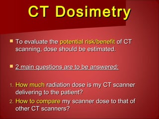
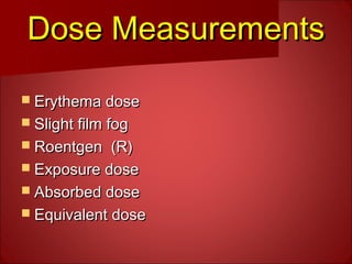
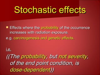
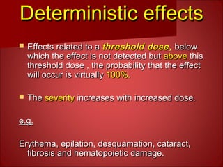
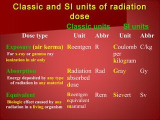
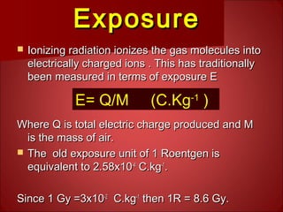
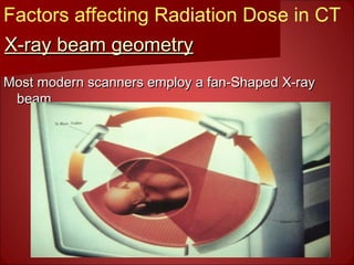
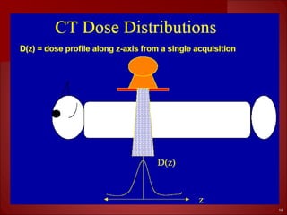
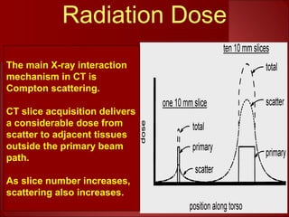
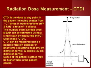
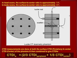
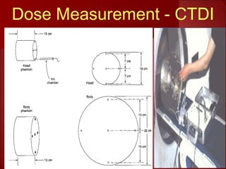
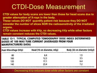
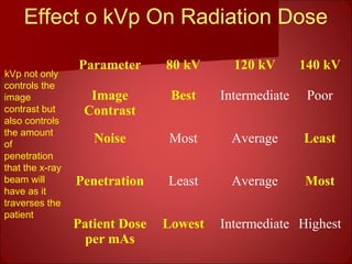
SlideShare a Scribd company logo
‫تعلم‬ ‫تكن‬ ‫مالم‬ ‫وعلمك‬‫تعلم‬ ‫تكن‬ ‫مالم‬ ‫وعلمك‬
‫ال‬ ‫فضل‬ ‫وكان‬‫ال‬ ‫فضل‬ ‫وكان‬
‫عظيما‬ ‫عليك‬‫عظيما‬ ‫عليك‬
Basics Of
Computed
Tomography (CT)
Lecture Ten
GGamalamal FFathallaathalla MM..
MMahdalyahdaly
gamal_mahdaly@hotmail.comgamal_mahdaly@hotmail.com
Radiation Safety and ProtectionRadiation Safety and Protection
The potentially harmful effects of ionizingThe potentially harmful effects of ionizing
radiation are either:radiation are either:
 Stochastic effectsStochastic effects
OrOr
 Deterministic effectsDeterministic effects
Stochastic effectsStochastic effects
 Effects where theEffects where the probabilityprobability of the occurrenceof the occurrence
increases with radiation exposure.increases with radiation exposure.
e.g.e.g. carcinogenesis and genetic effects.carcinogenesis and genetic effects.
i.e.i.e.
((The((The probabilityprobability, but, but not severitynot severity,,
of the end point condition, isof the end point condition, is
dose-dependentdose-dependent))))
Deterministic effectsDeterministic effects
 Effects related to aEffects related to a threshold dosethreshold dose,, belowbelow
which the effect is not detected butwhich the effect is not detected but aboveabove thisthis
threshold dose , the probability that the effectthreshold dose , the probability that the effect
will occur is virtuallywill occur is virtually 100%.100%.
 TheThe severityseverity increases with increased dose.increases with increased dose.
e.g.e.g.
Erythema, epilation, desquamation, cataract,Erythema, epilation, desquamation, cataract,
fibrosis and hematopoietic damage.fibrosis and hematopoietic damage.
CT DosimetryCT Dosimetry
 To evaluate theTo evaluate the potential risk/benefitpotential risk/benefit of CTof CT
scanning, dose should be estimated.scanning, dose should be estimated.
 2 main questions are to be answered:2 main questions are to be answered:
1.1. How muchHow much radiation dose is my CT scannerradiation dose is my CT scanner
delivering to the patient?delivering to the patient?
2.2. How to compareHow to compare my scanner dose to that ofmy scanner dose to that of
other CT scanners?other CT scanners?
Dose MeasurementsDose Measurements
 Erythema doseErythema dose
 Slight film fogSlight film fog
 Roentgen (R)Roentgen (R)
 Exposure doseExposure dose
 Absorbed doseAbsorbed dose
 Equivalent doseEquivalent dose
Classic and SI units of radiationClassic and SI units of radiation
dosedose
Dose type Unit Abbr Unit Abbr
Exposure (air kerma)
For x-ray or gamma ray
ionization in air only
Roentgen R Coulomb
per
kilogram
C/kg
Absorption
Energy deposited by any type
of radiation in any material
Radiation
absorbed
dose
Rad Gray Gy
Equivalent
Biologic effect caused by any
radiation in a living organism
Roentgen
equivalent
mammal
Rem Sievert Sv
Classic units SI units
ExposureExposure
 Ionizing radiation ionizes the gas molecules intoIonizing radiation ionizes the gas molecules into
electrically charged ions . This has traditionallyelectrically charged ions . This has traditionally
been measured in terms of exposure Ebeen measured in terms of exposure E
Where Q is total electric charge produced and MWhere Q is total electric charge produced and M
is the mass of air.is the mass of air.
 The old exposure unit of 1 Roentgen isThe old exposure unit of 1 Roentgen is
equivalent to 2.58x10equivalent to 2.58x10⁻⁴-4
C.kgC.kg-1-1
..
Since 1 Gy =3x10Since 1 Gy =3x10⁻²-2
C.kgC.kg-1-1
then 1R = 8.6 Gy.then 1R = 8.6 Gy.
E= Q/M (C.Kg-1
)
Absorbed dose (Gy)Absorbed dose (Gy)
Is the energy absorbed by exposed material
(air or tissue) in joules/kg
E absorbed by a mass M of tissue: E/M is the
the gray (Gy)
1 Gy = 1x10⁻³
J.g-1
Absorbed Dose = E/M (Gy)
Dose equivalent (Sievert)Dose equivalent (Sievert)
 Tissue damage due to different type ofTissue damage due to different type of
ionizing radiation ( Gamma, X, Beta, andionizing radiation ( Gamma, X, Beta, and
Alfa) varies considerably.Alfa) varies considerably.
 Dose equivalent allows for this byDose equivalent allows for this by
multiplying the absorbed dose (grays) bymultiplying the absorbed dose (grays) by
a weighting factor which depends on thea weighting factor which depends on the
type of radiation.type of radiation.
Grays x Q = Sieverts (Sv)
CT Radiation Dose
CT Radiation Dose has 3 unique features:
Axial CT image is very much collimated with minimal
scattered radiation.
Dose is evenly distributed ( in modern CTs) due to
rotational acquistion.
To achieve high contrast resolution, CT needs high SNR
which necessiates high Dose/ Volume i.e. high
technique.
PA Chest XR needs only 120 kVp, 2 mAs
Chest CT may need up to120 kVp, 200 mAs
X-ray beam geometryX-ray beam geometry
Most modern scanners employ a fan-Shaped X-rayMost modern scanners employ a fan-Shaped X-ray
beambeam
Factors affecting Radiation Dose in CT
The width of x-ray beam is viewed form sideThe width of x-ray beam is viewed form side
CollimatorCollimator
determines the width:determines the width:
An ideal dose distributionAn ideal dose distribution
along z axis is shown (B).along z axis is shown (B).
Actual bell-shaped doseActual bell-shaped dose
distribution curve (C).distribution curve (C).
A
B
C
A
Radiation Dose in CT
1616
1717
Radiation Dose
The main X-ray interaction
mechanism in CT is
Compton scattering.
CT slice acquisition delivers
a considerable dose from
scatter to adjacent tissues
outside the primary beam
path.
As slice number increases,
scattering also increases.
Methods of measuring patient doseMethods of measuring patient dose
Of the many dose measurement methods, weOf the many dose measurement methods, we
are going to consider only:are going to consider only:
 TheThe pencil ionizationpencil ionization chamberchamber method.method.
 CT dose indexCT dose index (CTDI) method.(CTDI) method.
 Multiple scan average doseMultiple scan average dose (MSAD)(MSAD)
method.method.
Radiation Dose Measurement - CTDI
CTDI is the dose to any point in
the patient including scatter from
7 CT slices in both directions (H/F
& F/H) ( a total of 14 slices).
The multiple scan average dose
MSAD can be estimated using a
single scan by measuring the CT
Dose Index (CTDI).
CTDI can be measured using a
pencil ionization chamber in
phantoms simulating head (16 cm
diameter acrylic) & bodies (32 cm
diameter acrylic.
Doses at the patient surface may
be higher than in the patient
center.
In head scans, the surface-to-center ratio is approximately 1:1.
In body scans, the surface-to-center ratio is approximately 2:1.
CTDI measurements are done at both the surface CTDI (Periphery) & center
CTDI (Center) of the phantom & then combined to give CTDIw
CTDIw = (2/3 CTDIperipheral + 1/3 CTDIcenter)
2121
Dose Measurement - CTDI
Dose indexDose index
 The CT dose index (CTDI) is mathematically defined as :The CT dose index (CTDI) is mathematically defined as :
 Where n is the number of distinct planes of the dataWhere n is the number of distinct planes of the data
collected during one revolution,collected during one revolution, sw is the slice width (insw is the slice width (in
mm), D(z) is the dose distribution, z is the dimensionmm), D(z) is the dose distribution, z is the dimension
along the patient’s axis. For axial CT scanners and spiralalong the patient’s axis. For axial CT scanners and spiral
CT scanners with single array of detectors, n = 1. ForCT scanners with single array of detectors, n = 1. For
multi slices CT scanner n is the number of activemulti slices CT scanner n is the number of active
detector rows.detector rows.
*The integral sign merely instructs the user to determine the area single curve (D(z).
*
Measuring the CTDIMeasuring the CTDI
 CTDI is measured using long cylindricalCTDI is measured using long cylindrical
ionization chamber and radiation doseionization chamber and radiation dose
from single slice.from single slice.
 The ionization chamber receives radiationThe ionization chamber receives radiation
from all parts of dose distribution D(z)from all parts of dose distribution D(z)
because its length is bigger than the widthbecause its length is bigger than the width
of the X-ray beam.of the X-ray beam.
 The total charge from the ionization chamber isThe total charge from the ionization chamber is
proportional to the integral in the CTDI definition.proportional to the integral in the CTDI definition.
•Where Q is the total charge collected during single scan and Cf is calibration
factor of ionization chamber.
•Because the ionization chamber measures the exposure and not the dose we, convert
Roentgen to cGy.
The integral in Equation is
numerically equal
to the area ( shaded region )
of the dose distribution
Note that the CTDI can be
increased by increasing the
area under the curve.
The area can be increased by
either :
1.increasing the intensity of
radiation, which
Raises top the of the curve.
Or
2. widening the beam ,
usually by opening the x-ray
collimator.
Multiple Scan Average DoseMultiple Scan Average Dose
 To measure radiation dose received byTo measure radiation dose received by
patient from a series of CT scans,patient from a series of CT scans,
between each two scans the patient isbetween each two scans the patient is
moved by a bed index distance, each slicemoved by a bed index distance, each slice
delivers its characteristic bell-shapeddelivers its characteristic bell-shaped
dose.dose.
 Finally, we have multiple consecutive bell-Finally, we have multiple consecutive bell-
shaped doses.shaped doses.
A series of 7spaced scans at a
constant bed index along the Z-
axis is acquired to produce 7 bell-
shaped dose distribution curves
(TOP).
Summation of these doses results
in the (BOTTOM) curve.
The total dose ( BOTTOM) curve
has peaks where the bell-shaped
curves overlap.
The dotted line through the total
dose curve is the multiple scan
average dose.
The MSAD is defined as the
average dose (at a particular depth
from the surface) resulting from a
large number of successive slices.
The MSAD refers to all the dose
delivered to the tissues including
dose due to scatter from all the
successive slices.
Multi-scan average doseMulti-scan average dose
(MSAD)(MSAD)
CTDI can be related to the MSAD by this equation
Where BI is the bed index or slice spacing (in mm), Sw is slice width (in
mm), and the number of active arrays of detectors.
CTDI-Dose Measurement

 Decreasing kVp reduces dose while other factors are constantDecreasing kVp reduces dose while other factors are constant
CTDI values for body scans are lower than those for head scans due to
greater attenuation of X-rays in the body.
These values DO NOT quantify patient risk because they DO NOT
consider the number of slices NOR the radiosensitivity of the irradiated
organs.
CTDI values increase with kVp, so decreasing kVp while other factors
remain constant reduces the CTDI values.
Effect o kVp On Radiation Dose
kVp not only
controls the
image
contrast but
also controls
the amount
of
penetration
that the x-ray
beam will
have as it
traverses the
patient
Parameter 80 kV 120 kV 140 kV
Image
Contrast
Best Intermediate Poor
Noise Most Average Least
Penetration Least Average Most
Patient Dose
per mAs
Lowest Intermediate Highest
Any Question???.
Take your Tiiime!!! .
Again Any Question???.
Otherwise, I’m going to ask!!!.
Should I Ask???.
Have a nice dayHave a nice day

More Related Content

What's hot

MDCT Principles and Applications- Avinesh Shrestha
MDCT Principles and Applications- Avinesh ShresthaMDCT Principles and Applications- Avinesh Shrestha
MDCT Principles and Applications- Avinesh Shrestha
Avinesh Shrestha
 
CT artifact
CT artifact CT artifact
CT artifact
Ganesan Yogananthem
 
CT ARTIFACTS.pptx
CT ARTIFACTS.pptxCT ARTIFACTS.pptx
CT ARTIFACTS.pptx
dypradio
 
CTDI (Computed Tomography Dose Index
CTDI (Computed Tomography Dose IndexCTDI (Computed Tomography Dose Index
CTDI (Computed Tomography Dose Index
Vivek Elangovan
 
Quality Assurance Programme in Computed Tomography
Quality Assurance Programme in Computed TomographyQuality Assurance Programme in Computed Tomography
Quality Assurance Programme in Computed Tomography
Ramzee Small
 
Ct tube and detectors
Ct tube and detectorsCt tube and detectors
Ct tube and detectors
sandip suman
 
Magnification(macro and micro radiography), distortion
Magnification(macro and micro radiography), distortionMagnification(macro and micro radiography), distortion
Magnification(macro and micro radiography), distortion
parthajyotidas11
 
MACRORADIOGRAPHY.pptx
MACRORADIOGRAPHY.pptxMACRORADIOGRAPHY.pptx
MACRORADIOGRAPHY.pptx
ThejaTej6
 
Ct quality control
Ct quality controlCt quality control
Ct quality control
SAJIDEJAZ1
 
MDCT (2)
MDCT (2)MDCT (2)
MDCT (2)
SHASHI BHUSHAN
 
DUAL ENERGY CT-1.pptx
DUAL ENERGY CT-1.pptxDUAL ENERGY CT-1.pptx
DUAL ENERGY CT-1.pptx
JyotiDalal22
 
Ct head protocols
Ct head protocolsCt head protocols
Ct head protocols
Maajid Mohi ud din
 
Image reconstrsuction in ct pdf
Image reconstrsuction in ct pdfImage reconstrsuction in ct pdf
Image reconstrsuction in ct pdf
mitians
 
Computed radiography
Computed radiographyComputed radiography
Computed radiography
Anjan Dangal
 
Multi slice ct ppt
Multi slice ct pptMulti slice ct ppt
Multi slice ct ppt
Anand Rk
 
CT numbers, window width and window level
CT numbers, window width and window levelCT numbers, window width and window level
CT numbers, window width and window level
Ganesan Yogananthem
 
Presentation1. ct physics.
Presentation1. ct physics.Presentation1. ct physics.
Presentation1. ct physics.
Abdellah Nazeer
 
CT radiation dose concepts and radiation dose optimization- Avinesh Shrestha
CT radiation dose concepts and radiation dose optimization- Avinesh Shrestha CT radiation dose concepts and radiation dose optimization- Avinesh Shrestha
CT radiation dose concepts and radiation dose optimization- Avinesh Shrestha
Avinesh Shrestha
 
DSA DEEPAK GUPTA
DSA  DEEPAK GUPTADSA  DEEPAK GUPTA
DSA DEEPAK GUPTA
DEEPAK
 
Digital breast tomosynthesis
Digital breast tomosynthesisDigital breast tomosynthesis
Digital breast tomosynthesis
Yashawant Yadav
 

What's hot (20)

MDCT Principles and Applications- Avinesh Shrestha
MDCT Principles and Applications- Avinesh ShresthaMDCT Principles and Applications- Avinesh Shrestha
MDCT Principles and Applications- Avinesh Shrestha
 
CT artifact
CT artifact CT artifact
CT artifact
 
CT ARTIFACTS.pptx
CT ARTIFACTS.pptxCT ARTIFACTS.pptx
CT ARTIFACTS.pptx
 
CTDI (Computed Tomography Dose Index
CTDI (Computed Tomography Dose IndexCTDI (Computed Tomography Dose Index
CTDI (Computed Tomography Dose Index
 
Quality Assurance Programme in Computed Tomography
Quality Assurance Programme in Computed TomographyQuality Assurance Programme in Computed Tomography
Quality Assurance Programme in Computed Tomography
 
Ct tube and detectors
Ct tube and detectorsCt tube and detectors
Ct tube and detectors
 
Magnification(macro and micro radiography), distortion
Magnification(macro and micro radiography), distortionMagnification(macro and micro radiography), distortion
Magnification(macro and micro radiography), distortion
 
MACRORADIOGRAPHY.pptx
MACRORADIOGRAPHY.pptxMACRORADIOGRAPHY.pptx
MACRORADIOGRAPHY.pptx
 
Ct quality control
Ct quality controlCt quality control
Ct quality control
 
MDCT (2)
MDCT (2)MDCT (2)
MDCT (2)
 
DUAL ENERGY CT-1.pptx
DUAL ENERGY CT-1.pptxDUAL ENERGY CT-1.pptx
DUAL ENERGY CT-1.pptx
 
Ct head protocols
Ct head protocolsCt head protocols
Ct head protocols
 
Image reconstrsuction in ct pdf
Image reconstrsuction in ct pdfImage reconstrsuction in ct pdf
Image reconstrsuction in ct pdf
 
Computed radiography
Computed radiographyComputed radiography
Computed radiography
 
Multi slice ct ppt
Multi slice ct pptMulti slice ct ppt
Multi slice ct ppt
 
CT numbers, window width and window level
CT numbers, window width and window levelCT numbers, window width and window level
CT numbers, window width and window level
 
Presentation1. ct physics.
Presentation1. ct physics.Presentation1. ct physics.
Presentation1. ct physics.
 
CT radiation dose concepts and radiation dose optimization- Avinesh Shrestha
CT radiation dose concepts and radiation dose optimization- Avinesh Shrestha CT radiation dose concepts and radiation dose optimization- Avinesh Shrestha
CT radiation dose concepts and radiation dose optimization- Avinesh Shrestha
 
DSA DEEPAK GUPTA
DSA  DEEPAK GUPTADSA  DEEPAK GUPTA
DSA DEEPAK GUPTA
 
Digital breast tomosynthesis
Digital breast tomosynthesisDigital breast tomosynthesis
Digital breast tomosynthesis
 

Viewers also liked

Units of radiation: roentgen, rad, gray, rem, sievert
Units of radiation: roentgen, rad, gray, rem, sievertUnits of radiation: roentgen, rad, gray, rem, sievert
Units of radiation: roentgen, rad, gray, rem, sievert
Jana Cosa Nostra
 
Patient radiation dose management
Patient  radiation dose managementPatient  radiation dose management
Patient radiation dose managementairwave12
 
8. ct dosimetrie, geleijns
8. ct dosimetrie, geleijns8. ct dosimetrie, geleijns
8. ct dosimetrie, geleijnsdrn
 
European guidance on estimating population doses from medical xray
European guidance on estimating population doses from medical xrayEuropean guidance on estimating population doses from medical xray
European guidance on estimating population doses from medical xrayAna Pires
 
Rsna2012 pediatric dosemapvstdphantom
Rsna2012 pediatric dosemapvstdphantomRsna2012 pediatric dosemapvstdphantom
Rsna2012 pediatric dosemapvstdphantomJeffYanof
 
Safety of ct scan.
Safety of ct scan.Safety of ct scan.
Safety of ct scan.
Hisham Aldabagh
 
01 radiation and its units
01 radiation and its units01 radiation and its units
01 radiation and its units
Syed Akram Kothwal
 
radIATION UNITS
radIATION UNITS radIATION UNITS
radIATION UNITS
SAlonii Chawla
 
Anatomia Rocca TC - CT temporal bone anatomy
Anatomia Rocca TC - CT temporal bone anatomyAnatomia Rocca TC - CT temporal bone anatomy
Anatomia Rocca TC - CT temporal bone anatomy
marinellaneri
 
SPECT/CT: HOW Much Radiation Dose CT Constitute
SPECT/CT: HOW Much Radiation Dose CT ConstituteSPECT/CT: HOW Much Radiation Dose CT Constitute
SPECT/CT: HOW Much Radiation Dose CT Constitute
Shahid Younas
 
Dose reduction technique in ct scan
Dose reduction technique in ct scanDose reduction technique in ct scan
Dose reduction technique in ct scan
Mohd Aiman Azmardi
 
Ct scan temporal bone Dr Zeeshan Ahmad
Ct scan temporal bone Dr Zeeshan AhmadCt scan temporal bone Dr Zeeshan Ahmad
Ct scan temporal bone Dr Zeeshan Ahmad
Dr Zeeshan Ahmad
 
Radiation units
Radiation unitsRadiation units
Radiation units
Dr Vijay Raturi
 
Ct temporal bone
Ct temporal bone Ct temporal bone
Ct temporal bone
Yasha Gupta
 
Computed tomography - radiation exposure
Computed tomography - radiation exposureComputed tomography - radiation exposure
Computed tomography - radiation exposure
Yan Vargas
 
Anatomy of temporal bone and it’s surgical importance
Anatomy of temporal bone and it’s surgical importanceAnatomy of temporal bone and it’s surgical importance
Anatomy of temporal bone and it’s surgical importance
sritama1988
 
CT Scan Vs MRI Scan
CT Scan Vs MRI ScanCT Scan Vs MRI Scan
CT Scan Vs MRI Scan
Ajay Nagisetti
 
Radiation safety
Radiation safetyRadiation safety
Radiation safetyairwave12
 

Viewers also liked (20)

Units of radiation: roentgen, rad, gray, rem, sievert
Units of radiation: roentgen, rad, gray, rem, sievertUnits of radiation: roentgen, rad, gray, rem, sievert
Units of radiation: roentgen, rad, gray, rem, sievert
 
Patient radiation dose management
Patient  radiation dose managementPatient  radiation dose management
Patient radiation dose management
 
8. ct dosimetrie, geleijns
8. ct dosimetrie, geleijns8. ct dosimetrie, geleijns
8. ct dosimetrie, geleijns
 
European guidance on estimating population doses from medical xray
European guidance on estimating population doses from medical xrayEuropean guidance on estimating population doses from medical xray
European guidance on estimating population doses from medical xray
 
Rsna2012 pediatric dosemapvstdphantom
Rsna2012 pediatric dosemapvstdphantomRsna2012 pediatric dosemapvstdphantom
Rsna2012 pediatric dosemapvstdphantom
 
Safety of ct scan.
Safety of ct scan.Safety of ct scan.
Safety of ct scan.
 
01 radiation and its units
01 radiation and its units01 radiation and its units
01 radiation and its units
 
radIATION UNITS
radIATION UNITS radIATION UNITS
radIATION UNITS
 
Dose reduction
Dose reductionDose reduction
Dose reduction
 
Anatomia Rocca TC - CT temporal bone anatomy
Anatomia Rocca TC - CT temporal bone anatomyAnatomia Rocca TC - CT temporal bone anatomy
Anatomia Rocca TC - CT temporal bone anatomy
 
SPECT/CT: HOW Much Radiation Dose CT Constitute
SPECT/CT: HOW Much Radiation Dose CT ConstituteSPECT/CT: HOW Much Radiation Dose CT Constitute
SPECT/CT: HOW Much Radiation Dose CT Constitute
 
Dose reduction technique in ct scan
Dose reduction technique in ct scanDose reduction technique in ct scan
Dose reduction technique in ct scan
 
Ct scan temporal bone Dr Zeeshan Ahmad
Ct scan temporal bone Dr Zeeshan AhmadCt scan temporal bone Dr Zeeshan Ahmad
Ct scan temporal bone Dr Zeeshan Ahmad
 
Radiation units
Radiation unitsRadiation units
Radiation units
 
Ct temporal bone
Ct temporal bone Ct temporal bone
Ct temporal bone
 
Computed tomography - radiation exposure
Computed tomography - radiation exposureComputed tomography - radiation exposure
Computed tomography - radiation exposure
 
Anatomy of temporal bone and it’s surgical importance
Anatomy of temporal bone and it’s surgical importanceAnatomy of temporal bone and it’s surgical importance
Anatomy of temporal bone and it’s surgical importance
 
Radiation units
Radiation unitsRadiation units
Radiation units
 
CT Scan Vs MRI Scan
CT Scan Vs MRI ScanCT Scan Vs MRI Scan
CT Scan Vs MRI Scan
 
Radiation safety
Radiation safetyRadiation safety
Radiation safety
 

Similar to Ct radiation dose & safety lecture 1

-Lecture 1.ppt
-Lecture 1.ppt-Lecture 1.ppt
-Lecture 1.ppt
Magde Gad
 
CT Dose Issues.pptx on the factors to be considered on radiation protection
CT Dose Issues.pptx on the factors to be considered on radiation protectionCT Dose Issues.pptx on the factors to be considered on radiation protection
CT Dose Issues.pptx on the factors to be considered on radiation protection
sanyengere
 
Basics of ct lecture 1
Basics of ct  lecture 1Basics of ct  lecture 1
Basics of ct lecture 1
Gamal Mahdaly
 
COMPUTED TOMOGRAPHY SCAN
COMPUTED TOMOGRAPHY SCANCOMPUTED TOMOGRAPHY SCAN
COMPUTED TOMOGRAPHY SCANShounak Nandi
 
Basics of CT- Lecture 1.ppt
Basics of CT- Lecture 1.pptBasics of CT- Lecture 1.ppt
Basics of CT- Lecture 1.ppt
Magde Gad
 
Radiation Safety In The Cath Lab
Radiation Safety In The Cath LabRadiation Safety In The Cath Lab
Radiation Safety In The Cath Lab
magdy elmasry
 
X ray tomography
X ray tomographyX ray tomography
Medical uses of ionising radiation
Medical uses of ionising radiationMedical uses of ionising radiation
Medical uses of ionising radiation
Amin Amin
 
medical equipment lec 6 CT scan
medical equipment lec 6 CT scanmedical equipment lec 6 CT scan
medical equipment lec 6 CT scan
cairo university
 
principals of computed tomography / dental implant courses
principals of computed tomography / dental implant coursesprincipals of computed tomography / dental implant courses
principals of computed tomography / dental implant courses
Indian dental academy
 
CT SCAN - 3 Marks - QUESTION AND ANSWERS
CT SCAN - 3 Marks - QUESTION AND ANSWERSCT SCAN - 3 Marks - QUESTION AND ANSWERS
CT SCAN - 3 Marks - QUESTION AND ANSWERS
Ganesan Yogananthem
 
CT scanner
CT scannerCT scanner
CT scanner
Sabari Kumar
 
Computed Tomography
Computed TomographyComputed Tomography
Computed Tomography
Sujan Poudel
 
computed Tomography
computed Tomographycomputed Tomography
Sandhya Dhyani (48437) CT SCAN AND MRI.pptx
Sandhya Dhyani (48437) CT SCAN AND MRI.pptxSandhya Dhyani (48437) CT SCAN AND MRI.pptx
Sandhya Dhyani (48437) CT SCAN AND MRI.pptx
GBPUAT Pantnagar
 
Basic principle of CT and generation of CT.pptx
Basic principle of CT and generation of CT.pptxBasic principle of CT and generation of CT.pptx
Basic principle of CT and generation of CT.pptx
sarita gaire
 
Patient data acquisition and treatment verification.pptx
Patient data acquisition and treatment verification.pptxPatient data acquisition and treatment verification.pptx
Patient data acquisition and treatment verification.pptx
Juliyet K Joy
 
L 7 ct physics
L 7 ct physics L 7 ct physics
L 7 ct physics
Shahid Younas
 
-Lecture 2.ppt
-Lecture 2.ppt-Lecture 2.ppt
-Lecture 2.ppt
Magde Gad
 
Ct Basics
Ct BasicsCt Basics
Ct Basics
Pramod Krishnan
 

Similar to Ct radiation dose & safety lecture 1 (20)

-Lecture 1.ppt
-Lecture 1.ppt-Lecture 1.ppt
-Lecture 1.ppt
 
CT Dose Issues.pptx on the factors to be considered on radiation protection
CT Dose Issues.pptx on the factors to be considered on radiation protectionCT Dose Issues.pptx on the factors to be considered on radiation protection
CT Dose Issues.pptx on the factors to be considered on radiation protection
 
Basics of ct lecture 1
Basics of ct  lecture 1Basics of ct  lecture 1
Basics of ct lecture 1
 
COMPUTED TOMOGRAPHY SCAN
COMPUTED TOMOGRAPHY SCANCOMPUTED TOMOGRAPHY SCAN
COMPUTED TOMOGRAPHY SCAN
 
Basics of CT- Lecture 1.ppt
Basics of CT- Lecture 1.pptBasics of CT- Lecture 1.ppt
Basics of CT- Lecture 1.ppt
 
Radiation Safety In The Cath Lab
Radiation Safety In The Cath LabRadiation Safety In The Cath Lab
Radiation Safety In The Cath Lab
 
X ray tomography
X ray tomographyX ray tomography
X ray tomography
 
Medical uses of ionising radiation
Medical uses of ionising radiationMedical uses of ionising radiation
Medical uses of ionising radiation
 
medical equipment lec 6 CT scan
medical equipment lec 6 CT scanmedical equipment lec 6 CT scan
medical equipment lec 6 CT scan
 
principals of computed tomography / dental implant courses
principals of computed tomography / dental implant coursesprincipals of computed tomography / dental implant courses
principals of computed tomography / dental implant courses
 
CT SCAN - 3 Marks - QUESTION AND ANSWERS
CT SCAN - 3 Marks - QUESTION AND ANSWERSCT SCAN - 3 Marks - QUESTION AND ANSWERS
CT SCAN - 3 Marks - QUESTION AND ANSWERS
 
CT scanner
CT scannerCT scanner
CT scanner
 
Computed Tomography
Computed TomographyComputed Tomography
Computed Tomography
 
computed Tomography
computed Tomographycomputed Tomography
computed Tomography
 
Sandhya Dhyani (48437) CT SCAN AND MRI.pptx
Sandhya Dhyani (48437) CT SCAN AND MRI.pptxSandhya Dhyani (48437) CT SCAN AND MRI.pptx
Sandhya Dhyani (48437) CT SCAN AND MRI.pptx
 
Basic principle of CT and generation of CT.pptx
Basic principle of CT and generation of CT.pptxBasic principle of CT and generation of CT.pptx
Basic principle of CT and generation of CT.pptx
 
Patient data acquisition and treatment verification.pptx
Patient data acquisition and treatment verification.pptxPatient data acquisition and treatment verification.pptx
Patient data acquisition and treatment verification.pptx
 
L 7 ct physics
L 7 ct physics L 7 ct physics
L 7 ct physics
 
-Lecture 2.ppt
-Lecture 2.ppt-Lecture 2.ppt
-Lecture 2.ppt
 
Ct Basics
Ct BasicsCt Basics
Ct Basics
 

Recently uploaded

Maxilla, Mandible & Hyoid Bone & Clinical Correlations by Dr. RIG.pptx
Maxilla, Mandible & Hyoid Bone & Clinical Correlations by Dr. RIG.pptxMaxilla, Mandible & Hyoid Bone & Clinical Correlations by Dr. RIG.pptx
Maxilla, Mandible & Hyoid Bone & Clinical Correlations by Dr. RIG.pptx
Dr. Rabia Inam Gandapore
 
TEST BANK for Operations Management, 14th Edition by William J. Stevenson, Ve...
TEST BANK for Operations Management, 14th Edition by William J. Stevenson, Ve...TEST BANK for Operations Management, 14th Edition by William J. Stevenson, Ve...
TEST BANK for Operations Management, 14th Edition by William J. Stevenson, Ve...
kevinkariuki227
 
Cervical & Brachial Plexus By Dr. RIG.pptx
Cervical & Brachial Plexus By Dr. RIG.pptxCervical & Brachial Plexus By Dr. RIG.pptx
Cervical & Brachial Plexus By Dr. RIG.pptx
Dr. Rabia Inam Gandapore
 
Alcohol_Dr. Jeenal Mistry MD Pharmacology.pdf
Alcohol_Dr. Jeenal Mistry MD Pharmacology.pdfAlcohol_Dr. Jeenal Mistry MD Pharmacology.pdf
Alcohol_Dr. Jeenal Mistry MD Pharmacology.pdf
Dr Jeenal Mistry
 
Tom Selleck Health: A Comprehensive Look at the Iconic Actor’s Wellness Journey
Tom Selleck Health: A Comprehensive Look at the Iconic Actor’s Wellness JourneyTom Selleck Health: A Comprehensive Look at the Iconic Actor’s Wellness Journey
Tom Selleck Health: A Comprehensive Look at the Iconic Actor’s Wellness Journey
greendigital
 
BRACHYTHERAPY OVERVIEW AND APPLICATORS
BRACHYTHERAPY OVERVIEW  AND  APPLICATORSBRACHYTHERAPY OVERVIEW  AND  APPLICATORS
BRACHYTHERAPY OVERVIEW AND APPLICATORS
Krishan Murari
 
heat stroke and heat exhaustion in children
heat stroke and heat exhaustion in childrenheat stroke and heat exhaustion in children
heat stroke and heat exhaustion in children
SumeraAhmad5
 
Antiulcer drugs Advance Pharmacology .pptx
Antiulcer drugs Advance Pharmacology .pptxAntiulcer drugs Advance Pharmacology .pptx
Antiulcer drugs Advance Pharmacology .pptx
Rohit chaurpagar
 
ARTHROLOGY PPT NCISM SYLLABUS AYURVEDA STUDENTS
ARTHROLOGY PPT NCISM SYLLABUS AYURVEDA STUDENTSARTHROLOGY PPT NCISM SYLLABUS AYURVEDA STUDENTS
ARTHROLOGY PPT NCISM SYLLABUS AYURVEDA STUDENTS
Dr. Vinay Pareek
 
Report Back from SGO 2024: What’s the Latest in Cervical Cancer?
Report Back from SGO 2024: What’s the Latest in Cervical Cancer?Report Back from SGO 2024: What’s the Latest in Cervical Cancer?
Report Back from SGO 2024: What’s the Latest in Cervical Cancer?
bkling
 
Flu Vaccine Alert in Bangalore Karnataka
Flu Vaccine Alert in Bangalore KarnatakaFlu Vaccine Alert in Bangalore Karnataka
Flu Vaccine Alert in Bangalore Karnataka
addon Scans
 
Triangles of Neck and Clinical Correlation by Dr. RIG.pptx
Triangles of Neck and Clinical Correlation by Dr. RIG.pptxTriangles of Neck and Clinical Correlation by Dr. RIG.pptx
Triangles of Neck and Clinical Correlation by Dr. RIG.pptx
Dr. Rabia Inam Gandapore
 
Surgical Site Infections, pathophysiology, and prevention.pptx
Surgical Site Infections, pathophysiology, and prevention.pptxSurgical Site Infections, pathophysiology, and prevention.pptx
Surgical Site Infections, pathophysiology, and prevention.pptx
jval Landero
 
Are There Any Natural Remedies To Treat Syphilis.pdf
Are There Any Natural Remedies To Treat Syphilis.pdfAre There Any Natural Remedies To Treat Syphilis.pdf
Are There Any Natural Remedies To Treat Syphilis.pdf
Little Cross Family Clinic
 
New Directions in Targeted Therapeutic Approaches for Older Adults With Mantl...
New Directions in Targeted Therapeutic Approaches for Older Adults With Mantl...New Directions in Targeted Therapeutic Approaches for Older Adults With Mantl...
New Directions in Targeted Therapeutic Approaches for Older Adults With Mantl...
i3 Health
 
Prix Galien International 2024 Forum Program
Prix Galien International 2024 Forum ProgramPrix Galien International 2024 Forum Program
Prix Galien International 2024 Forum Program
Levi Shapiro
 
HOT NEW PRODUCT! BIG SALES FAST SHIPPING NOW FROM CHINA!! EU KU DB BK substit...
HOT NEW PRODUCT! BIG SALES FAST SHIPPING NOW FROM CHINA!! EU KU DB BK substit...HOT NEW PRODUCT! BIG SALES FAST SHIPPING NOW FROM CHINA!! EU KU DB BK substit...
HOT NEW PRODUCT! BIG SALES FAST SHIPPING NOW FROM CHINA!! EU KU DB BK substit...
GL Anaacs
 
Charaka Samhita Sutra Sthana 9 Chapter khuddakachatuspadadhyaya
Charaka Samhita Sutra Sthana 9 Chapter khuddakachatuspadadhyayaCharaka Samhita Sutra Sthana 9 Chapter khuddakachatuspadadhyaya
Charaka Samhita Sutra Sthana 9 Chapter khuddakachatuspadadhyaya
Dr KHALID B.M
 
Evaluation of antidepressant activity of clitoris ternatea in animals
Evaluation of antidepressant activity of clitoris ternatea in animalsEvaluation of antidepressant activity of clitoris ternatea in animals
Evaluation of antidepressant activity of clitoris ternatea in animals
Shweta
 
Factory Supply Best Quality Pmk Oil CAS 28578–16–7 PMK Powder in Stock
Factory Supply Best Quality Pmk Oil CAS 28578–16–7 PMK Powder in StockFactory Supply Best Quality Pmk Oil CAS 28578–16–7 PMK Powder in Stock
Factory Supply Best Quality Pmk Oil CAS 28578–16–7 PMK Powder in Stock
rebeccabio
 

Recently uploaded (20)

Maxilla, Mandible & Hyoid Bone & Clinical Correlations by Dr. RIG.pptx
Maxilla, Mandible & Hyoid Bone & Clinical Correlations by Dr. RIG.pptxMaxilla, Mandible & Hyoid Bone & Clinical Correlations by Dr. RIG.pptx
Maxilla, Mandible & Hyoid Bone & Clinical Correlations by Dr. RIG.pptx
 
TEST BANK for Operations Management, 14th Edition by William J. Stevenson, Ve...
TEST BANK for Operations Management, 14th Edition by William J. Stevenson, Ve...TEST BANK for Operations Management, 14th Edition by William J. Stevenson, Ve...
TEST BANK for Operations Management, 14th Edition by William J. Stevenson, Ve...
 
Cervical & Brachial Plexus By Dr. RIG.pptx
Cervical & Brachial Plexus By Dr. RIG.pptxCervical & Brachial Plexus By Dr. RIG.pptx
Cervical & Brachial Plexus By Dr. RIG.pptx
 
Alcohol_Dr. Jeenal Mistry MD Pharmacology.pdf
Alcohol_Dr. Jeenal Mistry MD Pharmacology.pdfAlcohol_Dr. Jeenal Mistry MD Pharmacology.pdf
Alcohol_Dr. Jeenal Mistry MD Pharmacology.pdf
 
Tom Selleck Health: A Comprehensive Look at the Iconic Actor’s Wellness Journey
Tom Selleck Health: A Comprehensive Look at the Iconic Actor’s Wellness JourneyTom Selleck Health: A Comprehensive Look at the Iconic Actor’s Wellness Journey
Tom Selleck Health: A Comprehensive Look at the Iconic Actor’s Wellness Journey
 
BRACHYTHERAPY OVERVIEW AND APPLICATORS
BRACHYTHERAPY OVERVIEW  AND  APPLICATORSBRACHYTHERAPY OVERVIEW  AND  APPLICATORS
BRACHYTHERAPY OVERVIEW AND APPLICATORS
 
heat stroke and heat exhaustion in children
heat stroke and heat exhaustion in childrenheat stroke and heat exhaustion in children
heat stroke and heat exhaustion in children
 
Antiulcer drugs Advance Pharmacology .pptx
Antiulcer drugs Advance Pharmacology .pptxAntiulcer drugs Advance Pharmacology .pptx
Antiulcer drugs Advance Pharmacology .pptx
 
ARTHROLOGY PPT NCISM SYLLABUS AYURVEDA STUDENTS
ARTHROLOGY PPT NCISM SYLLABUS AYURVEDA STUDENTSARTHROLOGY PPT NCISM SYLLABUS AYURVEDA STUDENTS
ARTHROLOGY PPT NCISM SYLLABUS AYURVEDA STUDENTS
 
Report Back from SGO 2024: What’s the Latest in Cervical Cancer?
Report Back from SGO 2024: What’s the Latest in Cervical Cancer?Report Back from SGO 2024: What’s the Latest in Cervical Cancer?
Report Back from SGO 2024: What’s the Latest in Cervical Cancer?
 
Flu Vaccine Alert in Bangalore Karnataka
Flu Vaccine Alert in Bangalore KarnatakaFlu Vaccine Alert in Bangalore Karnataka
Flu Vaccine Alert in Bangalore Karnataka
 
Triangles of Neck and Clinical Correlation by Dr. RIG.pptx
Triangles of Neck and Clinical Correlation by Dr. RIG.pptxTriangles of Neck and Clinical Correlation by Dr. RIG.pptx
Triangles of Neck and Clinical Correlation by Dr. RIG.pptx
 
Surgical Site Infections, pathophysiology, and prevention.pptx
Surgical Site Infections, pathophysiology, and prevention.pptxSurgical Site Infections, pathophysiology, and prevention.pptx
Surgical Site Infections, pathophysiology, and prevention.pptx
 
Are There Any Natural Remedies To Treat Syphilis.pdf
Are There Any Natural Remedies To Treat Syphilis.pdfAre There Any Natural Remedies To Treat Syphilis.pdf
Are There Any Natural Remedies To Treat Syphilis.pdf
 
New Directions in Targeted Therapeutic Approaches for Older Adults With Mantl...
New Directions in Targeted Therapeutic Approaches for Older Adults With Mantl...New Directions in Targeted Therapeutic Approaches for Older Adults With Mantl...
New Directions in Targeted Therapeutic Approaches for Older Adults With Mantl...
 
Prix Galien International 2024 Forum Program
Prix Galien International 2024 Forum ProgramPrix Galien International 2024 Forum Program
Prix Galien International 2024 Forum Program
 
HOT NEW PRODUCT! BIG SALES FAST SHIPPING NOW FROM CHINA!! EU KU DB BK substit...
HOT NEW PRODUCT! BIG SALES FAST SHIPPING NOW FROM CHINA!! EU KU DB BK substit...HOT NEW PRODUCT! BIG SALES FAST SHIPPING NOW FROM CHINA!! EU KU DB BK substit...
HOT NEW PRODUCT! BIG SALES FAST SHIPPING NOW FROM CHINA!! EU KU DB BK substit...
 
Charaka Samhita Sutra Sthana 9 Chapter khuddakachatuspadadhyaya
Charaka Samhita Sutra Sthana 9 Chapter khuddakachatuspadadhyayaCharaka Samhita Sutra Sthana 9 Chapter khuddakachatuspadadhyaya
Charaka Samhita Sutra Sthana 9 Chapter khuddakachatuspadadhyaya
 
Evaluation of antidepressant activity of clitoris ternatea in animals
Evaluation of antidepressant activity of clitoris ternatea in animalsEvaluation of antidepressant activity of clitoris ternatea in animals
Evaluation of antidepressant activity of clitoris ternatea in animals
 
Factory Supply Best Quality Pmk Oil CAS 28578–16–7 PMK Powder in Stock
Factory Supply Best Quality Pmk Oil CAS 28578–16–7 PMK Powder in StockFactory Supply Best Quality Pmk Oil CAS 28578–16–7 PMK Powder in Stock
Factory Supply Best Quality Pmk Oil CAS 28578–16–7 PMK Powder in Stock
 

Ct radiation dose & safety lecture 1

  • 1. ‫تعلم‬ ‫تكن‬ ‫مالم‬ ‫وعلمك‬‫تعلم‬ ‫تكن‬ ‫مالم‬ ‫وعلمك‬ ‫ال‬ ‫فضل‬ ‫وكان‬‫ال‬ ‫فضل‬ ‫وكان‬ ‫عظيما‬ ‫عليك‬‫عظيما‬ ‫عليك‬
  • 2. Basics Of Computed Tomography (CT) Lecture Ten GGamalamal FFathallaathalla MM.. MMahdalyahdaly gamal_mahdaly@hotmail.comgamal_mahdaly@hotmail.com
  • 3. Radiation Safety and ProtectionRadiation Safety and Protection The potentially harmful effects of ionizingThe potentially harmful effects of ionizing radiation are either:radiation are either:  Stochastic effectsStochastic effects OrOr  Deterministic effectsDeterministic effects
  • 4. Stochastic effectsStochastic effects  Effects where theEffects where the probabilityprobability of the occurrenceof the occurrence increases with radiation exposure.increases with radiation exposure. e.g.e.g. carcinogenesis and genetic effects.carcinogenesis and genetic effects. i.e.i.e. ((The((The probabilityprobability, but, but not severitynot severity,, of the end point condition, isof the end point condition, is dose-dependentdose-dependent))))
  • 5. Deterministic effectsDeterministic effects  Effects related to aEffects related to a threshold dosethreshold dose,, belowbelow which the effect is not detected butwhich the effect is not detected but aboveabove thisthis threshold dose , the probability that the effectthreshold dose , the probability that the effect will occur is virtuallywill occur is virtually 100%.100%.  TheThe severityseverity increases with increased dose.increases with increased dose. e.g.e.g. Erythema, epilation, desquamation, cataract,Erythema, epilation, desquamation, cataract, fibrosis and hematopoietic damage.fibrosis and hematopoietic damage.
  • 6. CT DosimetryCT Dosimetry  To evaluate theTo evaluate the potential risk/benefitpotential risk/benefit of CTof CT scanning, dose should be estimated.scanning, dose should be estimated.  2 main questions are to be answered:2 main questions are to be answered: 1.1. How muchHow much radiation dose is my CT scannerradiation dose is my CT scanner delivering to the patient?delivering to the patient? 2.2. How to compareHow to compare my scanner dose to that ofmy scanner dose to that of other CT scanners?other CT scanners?
  • 7. Dose MeasurementsDose Measurements  Erythema doseErythema dose  Slight film fogSlight film fog  Roentgen (R)Roentgen (R)  Exposure doseExposure dose  Absorbed doseAbsorbed dose  Equivalent doseEquivalent dose
  • 8. Classic and SI units of radiationClassic and SI units of radiation dosedose Dose type Unit Abbr Unit Abbr Exposure (air kerma) For x-ray or gamma ray ionization in air only Roentgen R Coulomb per kilogram C/kg Absorption Energy deposited by any type of radiation in any material Radiation absorbed dose Rad Gray Gy Equivalent Biologic effect caused by any radiation in a living organism Roentgen equivalent mammal Rem Sievert Sv Classic units SI units
  • 9. ExposureExposure  Ionizing radiation ionizes the gas molecules intoIonizing radiation ionizes the gas molecules into electrically charged ions . This has traditionallyelectrically charged ions . This has traditionally been measured in terms of exposure Ebeen measured in terms of exposure E Where Q is total electric charge produced and MWhere Q is total electric charge produced and M is the mass of air.is the mass of air.  The old exposure unit of 1 Roentgen isThe old exposure unit of 1 Roentgen is equivalent to 2.58x10equivalent to 2.58x10⁻⁴-4 C.kgC.kg-1-1 .. Since 1 Gy =3x10Since 1 Gy =3x10⁻²-2 C.kgC.kg-1-1 then 1R = 8.6 Gy.then 1R = 8.6 Gy. E= Q/M (C.Kg-1 )
  • 10. Absorbed dose (Gy)Absorbed dose (Gy) Is the energy absorbed by exposed material (air or tissue) in joules/kg E absorbed by a mass M of tissue: E/M is the the gray (Gy) 1 Gy = 1x10⁻³ J.g-1 Absorbed Dose = E/M (Gy)
  • 11. Dose equivalent (Sievert)Dose equivalent (Sievert)  Tissue damage due to different type ofTissue damage due to different type of ionizing radiation ( Gamma, X, Beta, andionizing radiation ( Gamma, X, Beta, and Alfa) varies considerably.Alfa) varies considerably.  Dose equivalent allows for this byDose equivalent allows for this by multiplying the absorbed dose (grays) bymultiplying the absorbed dose (grays) by a weighting factor which depends on thea weighting factor which depends on the type of radiation.type of radiation. Grays x Q = Sieverts (Sv)
  • 12. CT Radiation Dose CT Radiation Dose has 3 unique features: Axial CT image is very much collimated with minimal scattered radiation. Dose is evenly distributed ( in modern CTs) due to rotational acquistion. To achieve high contrast resolution, CT needs high SNR which necessiates high Dose/ Volume i.e. high technique. PA Chest XR needs only 120 kVp, 2 mAs Chest CT may need up to120 kVp, 200 mAs
  • 13. X-ray beam geometryX-ray beam geometry Most modern scanners employ a fan-Shaped X-rayMost modern scanners employ a fan-Shaped X-ray beambeam Factors affecting Radiation Dose in CT
  • 14. The width of x-ray beam is viewed form sideThe width of x-ray beam is viewed form side CollimatorCollimator determines the width:determines the width: An ideal dose distributionAn ideal dose distribution along z axis is shown (B).along z axis is shown (B). Actual bell-shaped doseActual bell-shaped dose distribution curve (C).distribution curve (C). A B C A Radiation Dose in CT
  • 15.
  • 16. 1616
  • 17. 1717 Radiation Dose The main X-ray interaction mechanism in CT is Compton scattering. CT slice acquisition delivers a considerable dose from scatter to adjacent tissues outside the primary beam path. As slice number increases, scattering also increases.
  • 18. Methods of measuring patient doseMethods of measuring patient dose Of the many dose measurement methods, weOf the many dose measurement methods, we are going to consider only:are going to consider only:  TheThe pencil ionizationpencil ionization chamberchamber method.method.  CT dose indexCT dose index (CTDI) method.(CTDI) method.  Multiple scan average doseMultiple scan average dose (MSAD)(MSAD) method.method.
  • 19. Radiation Dose Measurement - CTDI CTDI is the dose to any point in the patient including scatter from 7 CT slices in both directions (H/F & F/H) ( a total of 14 slices). The multiple scan average dose MSAD can be estimated using a single scan by measuring the CT Dose Index (CTDI). CTDI can be measured using a pencil ionization chamber in phantoms simulating head (16 cm diameter acrylic) & bodies (32 cm diameter acrylic. Doses at the patient surface may be higher than in the patient center.
  • 20. In head scans, the surface-to-center ratio is approximately 1:1. In body scans, the surface-to-center ratio is approximately 2:1. CTDI measurements are done at both the surface CTDI (Periphery) & center CTDI (Center) of the phantom & then combined to give CTDIw CTDIw = (2/3 CTDIperipheral + 1/3 CTDIcenter)
  • 22. Dose indexDose index  The CT dose index (CTDI) is mathematically defined as :The CT dose index (CTDI) is mathematically defined as :  Where n is the number of distinct planes of the dataWhere n is the number of distinct planes of the data collected during one revolution,collected during one revolution, sw is the slice width (insw is the slice width (in mm), D(z) is the dose distribution, z is the dimensionmm), D(z) is the dose distribution, z is the dimension along the patient’s axis. For axial CT scanners and spiralalong the patient’s axis. For axial CT scanners and spiral CT scanners with single array of detectors, n = 1. ForCT scanners with single array of detectors, n = 1. For multi slices CT scanner n is the number of activemulti slices CT scanner n is the number of active detector rows.detector rows. *The integral sign merely instructs the user to determine the area single curve (D(z). *
  • 23. Measuring the CTDIMeasuring the CTDI  CTDI is measured using long cylindricalCTDI is measured using long cylindrical ionization chamber and radiation doseionization chamber and radiation dose from single slice.from single slice.  The ionization chamber receives radiationThe ionization chamber receives radiation from all parts of dose distribution D(z)from all parts of dose distribution D(z) because its length is bigger than the widthbecause its length is bigger than the width of the X-ray beam.of the X-ray beam.
  • 24.  The total charge from the ionization chamber isThe total charge from the ionization chamber is proportional to the integral in the CTDI definition.proportional to the integral in the CTDI definition. •Where Q is the total charge collected during single scan and Cf is calibration factor of ionization chamber. •Because the ionization chamber measures the exposure and not the dose we, convert Roentgen to cGy.
  • 25. The integral in Equation is numerically equal to the area ( shaded region ) of the dose distribution Note that the CTDI can be increased by increasing the area under the curve. The area can be increased by either : 1.increasing the intensity of radiation, which Raises top the of the curve. Or 2. widening the beam , usually by opening the x-ray collimator.
  • 26.
  • 27. Multiple Scan Average DoseMultiple Scan Average Dose  To measure radiation dose received byTo measure radiation dose received by patient from a series of CT scans,patient from a series of CT scans, between each two scans the patient isbetween each two scans the patient is moved by a bed index distance, each slicemoved by a bed index distance, each slice delivers its characteristic bell-shapeddelivers its characteristic bell-shaped dose.dose.  Finally, we have multiple consecutive bell-Finally, we have multiple consecutive bell- shaped doses.shaped doses.
  • 28. A series of 7spaced scans at a constant bed index along the Z- axis is acquired to produce 7 bell- shaped dose distribution curves (TOP). Summation of these doses results in the (BOTTOM) curve. The total dose ( BOTTOM) curve has peaks where the bell-shaped curves overlap. The dotted line through the total dose curve is the multiple scan average dose. The MSAD is defined as the average dose (at a particular depth from the surface) resulting from a large number of successive slices. The MSAD refers to all the dose delivered to the tissues including dose due to scatter from all the successive slices.
  • 29. Multi-scan average doseMulti-scan average dose (MSAD)(MSAD) CTDI can be related to the MSAD by this equation Where BI is the bed index or slice spacing (in mm), Sw is slice width (in mm), and the number of active arrays of detectors.
  • 30. CTDI-Dose Measurement   Decreasing kVp reduces dose while other factors are constantDecreasing kVp reduces dose while other factors are constant CTDI values for body scans are lower than those for head scans due to greater attenuation of X-rays in the body. These values DO NOT quantify patient risk because they DO NOT consider the number of slices NOR the radiosensitivity of the irradiated organs. CTDI values increase with kVp, so decreasing kVp while other factors remain constant reduces the CTDI values.
  • 31. Effect o kVp On Radiation Dose kVp not only controls the image contrast but also controls the amount of penetration that the x-ray beam will have as it traverses the patient Parameter 80 kV 120 kV 140 kV Image Contrast Best Intermediate Poor Noise Most Average Least Penetration Least Average Most Patient Dose per mAs Lowest Intermediate Highest
  • 32. Any Question???. Take your Tiiime!!! . Again Any Question???. Otherwise, I’m going to ask!!!. Should I Ask???.
  • 33. Have a nice dayHave a nice day