October 27, 2009MDCH/RSS CON Seminar
1
Computed Tomography
(CT)
Proposed Rules for Radiation Safety
John Ferris, Health Physicist
Radiation Safety Section
MI Dept Community Health
2.
October 27, 2009MDCH/RSS CON Seminar
2
CT – an introduction
"Computed tomography (CT)" means the production of a
tomogram by the acquisition and computer processing of x-ray
transmission data. Computed tomography includes the
capability of producing axial tomograms.
Computed axial tomography (CAT) scanning was invented by
Godfrey N. Hounsfield in 1972 and independently by Alan
Cormack in 1972.
Hounsfield’s CT scanner took several hours to acquire data
and days to reconstruct a single image from the data
3.
October 27, 2009MDCH/RSS CON Seminar
3
CT – an introduction
Siemens introduced the first commercial CT
system in May 1974 called the SIRETOM
First units could only image a patients head
These early units took several minutes to
acquire image data and several minutes to
reconstruct the data
4.
October 27, 2009MDCH/RSS CON Seminar
4
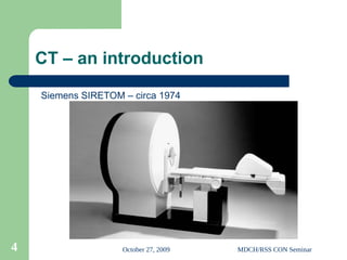
CT – an introduction
Siemens SIRETOM – circa 1974
5.
October 27, 2009MDCH/RSS CON Seminar
5
CT – an introduction
The latest multi-slice CT systems can image
an entire chest in less than ten seconds and
reconstructs the images in a similar time
period
Faster systems = higher volume of patients
Faster systems = development of new
techniques
6.
October 27, 2009MDCH/RSS CON Seminar
6
Radiation risk
Risk is proportional to absorbed dose
Risk is quantified by determining the
“effective dose” and is expressed as
millisievert (mSv)
Atomic bomb survivors who experienced
doses that were slightly higher than doses
encountered in CT demonstrated increased
cancers (5-20 mSv vs 1-10 mSv)
October 27, 2009MDCH/RSS CON Seminar
8
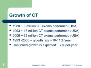
Growth of CT
1980 ~ 3 million CT exams performed (USA)
1993 ~ 18 million CT exams performed (USA)
2006 ~ 62 million CT exams performed (USA)
1993 -2006 – growth rate ~10-11%/year
Continued growth is expected ~ 7% per year
October 27, 2009MDCH/RSS CON Seminar
10
Medical dose - US population2
11.
October 27, 2009MDCH/RSS CON Seminar
11
Medical dose - US population2
12.
October 27, 2009MDCH/RSS CON Seminar
12
Average effective dose major sources (US)2
13.
October 27, 2009MDCH/RSS CON Seminar
13
Average effective dose all sources US2
14.
October 27, 2009MDCH/RSS CON Seminar
14
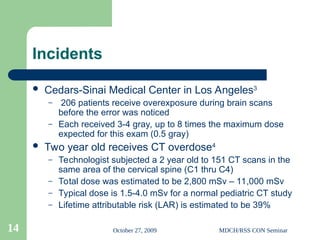
Incidents
Cedars-Sinai Medical Center in Los Angeles3
– 206 patients receive overexposure during brain scans
before the error was noticed
– Each received 3-4 gray, up to 8 times the maximum dose
expected for this exam (0.5 gray)
Two year old receives CT overdose4
– Technologist subjected a 2 year old to 151 CT scans in the
same area of the cervical spine (C1 thru C4)
– Total dose was estimated to be 2,800 mSv – 11,000 mSv
– Typical dose is 1.5-4.0 mSv for a normal pediatric CT study
– Lifetime attributable risk (LAR) is estimated to be 39%
15.
October 27, 2009MDCH/RSS CON Seminar
15
Why new CT rules
Current Ionizing Radiation Rules (1975)
Lack rules specifically regulating CT
Patient dose from CT >> regular x-ray exams
Higher dose = higher risk of cancer
Technological advances = increased use
Use of CT is expected to continue to grow
48% of all exposure comes from medical sources
49% of medical exposures comes from CT
16.
October 27, 2009MDCH/RSS CON Seminar
16
What rules are we proposing
Purpose/Scope/Exemptions
Personnel requirements
Equipment requirements
Quality control program
Facility design requirements
Surveys
17.
October 27, 2009MDCH/RSS CON Seminar
17
Sources
The Michigan Department of Community Health’s
Certificate of Need Review Standards for Computed Tomograp
hy (CT) Scanner Services
The Federal Performance Standards for Ionizing Radiation
Emitting Products, 21 C.F.R. §1020.33 “
Computed tomography (CT) equipment” (June 10, 2005)
The American College of Radiology’s
CT Accreditation Program Requirements
The Conference of Radiation Control Program Director’s
Suggested State Regulations for the Control of Radiation,
Part F X-ray in the Healing Arts.
Reviewing the proposed rules with interested stakeholders
18.
October 27, 2009MDCH/RSS CON Seminar
18
Exemptions
Purpose and scope
– Specify that the rules apply to all registrants who use CT
systems for the intentional exposure of humans for the
purpose of diagnostic imaging
– This would exempt PET/CT and SPECT/CT unless the CT
portion of the system is used for diagnostic imaging
Exemptions
– Specific exemption for CT systems with power ratings of 5
kilowatts or less
19.
October 27, 2009MDCH/RSS CON Seminar
19
Personnel requirements
Requirements for:
– Interpreting physicians
– Radiological technologists
– Medical physicist
20.
October 27, 2009MDCH/RSS CON Seminar
20
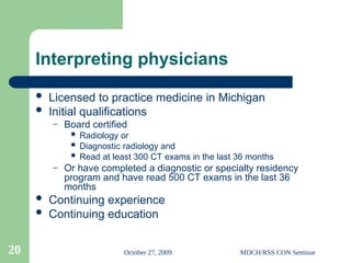
Interpreting physicians
Licensed to practice medicine in Michigan
Initial qualifications
– Board certified
Radiology or
Diagnostic radiology and
Read at least 300 CT exams in the last 36 months
– Or have completed a diagnostic or specialty residency
program and have read 500 CT exams in the last 36
months
Continuing experience
Continuing education
21.
October 27, 2009MDCH/RSS CON Seminar
21
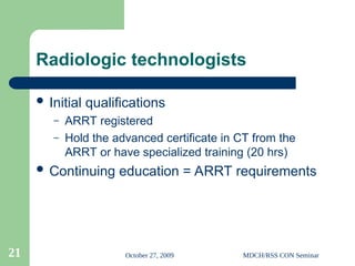
Radiologic technologists
Initial qualifications
– ARRT registered
– Hold the advanced certificate in CT from the
ARRT or have specialized training (20 hrs)
Continuing education = ARRT requirements
22.
October 27, 2009MDCH/RSS CON Seminar
22
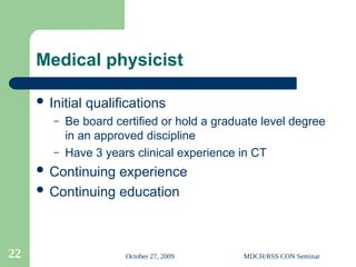
Medical physicist
Initial qualifications
– Be board certified or hold a graduate level degree
in an approved discipline
– Have 3 years clinical experience in CT
Continuing experience
Continuing education
23.
October 27, 2009MDCH/RSS CON Seminar
23
Equipment
Plan to adopt by reference the FDA’s CT
requirements for Computed tomography
– 21 C.F.R. §§1020.33 (June 10, 2005)
– Must be maintained in compliance with those
regulations
24.
October 27, 2009MDCH/RSS CON Seminar
24
Quality control program
Shall be established and implemented under
the supervision of the medical physicist (MP)
MP must perform an initial or acceptance test
of each CT system prior to use on patients
MP must perform an annual evaluation of the
CT system and quality control program
Facility conducts a continuous quality control
program designed/overseen by the MP
25.
October 27, 2009MDCH/RSS CON Seminar
25
Facility design requirements
Enclosure must meet the requirements of R325.5331
Operator must be able to operate the equipment
from a shielded position
Operator must be able to communicate with the
patient from the control panel
Operator must be able to see the patient from the
control panel
Electronic viewing systems must remain operational
or must be repaired before any further examinations
are performed
26.
October 27, 2009MDCH/RSS CON Seminar
26
Surveys
Requires a radiation shielding survey by a
medical physicist prior to use on patients for
newly installed systems or within 1 year for
existing systems
Medical physicist must provide a written
report to the facility
27.
October 27, 2009MDCH/RSS CON Seminar
27
References
1
- Average effective dose in millisieverts (mSv) as
compiled by Fred Mettler, Jr., et al., “Effective Doses
in Radiology and Diagnostic Nuclear Medicine: A
catelog,”Radiology Vol 248, No 1, pp 254-263, July
2008.
2
- NCRP Report 160, Ionizing Radiation Exposure of
the Population of the United States, 2009
3
– FDA Medical Devices Alerts and Notices
4
– “California technologist faces testimony in CT
overdose case”, AuntMinnie.com, September 18,
2009
28.
October 27, 2009MDCH/RSS CON Seminar
28
Contact Information
John Ferris, Jr., Health Physicist
Department of Community Health
Radiation Safety Section
www.michigan.gov/rss
jeferri@michigan.gov
Phone: 313 456-4660
Editor's Notes
#1 Welcome and Introduction
Some of you may know that MDCH, along with members of the Department of Environmental Quality (DEQ) are in the process of revising our entire Ionizing Radiation Rules. The revision process is necessary in order to become an agreement state. The proposed rules for CT we will discuss today is separate from that project. Hopefully we will be able to demonstrate why we feel it is important to go forward with these CT rules at this time.
#2 "Computed tomography (CT)" means the production of a tomogram by the acquisition and computer processing of x-ray transmission data. Computed tomography includes the capability of producing axial tomograms.
Computed axial tomography (CAT) scanning was invented by Godfrey N. Hounsfield in 1972 and independently by Alan Cormack in 1972.
Hounsfield’s CT scanner took several hours to acquire data and days to reconstruct a single image from the data
#3 About 2 years after CT was invented, Siemens introduced the first commercial CT system in May 1974 called the SIRETOM
First units could only image a patients head
These early units took several minutes to acquire image data and several minutes to reconstruct the data
#5 Today, due to several technological advances, the latest multi-slice/multi-detector systems can image and reconstruct data in just a few seconds.
These faster more technologically advanced systems have lead to a higher volume of patients being imaged. One, because they are simply faster and allow more patients to be imaged in a given amount of time and two, advances in technology have lead to the development of new imaging techniques and an ever expanding use of CT.
Three of the major advances include:
1 – Slip-ring technology – allows for a continuous rotation of the x-ray tube and other components of the system
2 – Helical scanning – allows for a rapid acquisition of a volume set - the patient is moved through the gantry during the scan – this was made possible by the invention of slip-ring technology
3 – Multi-detector CT (MDCT) – allows for the acquisition of a larger volume of data (large number of thin slices) during a single rotation of the x-ray tube
#6 In radiation protection, it is commonly assumed that the risk for adverse health effects is proportional to the amount of radiation dose absorbed. The amount of dose received is dependent on the type of examination performed.
Risk is usually quantified by estimating the “effective dose” to partial or whole-body irradiation from or between different sources or exams. The quantity most relevant for assessing this risk is the “effective dose”. Effective dose is evaluated in units of millisieverts (mSv).
sievert (Sv): The special name (in the SI system) for the unit of equivalent dose and effective dose (or effective dose equivalent); 1 Sv =1 J/kg. 1 Sv = 100 rem (see rem).
Effective dose from diagnostic CT procedures is estimated to be 1-10 millisieverts. This range is not much lower than the lowest doses of 5-20 mSv received by some of the Japanese survivors of the atomic bombs. These survivors, who are estimated to have experienced doses only slightly larger than those encountered in CT, have demonstrated a small but increased radiation related excess relative risk for cancer mortality.
Estimates of the effective dose from diagnostic procedures can vary by a factor of 10 or more depending on the type of CT procedure, patient size, the CT system and its operating technique.
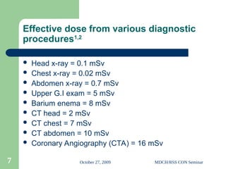
#7 The following slide shows typical effective dose levels for various diagnostic procedures.
It should be noted that effective doses for most CT studies are significantly higher than for conventional x-rays.
Dose from an abdomen CT exam is approximately 14 times higher than for an abdomen x-ray.
Dose from a chest CT exam is approximately 350 times higher than that received from a chest x-ray
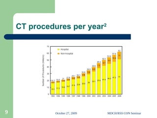
#8 Since the first commercial use of CT we have seen a rapid and huge increase in the number of CT procedures being performed in the US.
In 1980 there were about 3 million CT exams performed in the United States.
By 1993 that had grown to over 18 million and by 2006 it was over 62 million.
On average the use of CT increased between 10-11% per year from 1993 to 2006.
It is expected that use of CT will continue to grow at about 7% per year.
#9 This chart shows the growth in the use of CT over the last one and a half decades. Much of this growth is attributed to the technological improvements made in CT
1 – Slip-ring technology – allows for a continuous rotation of the x-ray tube and other components
2 – Helical scanning – allows for a rapid acquisition of a volume set - the patient to be moved through the gantry during the scan – this was made possible by the invention of slip-ring technology
3 – Mulitdetector CT (MDCT) – allows for the acquisition of larger volume (large number of thin slices ) during a single rotation of the x-ray tube
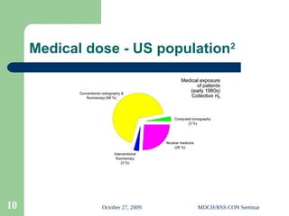
#10 From a medical perspective, we can look at the various imaging modalities to see where the greatest risk is (the areas with the highest percentage of dose).
From this chart we can see that back in the early 1980s, conventional radiography and fluoroscopy contributed about 68% of the dose, while CT contributed about 3%.
HE – Effective dose equivalent as a percentage of total effective dose for the US population in early 1980s in millisieverts
effective dose equivalent (HE): The sum over specified organs and
tissues of the products of the mean dose equivalent in a tissue (HT)
and the weighting factor for that tissue or organ (wT):
. The formulation differs from ICRP (1991) and is defined in ICRP
(1977). Now superseded in ICRP and NCRP recommendations by effective
dose, but the formulation is still in use by most federal and state
agencies.
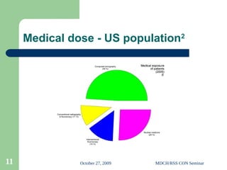
#11 Looking at the same type of data in 2006, we can see the picture has changed. Now CT contributes almost ½ of all medical dose, while contributions from conventional radiography and fluoroscopy dropped to about 11%. There was also an increase in dose from interventional fluoroscopy which went from 3% to 14%. Contributions from nuclear medicine procedures remained about the same.
S – Collective effective dose as a percentage of total collective effective dose in person-sieverts
collective effective dose (S) (person-Sv): Most frequently the product
of the mean effective dose for a population and the number of persons
in the population, but, more precisely, and preferably, the sum of all
individual effective doses in the population of concern.
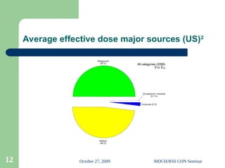
#12 This chart illustrates the average effective dose from all sources. While background radiation contributes about 50%, contributions from medical imaging is right behind at about 48%.
#13 This chart breaks the previous chart down into specific sources.
Of particular note is the observation that while the number of CT scans is only 17 % of the total procedures for the medical category, CT contributes nearly one-half (49 %) of all the dose for medical exposure, and nearly one-quarter (24%) of the dose for all sources of exposure.
#14 Earlier we talked about the higher risk from CT examination. Lately, there have been two reported incidents involving CT imaging that resulted in patients receiving significant overexposures.
The first incident:
Cedar-Sinai: In this case someone changed the imaging parameters for brain perfusion studies. For over eighteen months the error went unnoticed by the radiologists, technologists, and medical physicists. It took a patient complaining about hair loss, before the facility realized the error. From February 2008 until August 2009 about 206 patients underwent this study and received 6-8 times the normal dose they should have.
The second incident:
Two year old: In January 2008, a technologist subjected a 23 month old patient to a total of 151 scans at the same location while attempting to perform a CT scan of the cervical spine. Estimates of the patients total dose range from 2,800 mSv to as high as 11,000 mSv. The typical dose for this type of exam is usually 1.5-4.0 mSv. According to the hospital’s medial physicist, the child has a lifetime increased risk of a fatal cancer of 39%.
#15 Current IRR took effect in 1975 at about the time diagnostic CT imaging was just getting started and subsequently Michigan has no specific regulations governing the use of CT in diagnostic imaging.
Patient dose is much higher than from conventional x-rays.
Conventional thinking is that higher dose leads to higher risk of having cancer
Over the years, technological advances in CT have allowed for higher patient volumes to be imaged and have yielded many new clinical protocols that resulted in an increasing number of CT exams each year. Higher volumes and higher number of procedures have increased the collective effective dose to the population.
It is expected that the number of CT exams will continue to grow at about 7% per year.
48% of all exposure comes from medical sources
49% of medical exposure comes from CT
CT contributes about 24% of all exposure
#16 The major areas we plan to include in the new CT rules are:
Purpose/Scope/Exemptions – The current policy for regulatory rule specifies that each rule set should have a purpose and scope that describe who the rules are for and what uses they will regulate and if there are any exemptions.
Personnel requirements - We plan to include necessary personnel requirements.
There will also be requirements for
Equipment
Quality control
Facility design
And
Radiation surveys
#17 In preparing our proposed rules, we used several sources for guidance.
They include:
The Certificate of Need Review Standards for Computed Tomography (CT) Scanner Services
The Federal Performance Standards for “Computed tomography (CT) equipment”.
The American College of Radiology’s CT Accreditation Program Requirements
The Conference of Radiation Control Program Director’s Suggested State Regulations for the Control of Radiation, Part F X-ray in the Healing Arts
And
We are reviewing the proposed rules with interested stakeholders
#18 The rules in a little more detail:
Read this slide
R325.5701. Purpose and scope.
Rule 701. (1) This part establishes requirements governing the use of computed tomography (CT) systems in the healing arts.
(2) This part applies to all registrants who use CT systems for the intentional exposure of humans for the purpose of diagnostic imaging.
(3) CT systems with power ratings of 5 kilowatts or less are exempt from this part. CT scanners used only for attenuation corrections and anatomical markers are also exempt from this part.
(4) In addition to the requirements of this part, all registrants are subject to the applicable provisions of R325.5001 to R325.5665.
#19 Personnel requirements will cover interpreting physicians, radiological technologists, and medical physicists.
#20 Read slide
R325.5705. Interpreting physicians.
Rule 705. The following requirements apply to all physicians involved in the interpretation of CT images.
(a) Initial qualifications. Before beginning to interpret CT examinations independently, a physician shall be licensed under article 15 of the act and shall meet one of the following:
(i) Be board certified in radiology or diagnostic radiology by the American board of radiologists, the American osteopathic board of radiology, the royal college of physicians and surgeons of Canada, or le college des medicins du Quebec and have interpreted or reviewed 300 CT examinations in the past 36 months; or
(ii) Have completed an accredited diagnostic radiology residency and have interpreted or reviewed 500 CT examinations in the past 36 months under supervision; or
(iii) Have completed an accredited specialty residency, have a combined total of 200 hours of category I continuing medical education (CME) in the performance of and interpretation of CT in the subspecialty where CT reading occurs, and have interpreted or reviewed 500 CT examinations in the past 36 months under supervision.
(b) Continuing experience. Following the second anniversary date in which the requirements of subrule (a) of this rule were completed, the interpreting physician shall have interpreted or multi-read at least an average of 9 CT examinations per month over the prior 24-month period. This equates to interpreting at least 216 CT examinations in the prior 24-month period.
(c) Continuing education. Following the third anniversary date in which the requirements of subrule (a) of this rule were completed, the interpreting physician shall have earned at least 15 continuing medical education units in CT, at least half of which shall be category I, in the prior 36-month period.
#21 Read slide
R325.5707. Radiologic technologists.
Rule 707. The following requirements apply to all radiologic technologists involved in the operation of CT systems.
(a) Initial qualifications. Before beginning to perform CT examinations independently, the technologist shall:
(i) Be currently registered by the American registry of radiological technologists (ARRT); and
(ii) Document at least 20 hours of training and experience in operating CT equipment, radiation physics, and radiation protection or have the advanced certification in computed tomography from the ARRT.
(b) Continuing education. The technologist shall be in compliance with the ARRT requirements for continuing education appropriate to his or her practice needs, which are 24 credits in a 2-year period.
#22 Read slide
R325.5709. Medical physicist.
Rule 709. Each registrant with one or more CT systems shall employ or contract with a medical physicist to review the quality and safety of the operation of the CT scanner. The medical physicist shall meet the following:
(a) Initial qualifications.
Be certified in diagnostic radiological physics or radiological physics by the American board of radiology or by the Canadian college of physics in medicine; or
Have a graduate degree in medical physics, radiological physics, physics, or other relevant physical science or engineering discipline from an accredited institution and have coursework in the biological sciences with at least 1 course in biology or radiation biology and 1 course in anatomy, physiology, or similar topics related to the practice of medical physics.
Initial Experience. Have 3 years documented experience in a clinical CT environment.
Continuing experience. Following the second anniversary date of when the requirements of subrules (a) and (b) of this rule were completed, the medical physicist shall have surveyed at least 2 CT units in the prior 24-month period.
(d) Continuing education. Following the third anniversary date in which the requirements of subrules (a) and (b) of this rule were completed, the medical physicist shall have earned at least 15 continuing medical education units in CT, at least half of which shall be category I, in the prior 36-month period.
#23 Read slide
R325.5711. Equipment requirements.
Rule 711. (1) The regulations are adopted by reference. Copies of these regulations are available at no cost from the Radiation Safety Section, Michigan Department of Community Health, P.O. Box 30664, Lansing, Michigan 48909 and from the Center for Devices and Radiological Health, U.S. Food and Drug Administration, 5600 Fishers Lane, Rockville, Maryland 20857.
(2) CT equipment shall be maintained in compliance with the requirements of subrule (1) of this rule.
#24 Read slide
R325.5713. Quality control program.
Rule 713. (1) A quality control program shall be established and implemented under the supervision of the medical physicist.
(2) Initial performance testing of the CT system shall be completed by the medical physicist before use on human patients.
(3) Measurements of the radiation output of a CT x-ray system shall be performed with a calibrated dosimetry system. The dosimetry system shall have been calibrated within the preceding 24 months and the calibration shall be traceable to a national standard. During the calendar year in which the dosimetry system is not calibrated, an inter-comparison to a system calibrated within the previous 12 months shall be performed.
R325.5717. Continuous quality control.
Rule 717. (1) The CT facility shall establish a continuous quality control program with the assistance of the medical physicist. The medical physicist shall determine the frequency of each test and who should perform the test. An on-site radiological technologist should be identified to be responsible for conducting routine quality control.
(2) The quality control tests shall be performed in accordance with written procedures and methods. If the results of a quality control test fall outside of the control limits, corrective action shall be taken.
#25 Read slide
R325.5721. Facility design requirements
Rule 721. (1) A CT equipment enclosure shall be in compliance with the requirements of R325.5331.
(2) The operator shall be able to operate the equipment from a shielded position.
(3) Provision shall be made for two-way aural communication between the patient and the operator at the control panel.
(4) Windows, mirrors, closed-circuit television, or an equivalent shall be provided to permit continuous observation of the patient during irradiation and shall be so located that the operator can observe the patient from the control panel.
(5) If the primary viewing system is by electronic means and that system stops functioning properly, the system must be replaced or paired before any further examinations are performed.
#26 Read slide
R325.5723. Surveys.
Rule 723. (1) All CT x-ray systems installed after [insert effective date of the regulations] shall have a survey as defined in R325.5221 made initially before use on human patients. Those systems not previously surveyed shall have a survey made within one year of [insert effective date of the regulations]. In addition, such surveys shall be done after any change in the facility or equipment which might cause a significant increase in radiation hazard. The surveys required by this subrule shall be made by, or under the direct supervision of, a medical physicist.
(2) The registrant shall obtain a written report of the survey from the medical physicist, and a copy of the report shall be made available to the department upon request.