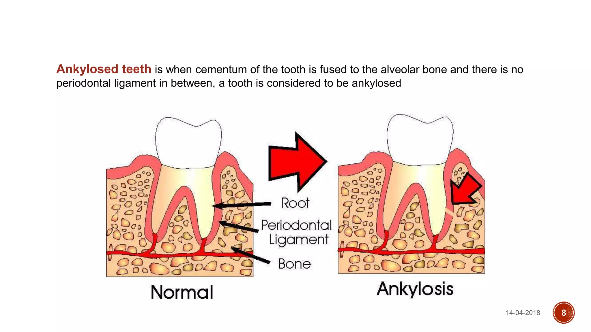
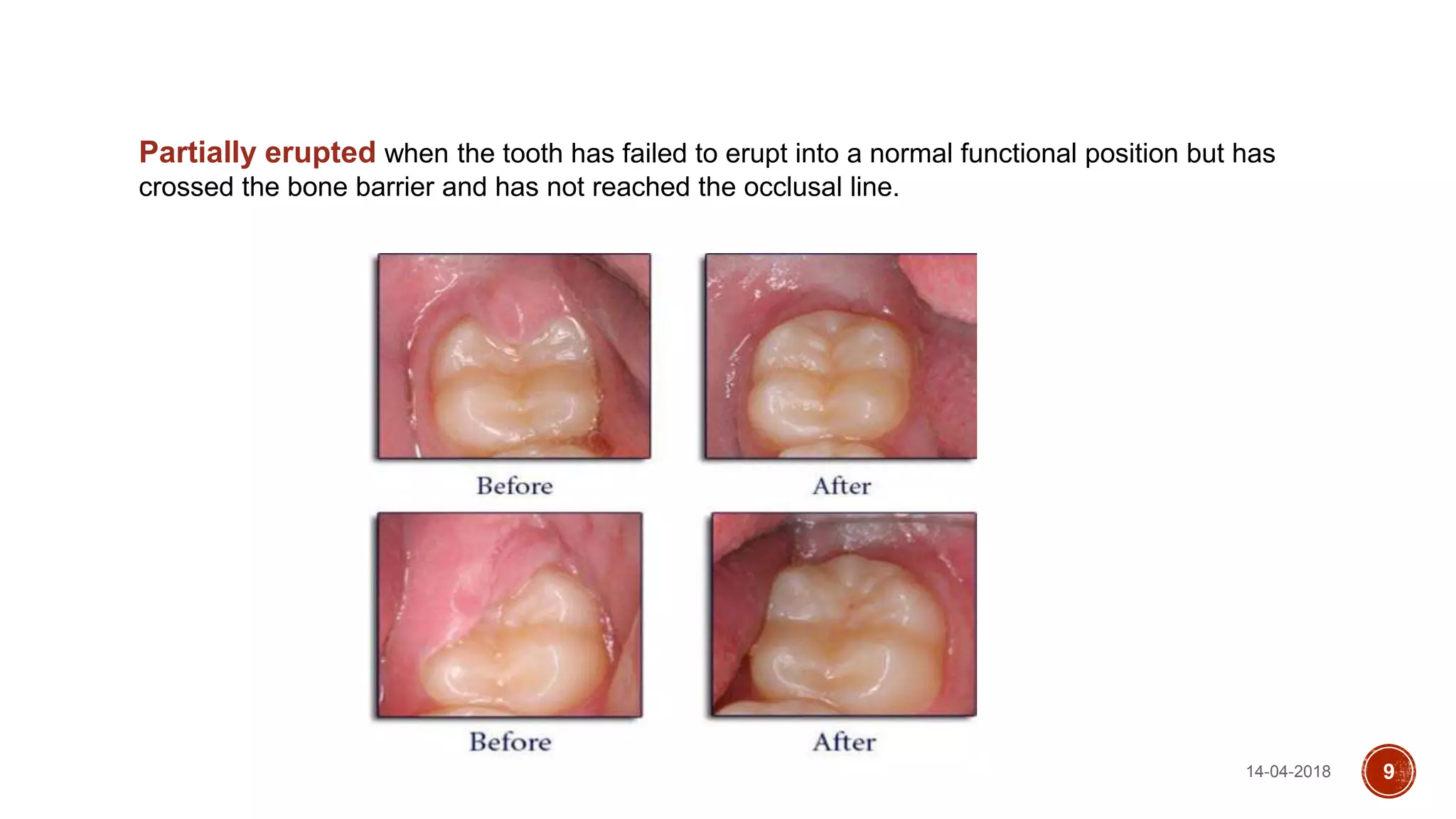
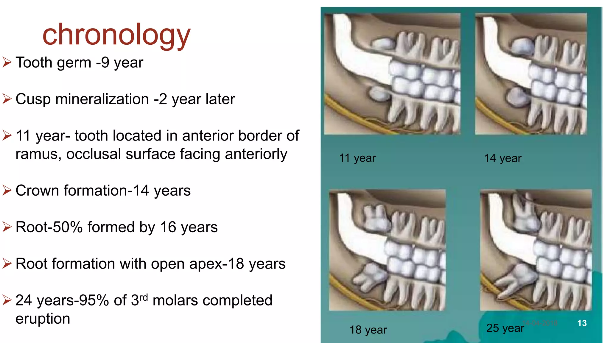
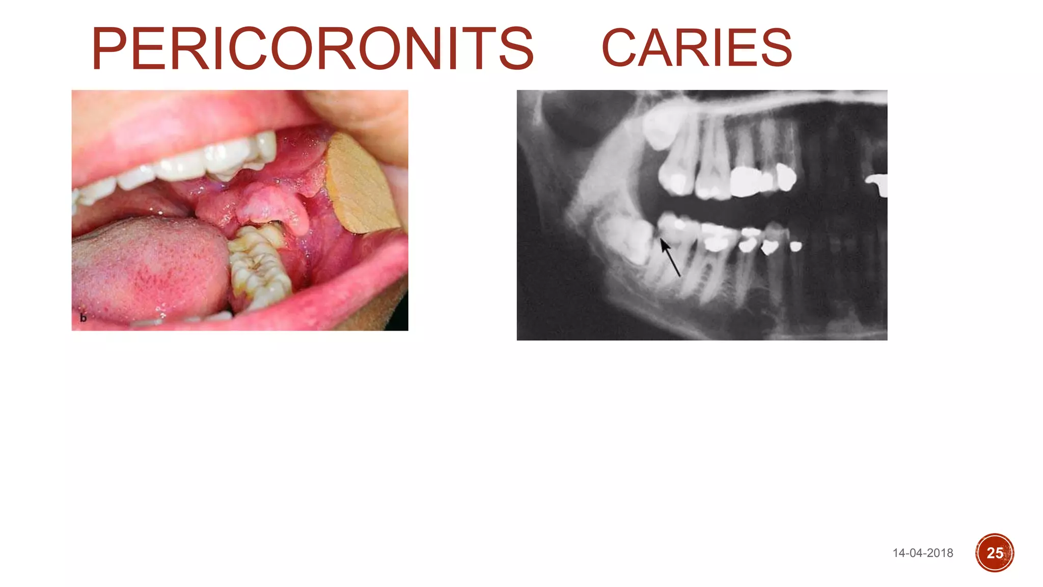
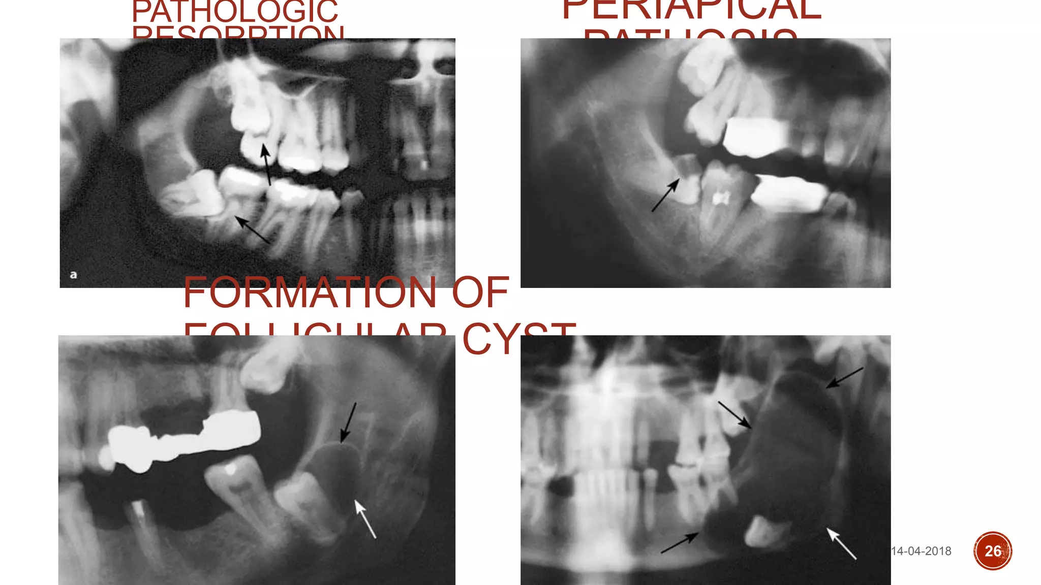
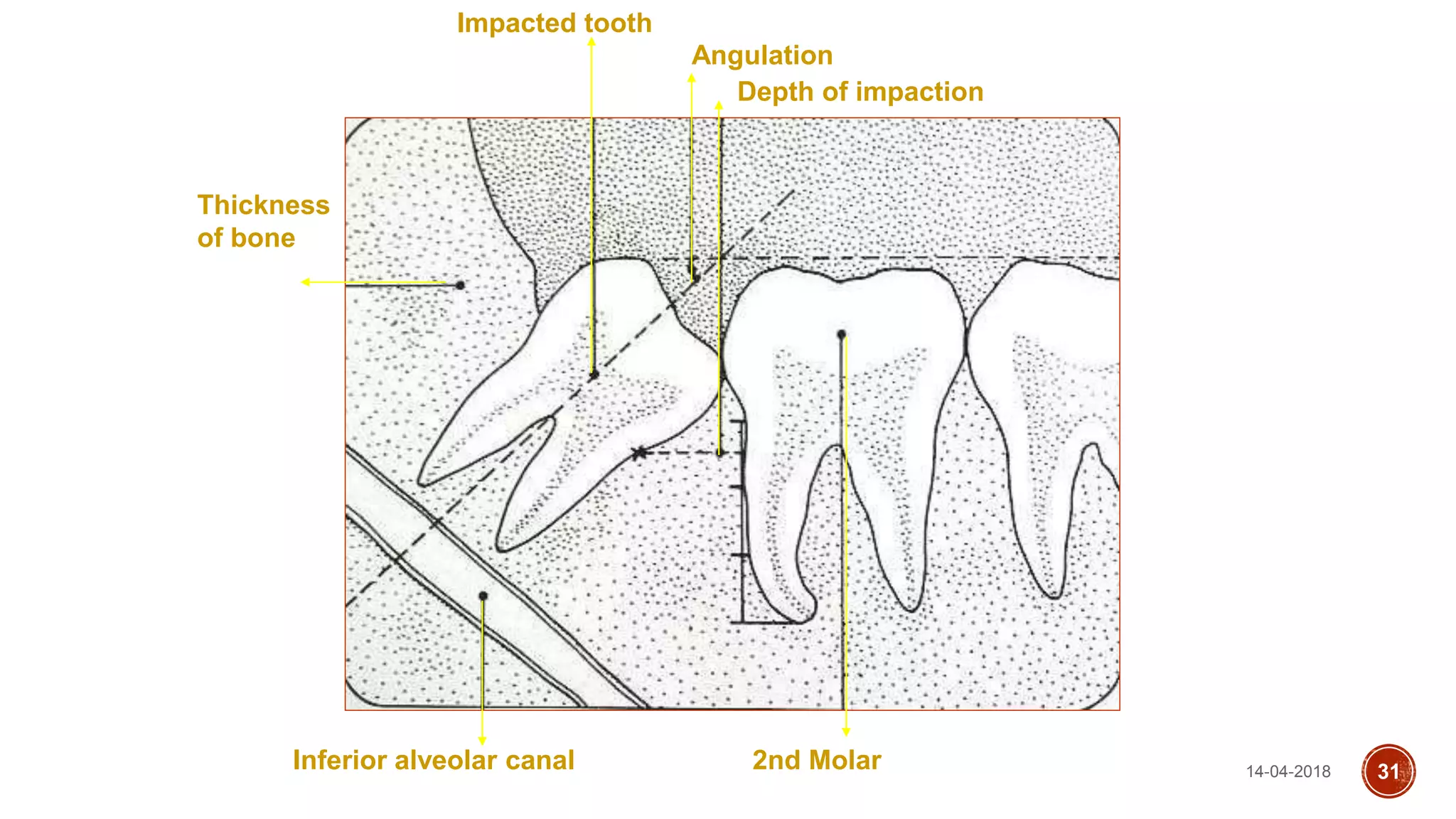
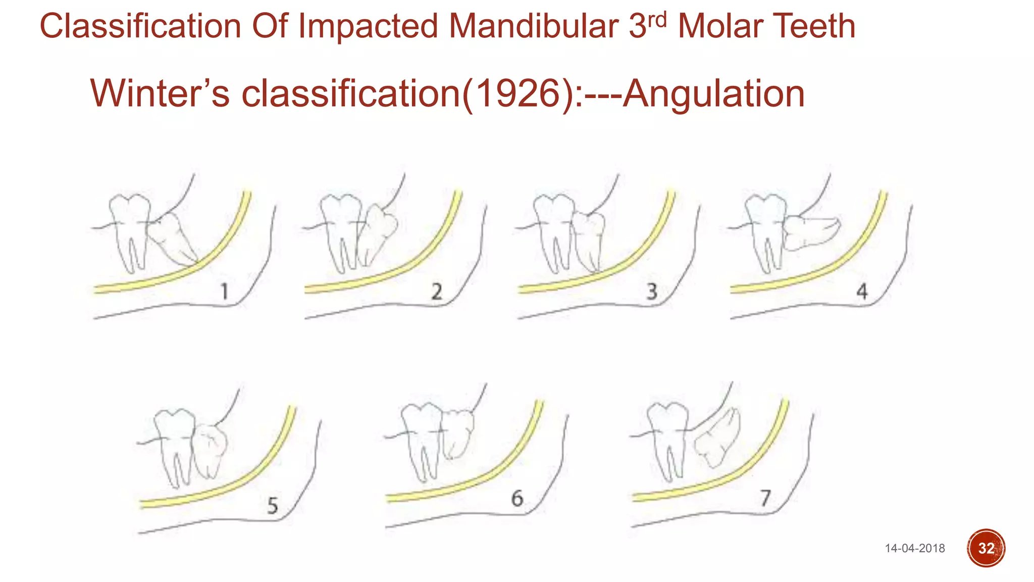
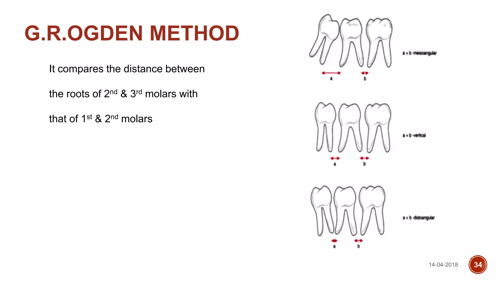
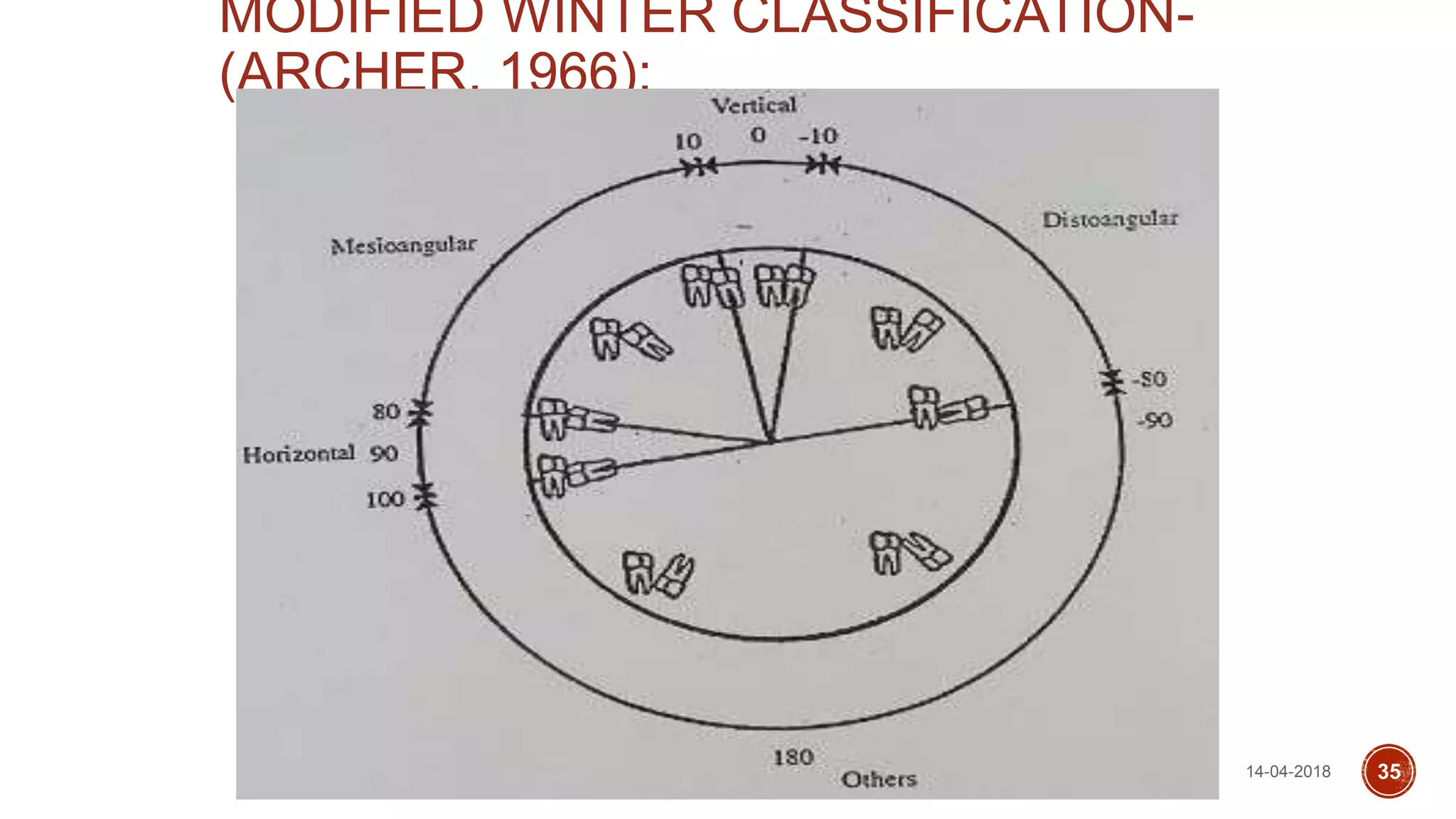
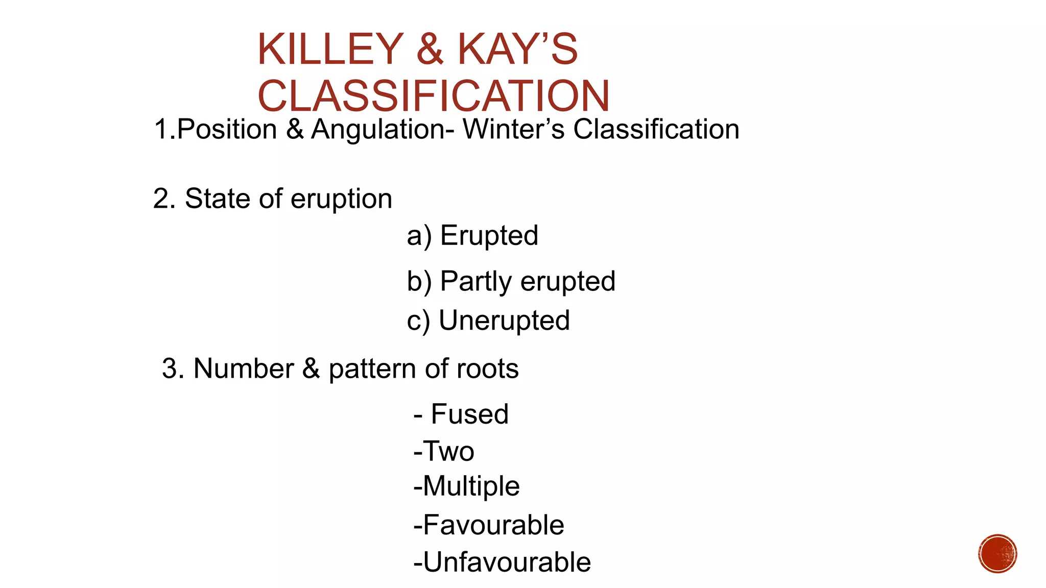
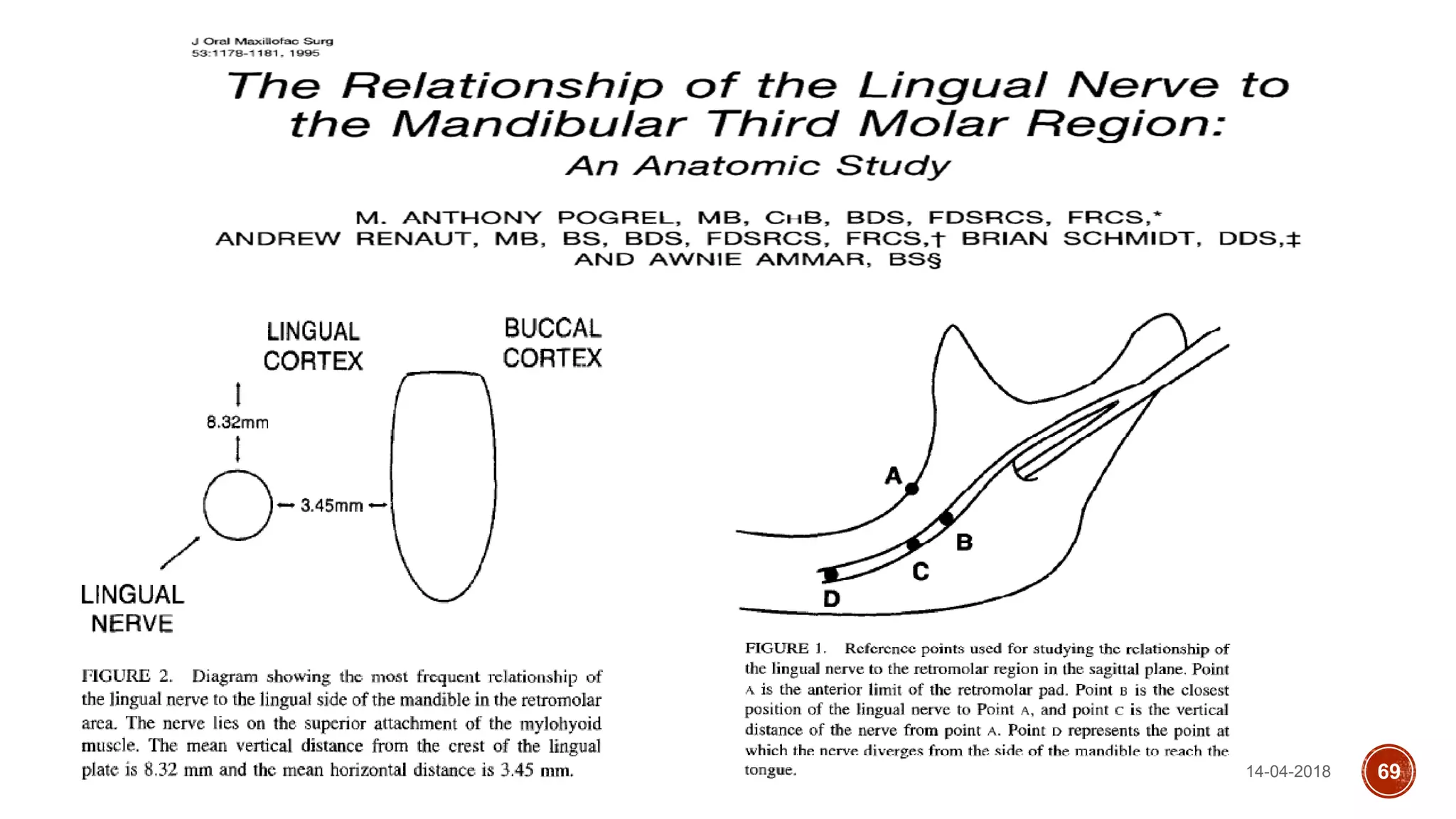
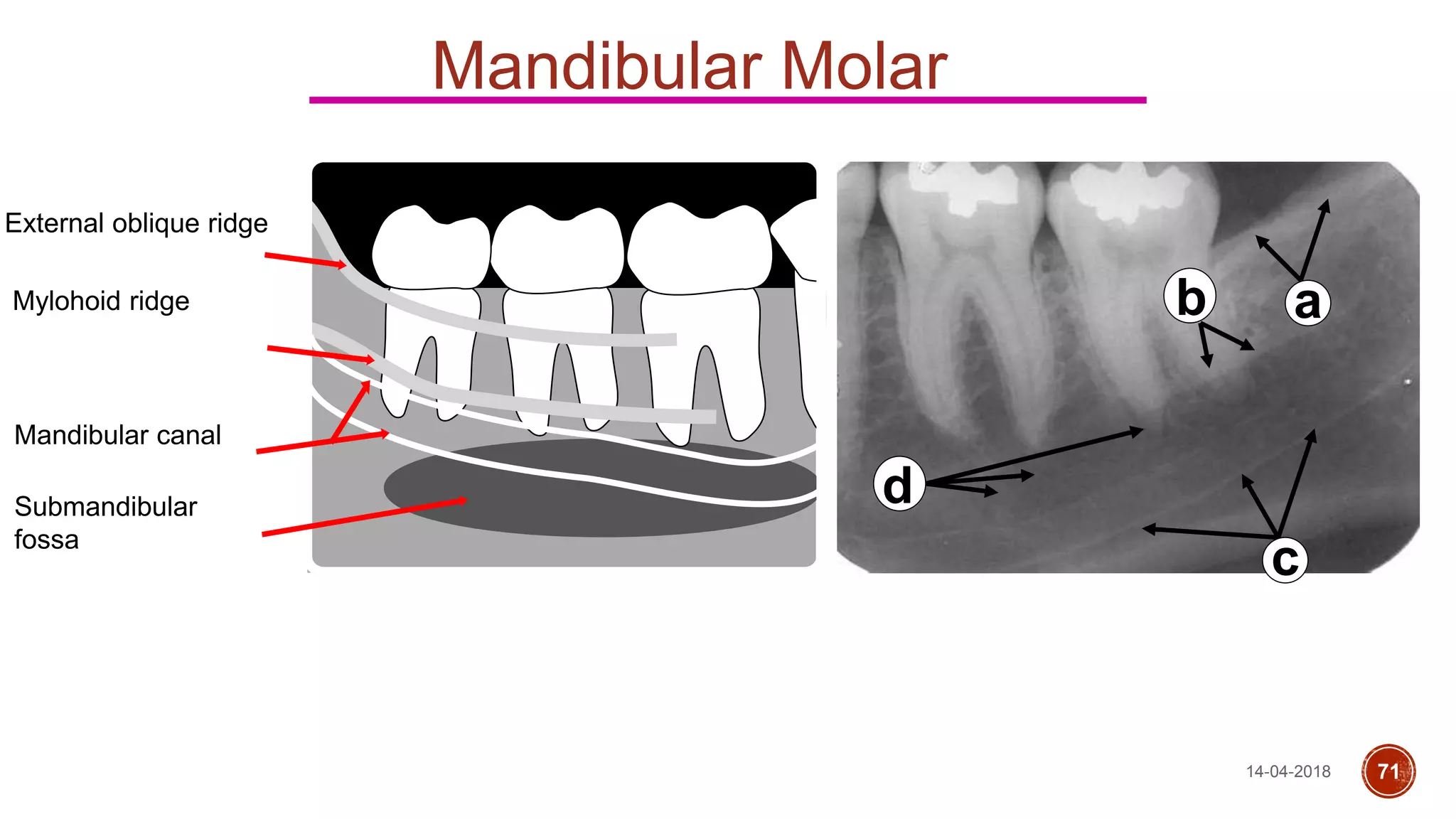
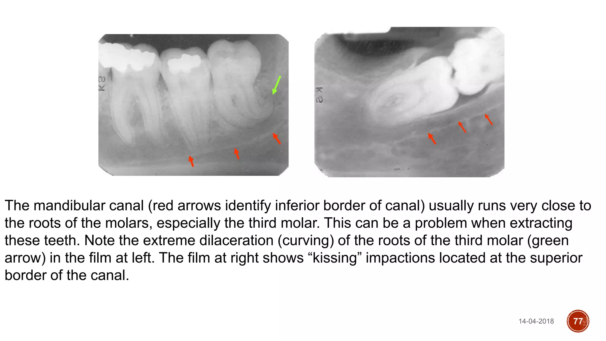
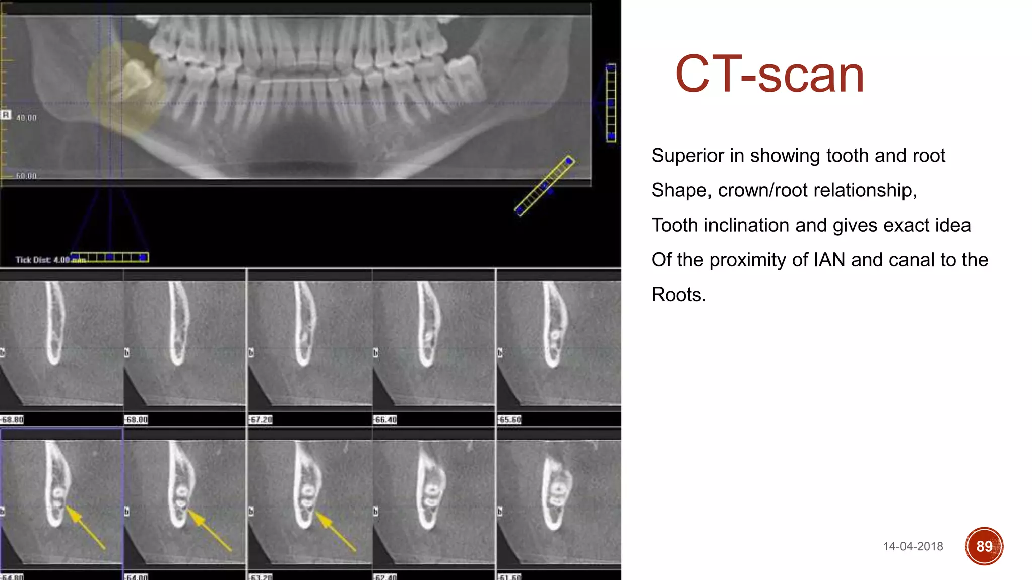
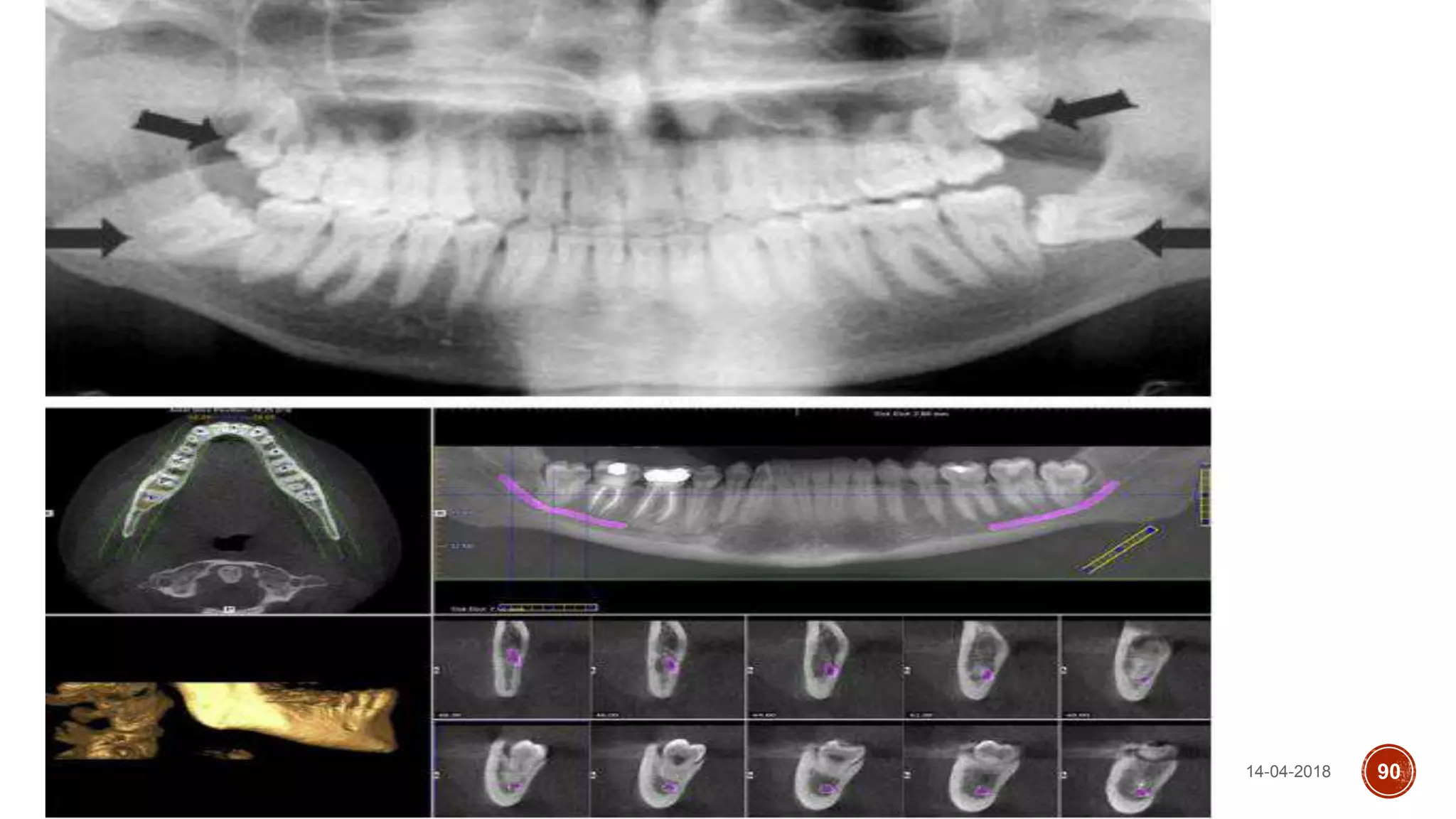
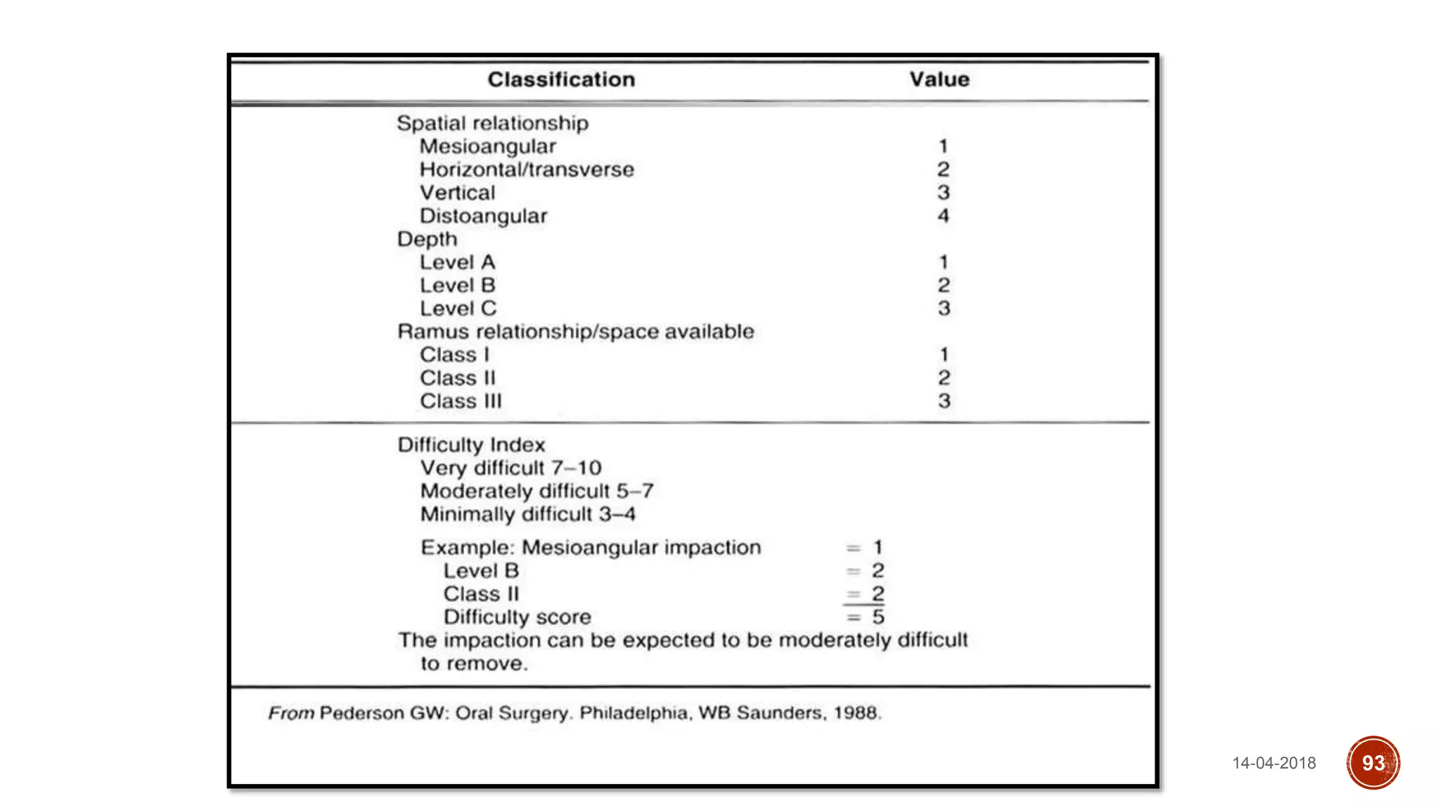
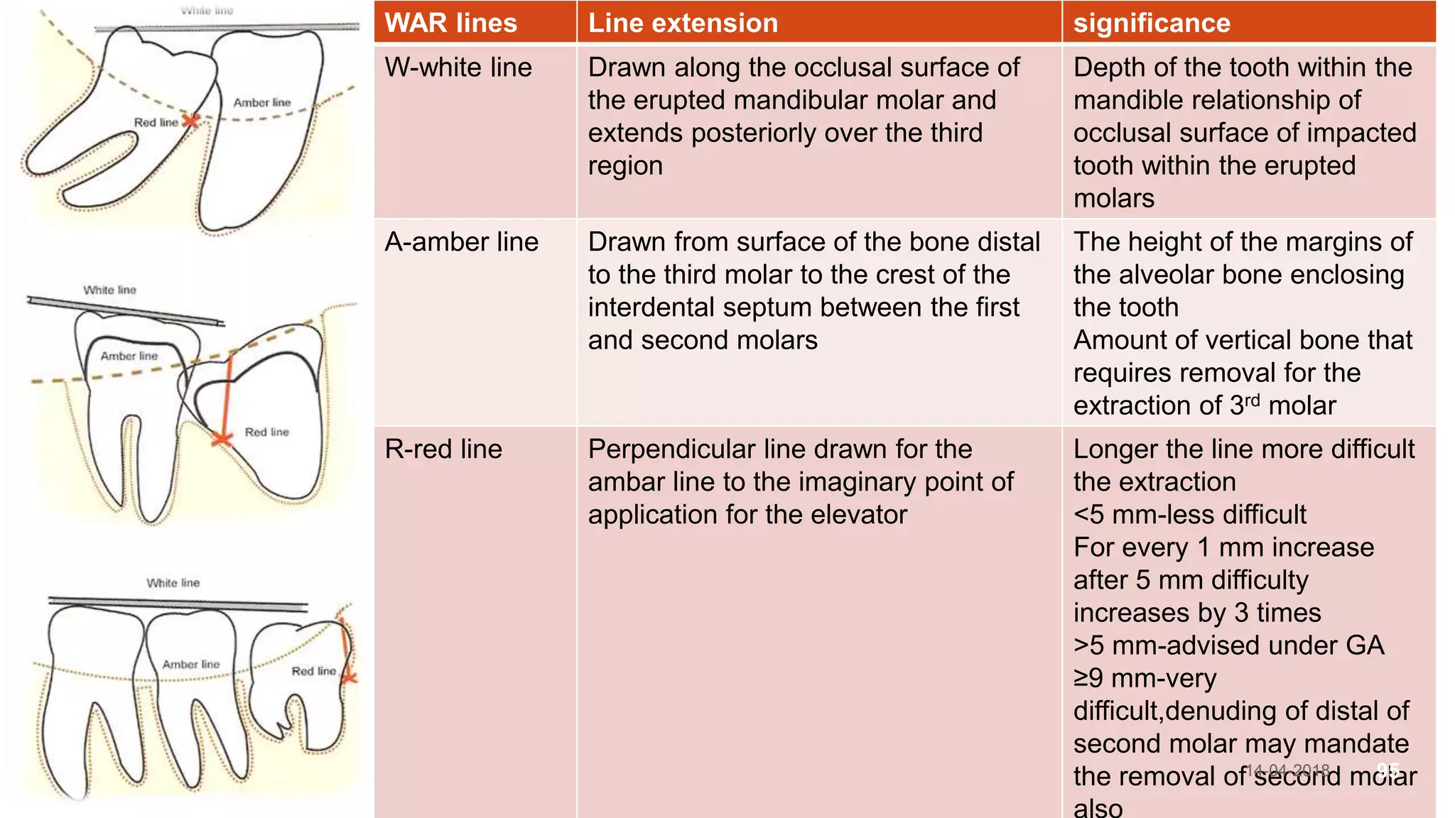
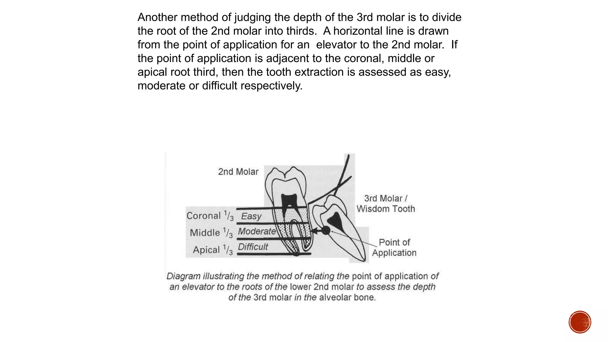
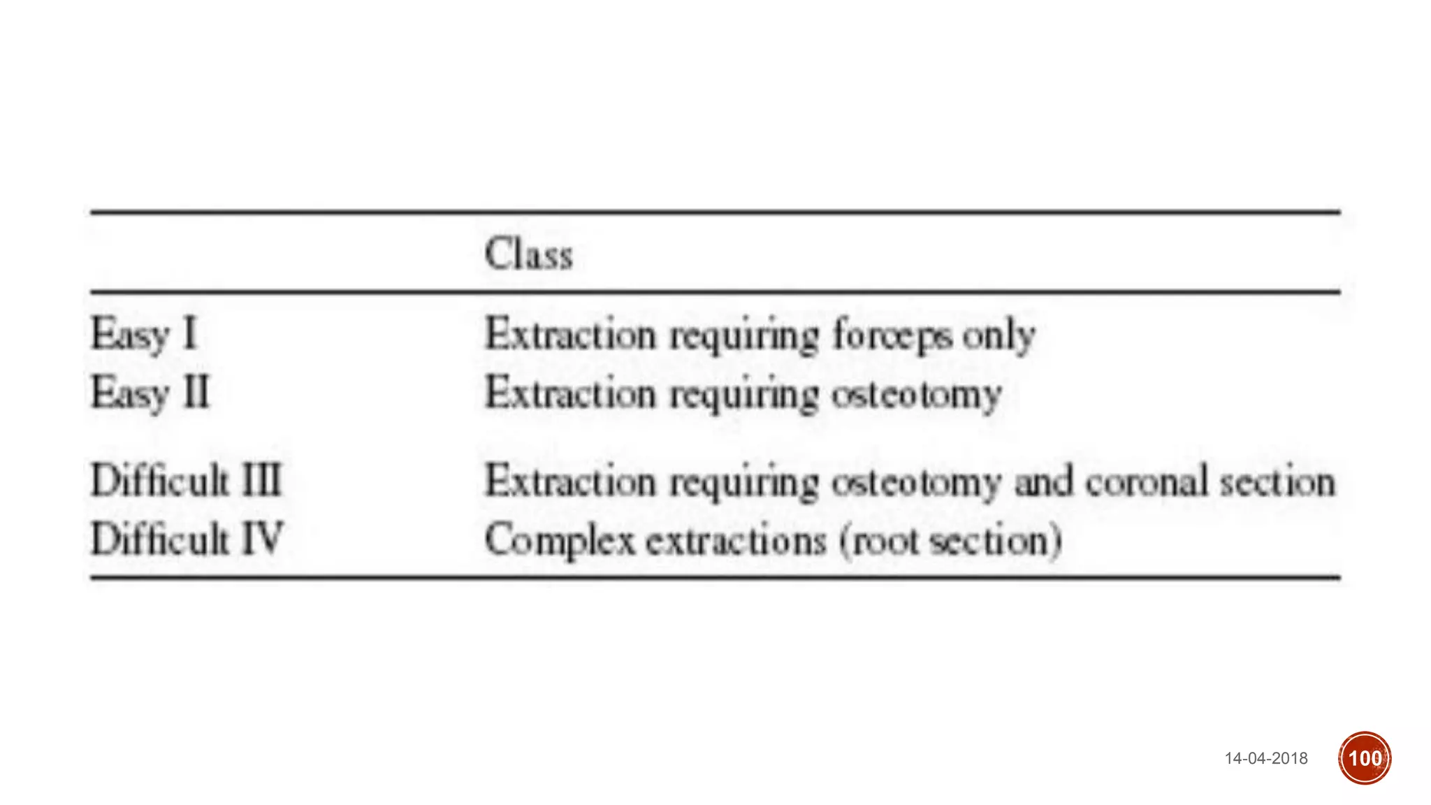
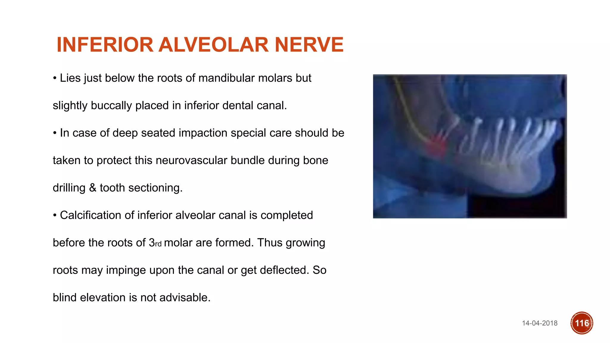
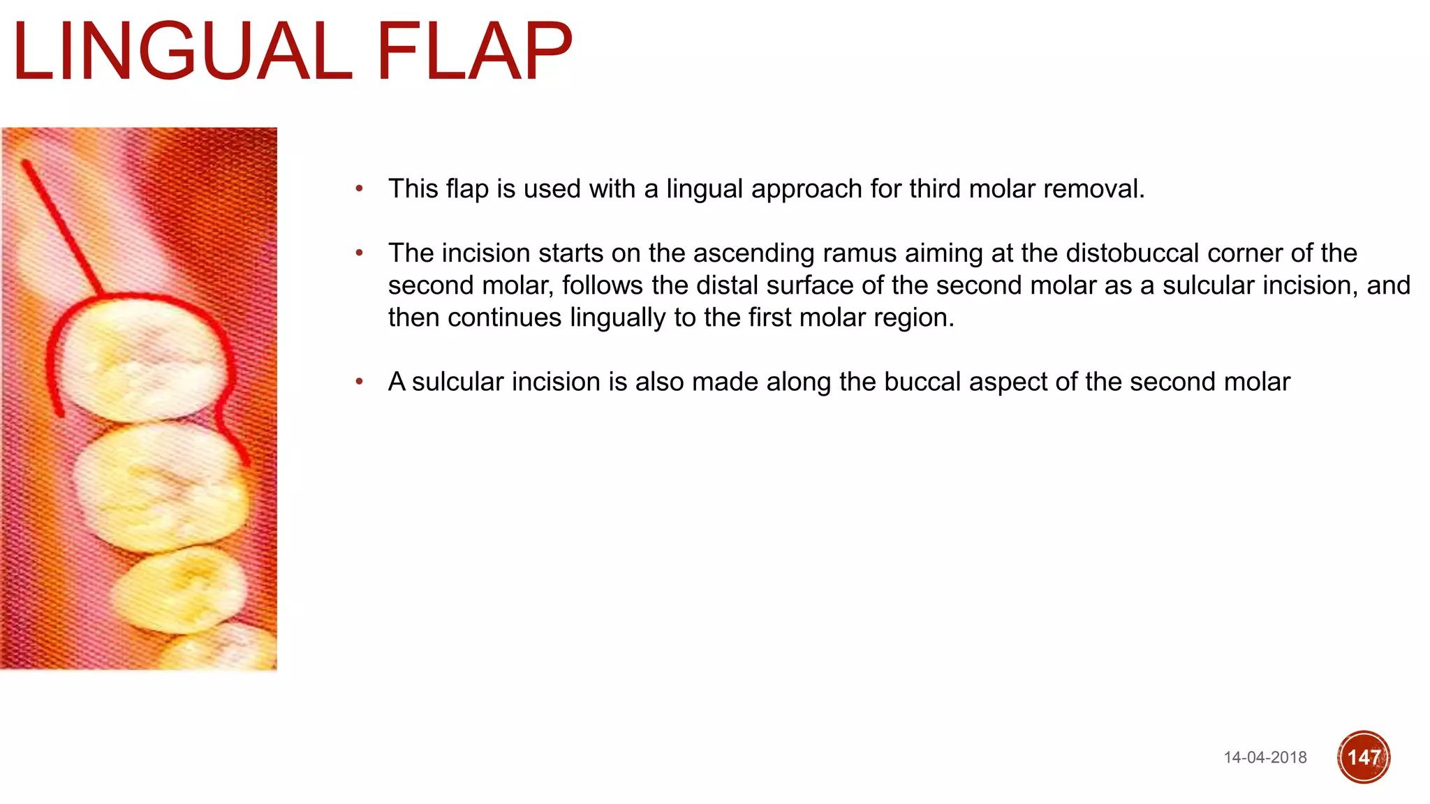
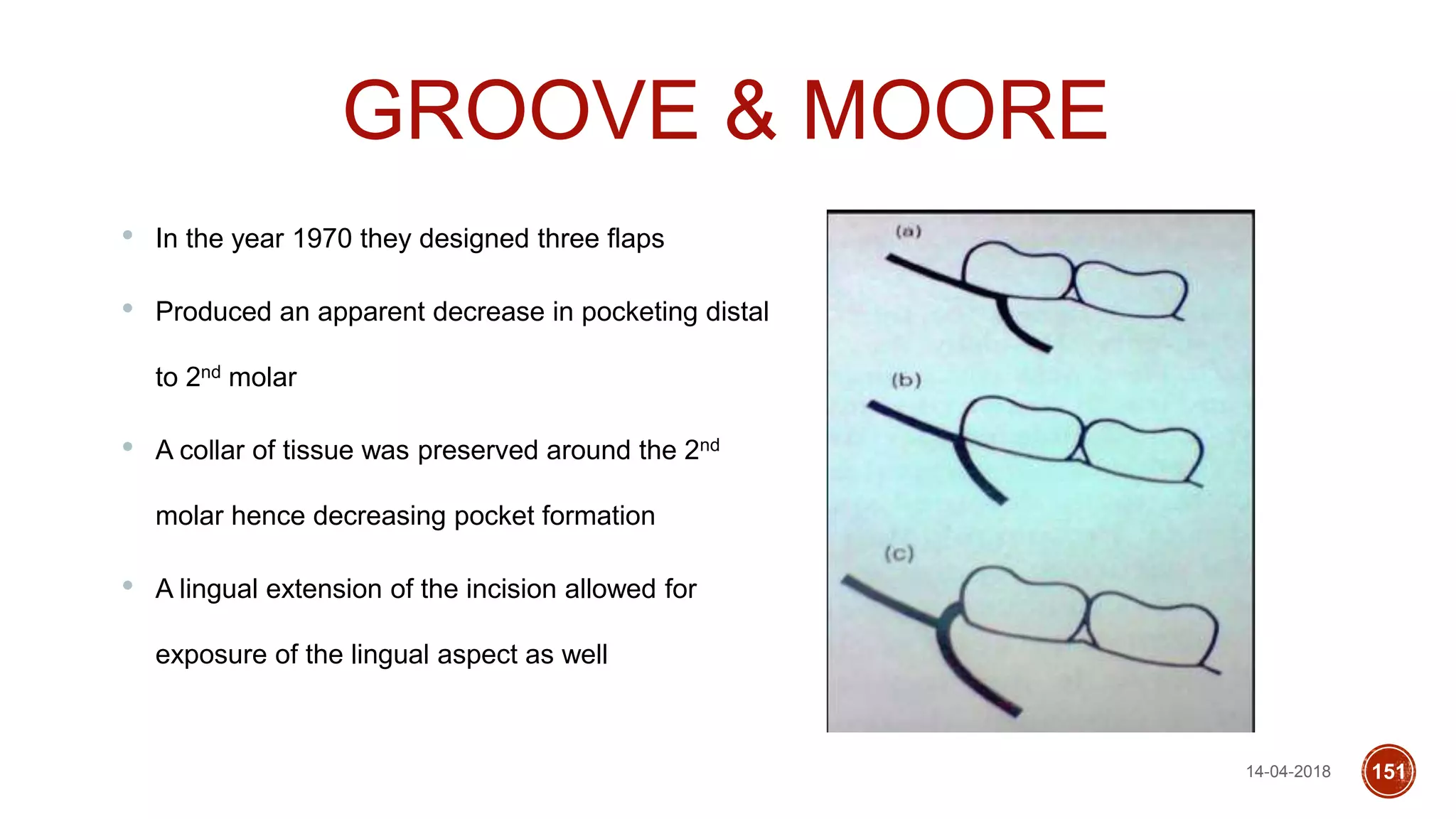
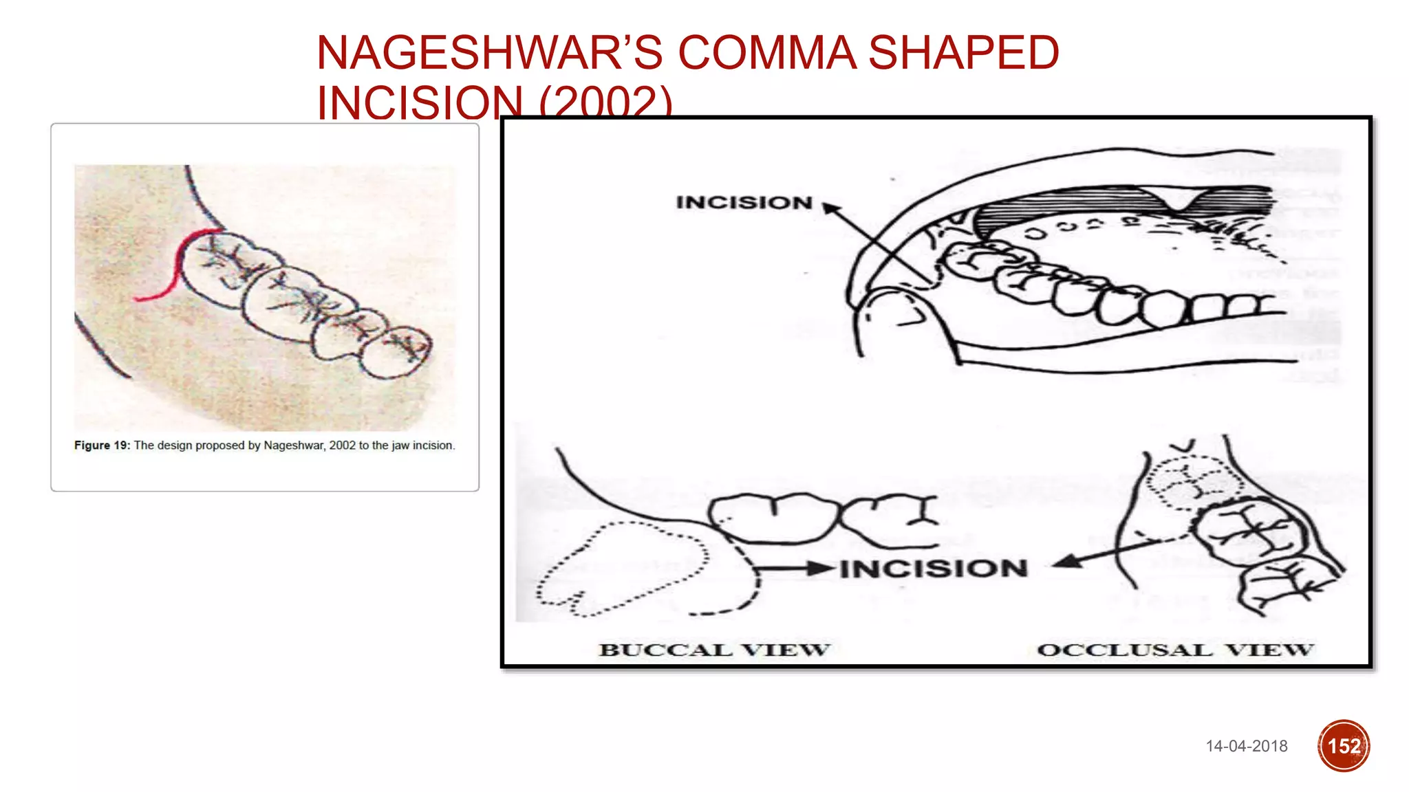
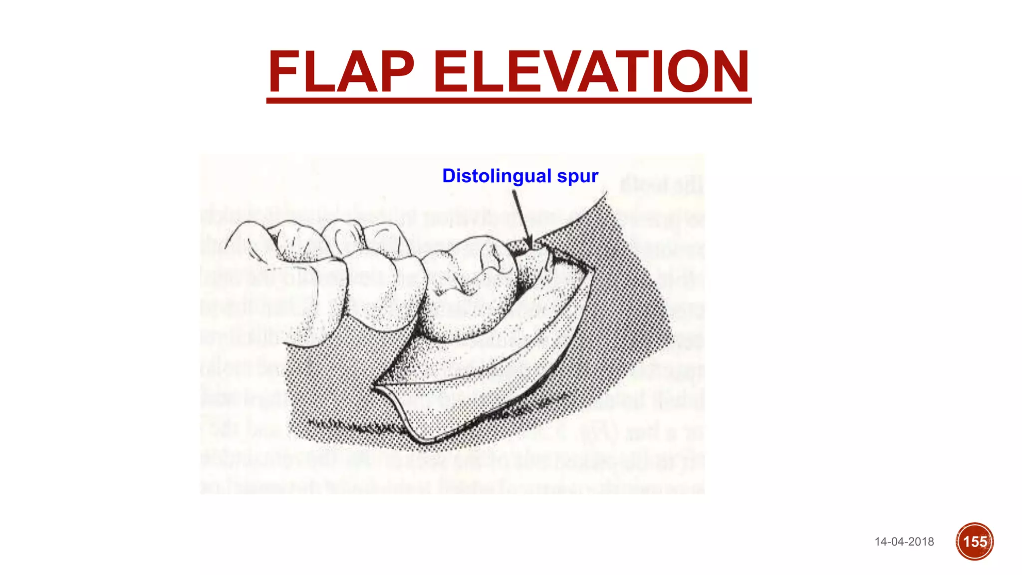
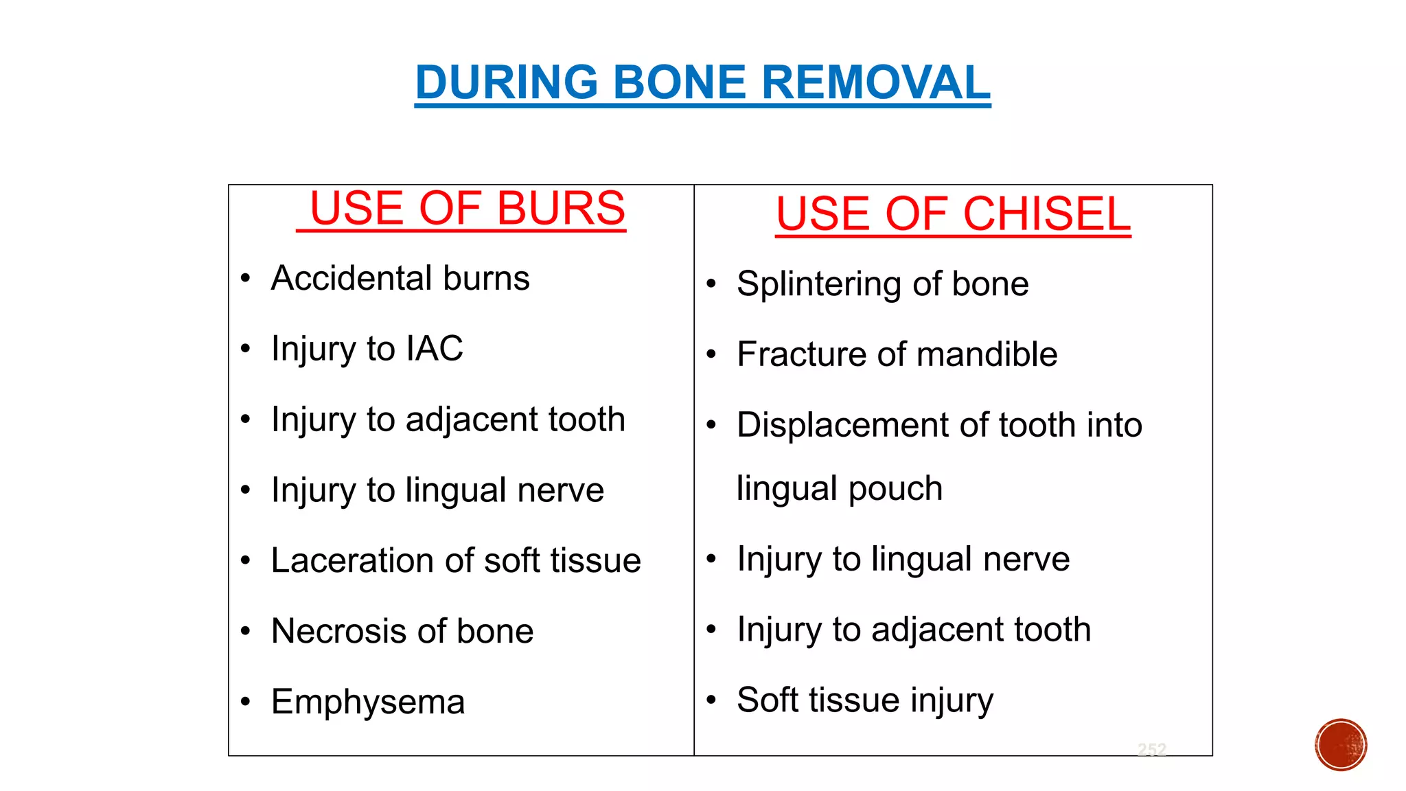
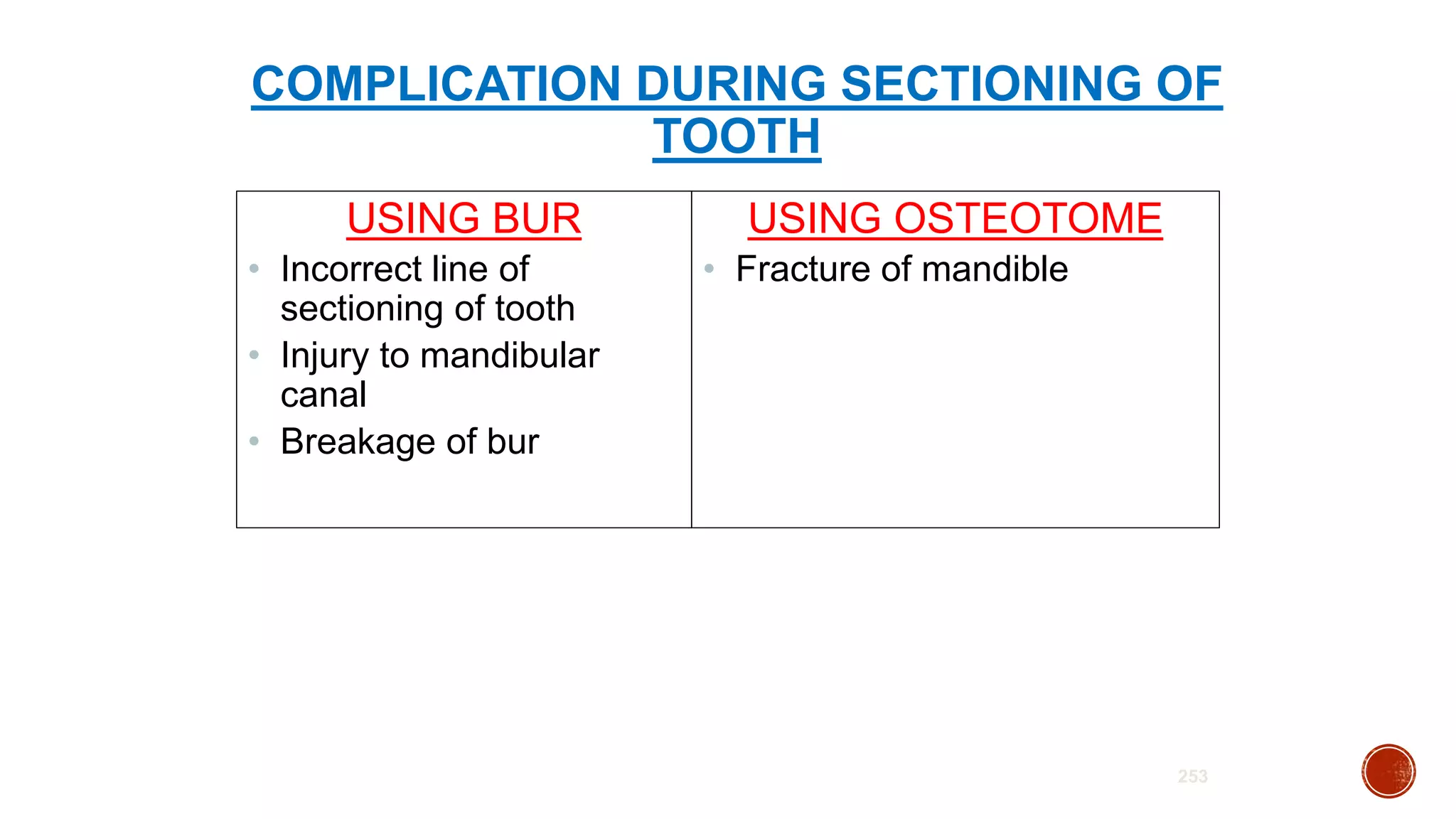
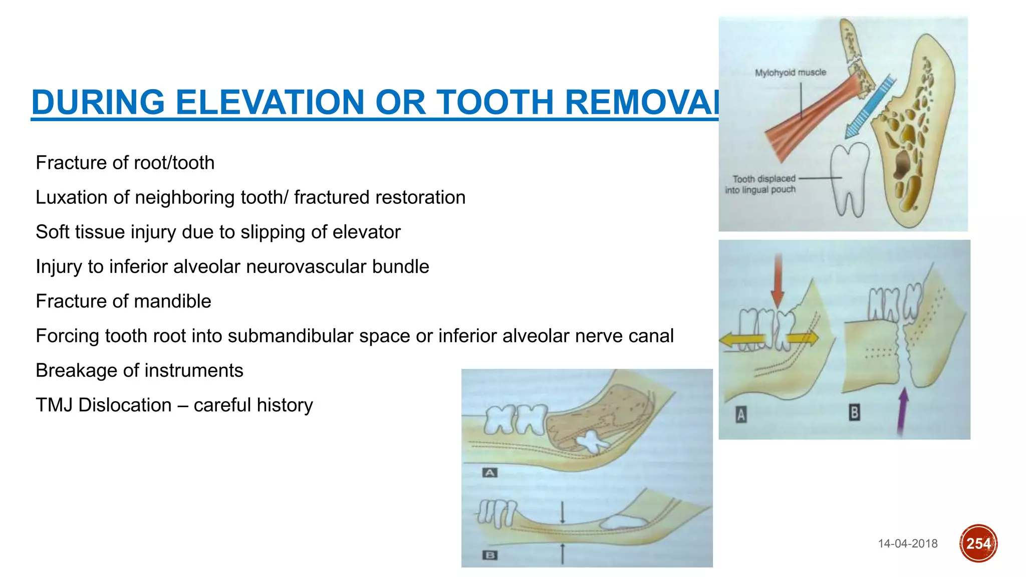
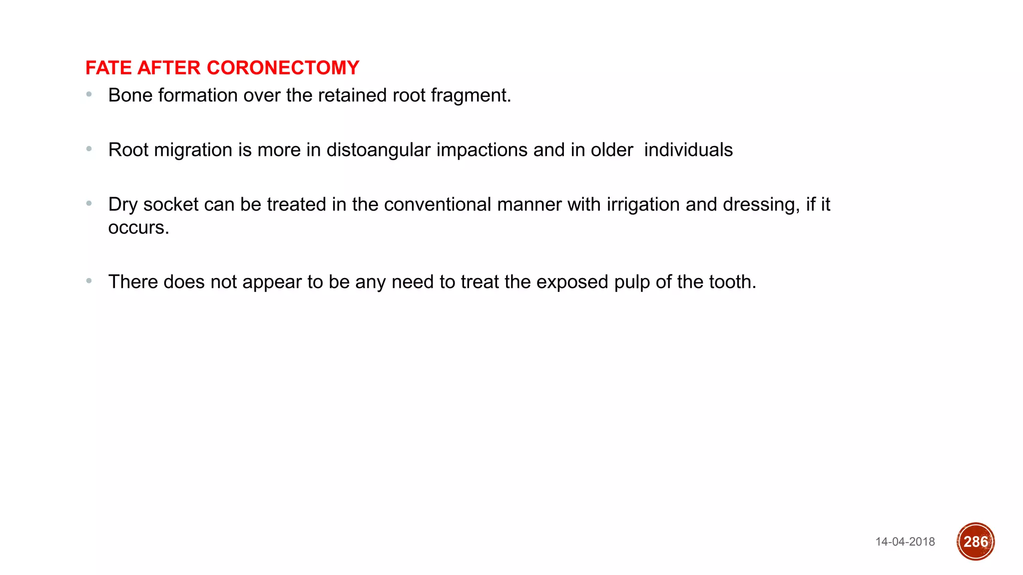
This document discusses impacted teeth, specifically focusing on impacted third molars. It begins with definitions of impacted teeth and provides the etymology and theories of tooth impaction. Local and systemic causes of impaction are described. Surgical anatomy of impacted third molars is reviewed, along with classifications of impacted mandibular third molars. Indications and contraindications for removal are outlined. Complications are briefly mentioned. The document is intended as a reference for oral and maxillofacial surgeons regarding the management of impacted third molars.
orthognathic surgery is very intresting and well knowing branch in oral surgery ....this presentation is dealing with jaw correction surgery in upper jaw.
Dr. Ahmed M. Adawy, Professor Emeritus, Dep. Oral & Maxillofacial Surgery. Former Dean, Faculty of Dental Medicine, Al-Azhar University. Mandibular angle fractures account for 23% to 42% of all facial fractures. Fracture of mandibular angle can be classified as (A) Vertical favorable or unfavorable, (B) Horizontally favorable of unfavorable. Traditionally, mandibular angle fractures have been treated with either closed reduction and inter-maxillary fixation or open reduction and internal fixation with or without inter-maxillary fixation. Patients treated with inter-maxillary fixation have a restricted airway and loose excess weight. Rigid internal fixation and early return to function have eliminated the use of wire osteosenthysis and prolonged use of inter-maxillary fixation. The principal of rigid fixation, however, have inherent set of disadvantages including damage to the inferior alveolar nerve and the marginal mandibular branch of facial nerve. Postoperative malocclusion rates are also high. With the introduction of semi-rigid technique fracture of the mandibular angle could be treated according to Champy’s Ideal lines of osteosenthysis. The technique involves placement of a single monocortial miniplate on the superior border of the mandible. However, some studies suggested using a second miniplate along the inferior border. Wether one or two miniplates should be used is still debatable. The application of 3D plates may provide additional stability in 3 dimension and good resistance against torque forces.
Arthrocentesis of the temporomandibular jointAhmed Adawy
Dr. Ahmed M. Adawy, Professor Emeritus, Dep. Oral & Maxillofacial Surgery. Former Dean, Faculty of Dental Medicine, Al-Azhar University. Arthrocentesis of the temporomandibular joint refers to lavage of the upper joint space, hydraulic pressure and manipulation to release adhesions of the “anchored disc phenomenon” and improve motion. The technique of arthrocentesis is discussed together with the indications and contraindications of the procedure. Further, the presentation includes modifications of the standard technique.
Classification of Impaction and Methods & Techniques of Third molar/Manidibular impaction removal,Flap designs of impaction removal techniques and more
Wisdom teeth are the third and last molars on each side of the upper and lower jaws. They are also the final teeth to erupt; they usually appear when a person is in their late teens or early twenties
Orthodontics problems, first permanent molars formation eruption and extraction Lama K Banna
Orthodontics problems, first permanent molars formation, eruption, and extraction.
Al Azhar University
Faculty of oral and dental medicine
fifth-year second semester
By Lama El Banna
Canine Impaction and Its Importance in OrthodonticsAnalhaq Shaikh
Canine Impaction, Its Importance in Orthodontics, Etiology, Diagnosis and Management.
by Dr Analhaq Shaikh, 2nd year Postgraduate student, Sharavathi Dental College and Hospital, Shimoga, Karnataka
Canine Impaction can also be termed as Shy Canine.
Detailed description on management of impacted maxillary and mandibular third molars. Surgical approaches and complications are also discussed in details.
Autotransplantation of Tooth in Children with Mixed DentitionAbu-Hussein Muhamad
Autotransplantation of tooth in children is the surgical movement of a tooth from one place in the mouth to another
in the similar individual. Once thought to be uncertain, autotransplantation has achieved high success rates and is an
outstanding option for tooth replacement in children. Although the indications for autotransplantation are narrow, careful
patient assortment coupled with a suitable method can lead to exceptional esthetic and useful results. One benefit of
this procedure is that placement of an implant-supported prosthesis or other form of prosthetic tooth replacement is
not needed. A review of the recommended surgical technique as well as success rates is also discussed.
Managing congenitally missing lateral incisors with single tooth implants Abu-Hussein Muhamad
Congenitally missing teeth are frequently presented to the dentist. Interdisciplinary approach may be needed for the proper treatment plan. Several treatment
options exist for the replacement of congenitally missing lateral incisors. These options include canine substitution, resin bonded fixed partial dentures, cantilevered
fixed partial dentures, conventional fixed partial dentures and single tooth implants. Depending on which treatment option is chosen, a specific criterion has to be
addressed. Interdisciplinary treatment plays a vital role to achieve an excellent, esthetic result for a most predictable outcome. This paper describes the therapeutic use
of osseointegrated implants to replace congenitally missing upper lateral incisors. Highlighting the importance of the Orthodontic/Restorative interface.
Esthetic Management of Congenitally Missing Lateral Incisors With Single Toot...Abu-Hussein Muhamad
Congenitally missing teeth are frequently presented to the dentist. Interdisciplinary approach may be needed for the proper treatment plan. Several treatment options exist for the replacement of congenitally missing lateral incisors.This case report addresses the fundamental considerations related to replacement of a congenitally missing lateral incisor by a team approach.
Teeth in The Line of Mandibular FracturesAhmed Adawy
Dr. Ahmed M. Adawy, Professor Emeritus, Dep. Oral & Maxillofacial Surgery. Former Dean, Faculty of Dental Medicine, Al-Azhar University. Approximately 60% of fractures of the mandible occur in the teeth bearing area. Incisors and third molars are the most commonly involved teeth on the fracture lines. The damaged to the tooth involved at the fracture site may include exposure of the root surface subluxation, avulsion or root fracture. This may lead to the vitalization, consequent infection and complicated healing of the fraction. Wether to remove or preserve the tooth in line of fraction is discussed. Certain guidelines have been suggested.
New Directions in Targeted Therapeutic Approaches for Older Adults With Mantl...i3 Health
i3 Health is pleased to make the speaker slides from this activity available for use as a non-accredited self-study or teaching resource.
This slide deck presented by Dr. Kami Maddocks, Professor-Clinical in the Division of Hematology and
Associate Division Director for Ambulatory Operations
The Ohio State University Comprehensive Cancer Center, will provide insight into new directions in targeted therapeutic approaches for older adults with mantle cell lymphoma.
STATEMENT OF NEED
Mantle cell lymphoma (MCL) is a rare, aggressive B-cell non-Hodgkin lymphoma (NHL) accounting for 5% to 7% of all lymphomas. Its prognosis ranges from indolent disease that does not require treatment for years to very aggressive disease, which is associated with poor survival (Silkenstedt et al, 2021). Typically, MCL is diagnosed at advanced stage and in older patients who cannot tolerate intensive therapy (NCCN, 2022). Although recent advances have slightly increased remission rates, recurrence and relapse remain very common, leading to a median overall survival between 3 and 6 years (LLS, 2021). Though there are several effective options, progress is still needed towards establishing an accepted frontline approach for MCL (Castellino et al, 2022). Treatment selection and management of MCL are complicated by the heterogeneity of prognosis, advanced age and comorbidities of patients, and lack of an established standard approach for treatment, making it vital that clinicians be familiar with the latest research and advances in this area. In this activity chaired by Michael Wang, MD, Professor in the Department of Lymphoma & Myeloma at MD Anderson Cancer Center, expert faculty will discuss prognostic factors informing treatment, the promising results of recent trials in new therapeutic approaches, and the implications of treatment resistance in therapeutic selection for MCL.
Target Audience
Hematology/oncology fellows, attending faculty, and other health care professionals involved in the treatment of patients with mantle cell lymphoma (MCL).
Learning Objectives
1.) Identify clinical and biological prognostic factors that can guide treatment decision making for older adults with MCL
2.) Evaluate emerging data on targeted therapeutic approaches for treatment-naive and relapsed/refractory MCL and their applicability to older adults
3.) Assess mechanisms of resistance to targeted therapies for MCL and their implications for treatment selection
Pulmonary Thromboembolism - etilogy, types, medical- Surgical and nursing man...VarunMahajani
Disruption of blood supply to lung alveoli due to blockage of one or more pulmonary blood vessels is called as Pulmonary thromboembolism. In this presentation we will discuss its causes, types and its management in depth.
Prix Galien International 2024 Forum ProgramLevi Shapiro
June 20, 2024, Prix Galien International and Jerusalem Ethics Forum in ROME. Detailed agenda including panels:
- ADVANCES IN CARDIOLOGY: A NEW PARADIGM IS COMING
- WOMEN’S HEALTH: FERTILITY PRESERVATION
- WHAT’S NEW IN THE TREATMENT OF INFECTIOUS,
ONCOLOGICAL AND INFLAMMATORY SKIN DISEASES?
- ARTIFICIAL INTELLIGENCE AND ETHICS
- GENE THERAPY
- BEYOND BORDERS: GLOBAL INITIATIVES FOR DEMOCRATIZING LIFE SCIENCE TECHNOLOGIES AND PROMOTING ACCESS TO HEALTHCARE
- ETHICAL CHALLENGES IN LIFE SCIENCES
- Prix Galien International Awards Ceremony
- Video recording of this lecture in English language: https://youtu.be/lK81BzxMqdo
- Video recording of this lecture in Arabic language: https://youtu.be/Ve4P0COk9OI
- Link to download the book free: https://nephrotube.blogspot.com/p/nephrotube-nephrology-books.html
- Link to NephroTube website: www.NephroTube.com
- Link to NephroTube social media accounts: https://nephrotube.blogspot.com/p/join-nephrotube-on-social-media.html
The prostate is an exocrine gland of the male mammalian reproductive system
It is a walnut-sized gland that forms part of the male reproductive system and is located in front of the rectum and just below the urinary bladder
Function is to store and secrete a clear, slightly alkaline fluid that constitutes 10-30% of the volume of the seminal fluid that along with the spermatozoa, constitutes semen
A healthy human prostate measures (4cm-vertical, by 3cm-horizontal, 2cm ant-post ).
It surrounds the urethra just below the urinary bladder. It has anterior, median, posterior and two lateral lobes
It’s work is regulated by androgens which are responsible for male sex characteristics
Generalised disease of the prostate due to hormonal derangement which leads to non malignant enlargement of the gland (increase in the number of epithelial cells and stromal tissue)to cause compression of the urethra leading to symptoms (LUTS
Lung Cancer: Artificial Intelligence, Synergetics, Complex System Analysis, S...Oleg Kshivets
RESULTS: Overall life span (LS) was 2252.1±1742.5 days and cumulative 5-year survival (5YS) reached 73.2%, 10 years – 64.8%, 20 years – 42.5%. 513 LCP lived more than 5 years (LS=3124.6±1525.6 days), 148 LCP – more than 10 years (LS=5054.4±1504.1 days).199 LCP died because of LC (LS=562.7±374.5 days). 5YS of LCP after bi/lobectomies was significantly superior in comparison with LCP after pneumonectomies (78.1% vs.63.7%, P=0.00001 by log-rank test). AT significantly improved 5YS (66.3% vs. 34.8%) (P=0.00000 by log-rank test) only for LCP with N1-2. Cox modeling displayed that 5YS of LCP significantly depended on: phase transition (PT) early-invasive LC in terms of synergetics, PT N0—N12, cell ratio factors (ratio between cancer cells- CC and blood cells subpopulations), G1-3, histology, glucose, AT, blood cell circuit, prothrombin index, heparin tolerance, recalcification time (P=0.000-0.038). Neural networks, genetic algorithm selection and bootstrap simulation revealed relationships between 5YS and PT early-invasive LC (rank=1), PT N0—N12 (rank=2), thrombocytes/CC (3), erythrocytes/CC (4), eosinophils/CC (5), healthy cells/CC (6), lymphocytes/CC (7), segmented neutrophils/CC (8), stick neutrophils/CC (9), monocytes/CC (10); leucocytes/CC (11). Correct prediction of 5YS was 100% by neural networks computing (area under ROC curve=1.0; error=0.0).
CONCLUSIONS: 5YS of LCP after radical procedures significantly depended on: 1) PT early-invasive cancer; 2) PT N0--N12; 3) cell ratio factors; 4) blood cell circuit; 5) biochemical factors; 6) hemostasis system; 7) AT; 8) LC characteristics; 9) LC cell dynamics; 10) surgery type: lobectomy/pneumonectomy; 11) anthropometric data. Optimal diagnosis and treatment strategies for LC are: 1) screening and early detection of LC; 2) availability of experienced thoracic surgeons because of complexity of radical procedures; 3) aggressive en block surgery and adequate lymph node dissection for completeness; 4) precise prediction; 5) adjuvant chemoimmunoradiotherapy for LCP with unfavorable prognosis.
Title: Sense of Smell
Presenter: Dr. Faiza, Assistant Professor of Physiology
Qualifications:
MBBS (Best Graduate, AIMC Lahore)
FCPS Physiology
ICMT, CHPE, DHPE (STMU)
MPH (GC University, Faisalabad)
MBA (Virtual University of Pakistan)
Learning Objectives:
Describe the primary categories of smells and the concept of odor blindness.
Explain the structure and location of the olfactory membrane and mucosa, including the types and roles of cells involved in olfaction.
Describe the pathway and mechanisms of olfactory signal transmission from the olfactory receptors to the brain.
Illustrate the biochemical cascade triggered by odorant binding to olfactory receptors, including the role of G-proteins and second messengers in generating an action potential.
Identify different types of olfactory disorders such as anosmia, hyposmia, hyperosmia, and dysosmia, including their potential causes.
Key Topics:
Olfactory Genes:
3% of the human genome accounts for olfactory genes.
400 genes for odorant receptors.
Olfactory Membrane:
Located in the superior part of the nasal cavity.
Medially: Folds downward along the superior septum.
Laterally: Folds over the superior turbinate and upper surface of the middle turbinate.
Total surface area: 5-10 square centimeters.
Olfactory Mucosa:
Olfactory Cells: Bipolar nerve cells derived from the CNS (100 million), with 4-25 olfactory cilia per cell.
Sustentacular Cells: Produce mucus and maintain ionic and molecular environment.
Basal Cells: Replace worn-out olfactory cells with an average lifespan of 1-2 months.
Bowman’s Gland: Secretes mucus.
Stimulation of Olfactory Cells:
Odorant dissolves in mucus and attaches to receptors on olfactory cilia.
Involves a cascade effect through G-proteins and second messengers, leading to depolarization and action potential generation in the olfactory nerve.
Quality of a Good Odorant:
Small (3-20 Carbon atoms), volatile, water-soluble, and lipid-soluble.
Facilitated by odorant-binding proteins in mucus.
Membrane Potential and Action Potential:
Resting membrane potential: -55mV.
Action potential frequency in the olfactory nerve increases with odorant strength.
Adaptation Towards the Sense of Smell:
Rapid adaptation within the first second, with further slow adaptation.
Psychological adaptation greater than receptor adaptation, involving feedback inhibition from the central nervous system.
Primary Sensations of Smell:
Camphoraceous, Musky, Floral, Pepperminty, Ethereal, Pungent, Putrid.
Odor Detection Threshold:
Examples: Hydrogen sulfide (0.0005 ppm), Methyl-mercaptan (0.002 ppm).
Some toxic substances are odorless at lethal concentrations.
Characteristics of Smell:
Odor blindness for single substances due to lack of appropriate receptor protein.
Behavioral and emotional influences of smell.
Transmission of Olfactory Signals:
From olfactory cells to glomeruli in the olfactory bulb, involving lateral inhibition.
Primitive, less old, and new olfactory systems with different path
micro teaching on communication m.sc nursing.pdfAnurag Sharma
Microteaching is a unique model of practice teaching. It is a viable instrument for the. desired change in the teaching behavior or the behavior potential which, in specified types of real. classroom situations, tends to facilitate the achievement of specified types of objectives.
Flu Vaccine Alert in Bangalore Karnatakaaddon Scans
As flu season approaches, health officials in Bangalore, Karnataka, are urging residents to get their flu vaccinations. The seasonal flu, while common, can lead to severe health complications, particularly for vulnerable populations such as young children, the elderly, and those with underlying health conditions.
Dr. Vidisha Kumari, a leading epidemiologist in Bangalore, emphasizes the importance of getting vaccinated. "The flu vaccine is our best defense against the influenza virus. It not only protects individuals but also helps prevent the spread of the virus in our communities," he says.
This year, the flu season is expected to coincide with a potential increase in other respiratory illnesses. The Karnataka Health Department has launched an awareness campaign highlighting the significance of flu vaccinations. They have set up multiple vaccination centers across Bangalore, making it convenient for residents to receive their shots.
To encourage widespread vaccination, the government is also collaborating with local schools, workplaces, and community centers to facilitate vaccination drives. Special attention is being given to ensuring that the vaccine is accessible to all, including marginalized communities who may have limited access to healthcare.
Residents are reminded that the flu vaccine is safe and effective. Common side effects are mild and may include soreness at the injection site, mild fever, or muscle aches. These side effects are generally short-lived and far less severe than the flu itself.
Healthcare providers are also stressing the importance of continuing COVID-19 precautions. Wearing masks, practicing good hand hygiene, and maintaining social distancing are still crucial, especially in crowded places.
Protect yourself and your loved ones by getting vaccinated. Together, we can help keep Bangalore healthy and safe this flu season. For more information on vaccination centers and schedules, residents can visit the Karnataka Health Department’s official website or follow their social media pages.
Stay informed, stay safe, and get your flu shot today!
These lecture slides, by Dr Sidra Arshad, offer a quick overview of physiological basis of a normal electrocardiogram.
Learning objectives:
1. Define an electrocardiogram (ECG) and electrocardiography
2. Describe how dipoles generated by the heart produce the waveforms of the ECG
3. Describe the components of a normal electrocardiogram of a typical bipolar leads (limb II)
4. Differentiate between intervals and segments
5. Enlist some common indications for obtaining an ECG
Study Resources:
1. Chapter 11, Guyton and Hall Textbook of Medical Physiology, 14th edition
2. Chapter 9, Human Physiology - From Cells to Systems, Lauralee Sherwood, 9th edition
3. Chapter 29, Ganong’s Review of Medical Physiology, 26th edition
4. Electrocardiogram, StatPearls - https://www.ncbi.nlm.nih.gov/books/NBK549803/
5. ECG in Medical Practice by ABM Abdullah, 4th edition
6. ECG Basics, http://www.nataliescasebook.com/tag/e-c-g-basics
HOT NEW PRODUCT! BIG SALES FAST SHIPPING NOW FROM CHINA!! EU KU DB BK substit...GL Anaacs
Contact us if you are interested:
Email / Skype : kefaya1771@gmail.com
Threema: PXHY5PDH
New BATCH Ku !!! MUCH IN DEMAND FAST SALE EVERY BATCH HAPPY GOOD EFFECT BIG BATCH !
Contact me on Threema or skype to start big business!!
Hot-sale products:
NEW HOT EUTYLONE WHITE CRYSTAL!!
5cl-adba precursor (semi finished )
5cl-adba raw materials
ADBB precursor (semi finished )
ADBB raw materials
APVP powder
5fadb/4f-adb
Jwh018 / Jwh210
Eutylone crystal
Protonitazene (hydrochloride) CAS: 119276-01-6
Flubrotizolam CAS: 57801-95-3
Metonitazene CAS: 14680-51-4
Payment terms: Western Union,MoneyGram,Bitcoin or USDT.
Deliver Time: Usually 7-15days
Shipping method: FedEx, TNT, DHL,UPS etc.Our deliveries are 100% safe, fast, reliable and discreet.
Samples will be sent for your evaluation!If you are interested in, please contact me, let's talk details.
We specializes in exporting high quality Research chemical, medical intermediate, Pharmaceutical chemicals and so on. Products are exported to USA, Canada, France, Korea, Japan,Russia, Southeast Asia and other countries.
Explore natural remedies for syphilis treatment in Singapore. Discover alternative therapies, herbal remedies, and lifestyle changes that may complement conventional treatments. Learn about holistic approaches to managing syphilis symptoms and supporting overall health.
3. INTRODUCTION
The management of impacted teeth is a basic component of most oral and maxillofacial surgery
practices.
Although the majority of impacted teeth are third molars, any other tooth may be impacted.
The usual care for impacted third molars is removal; however, the care for impacted teeth other than third
molars may include exposure (with or without attachment of an orthodontic bracket), uprighting,
transplantation, or removal. These teeth often pose challenges in treatment planning and surgical care
14-04-2018 3
4. ETYMOLOGY
They are generally thought to be called wisdom teeth because they appear so late – much later than the
other teeth, at an age where people are presumably "wiser" than as a child, when the other teeth erupt.
The term probably came as a translation of the Latin dens sapientiae.
Wisdom teeth usually appear between the ages of 17 and 25. a time of life that has been called the
"Age of Wisdom”.
14-04-2018 4
5. Impacted tooth is a tooth which is completely or partially unerupted and is
positioned against another tooth, bone or soft tissue so that its further eruption is
unlikely, described according to its anatomic position.
14-04-2018 5
6. 14-04-2018 6
Malposed tooth is tooth, unerupted / erupted which is in an abnormal position in the
maxilla/mandible
8. 14-04-2018 8
Ankylosed teeth is when cementum of the tooth is fused to the alveolar bone and there is no
periodontal ligament in between, a tooth is considered to be ankylosed
9. 14-04-2018 9
Partially erupted when the tooth has failed to erupt into a normal functional position but has
crossed the bone barrier and has not reached the occlusal line.
11. DEFINITION
BY ARCHER –
“A tooth which is completely or partially unerupted beyond its chronologic age & is positioned against
another tooth, bone or soft tissue, so that further eruption is not possible is known as impaction”
ANDERSSON (1997) , FONSECA (2000) ,PETERSON & LASKIN
Impaction is defined as a cessation of eruption of a tooth caused by a clinically or radio graphically
detectable physical barrier in the eruption path or by an ectopic position of the tooth.
SHAFER-
Impacted teeth are those prevented from erupting by some physical barrier in the eruption path
14-04-2018 11
12. 14-04-2018 12
AMERICAN SOCIETY OF ORAL SURGEONS 1971
A tooth which is completely or partially unerupted and is positioned against another tooth, bone or soft
tissue so that its further eruption is unlikely, described according to its anatomic position.
LYTLE: --
Impacted tooth is one that has failed to erupt into normal functional position beyond the time usually
expected for such experience. Eruption is prevented by adjacent hard or soft tissue including
tooth, bone, of dense soft tissues.
13. 11 year 14 year
18 year 25 year
Tooth germ -9 year
Cusp mineralization -2 year later
11 year- tooth located in anterior border of
ramus, occlusal surface facing anteriorly
Crown formation-14 years
Root-50% formed by 16 years
Root formation with open apex-18 years
24 years-95% of 3rd molars completed
eruption
chronology
14-04-2018 13
14. THEORIES OF IMPACTION
(ACCORDING TO DURBECK)
Phylogenic Theory
Mendelian Theory
Endocrine Theory
Orthodontic Theory
Skeletal Theory
Anthropological Theory
14-04-2018 14
www.indianjournals.com/%2Fijor.aspx?target=ijor:rjpt&volume=7
15. PHYLOGENIC THEORY
Nature tries to eliminate that which is not used.
Third molar occupies an abnormal position and may be considered as a vestigial
organ without purpose or function
• Individual receives small jaw from one parent and complement of large teeth from another parent.
Mendelian Theory
14-04-2018 15
16. ENDOCRINE THEORY
Lack of growth of the jaws is due to the lack of the function of the anterior lobe of the pituitary
gland with the result that sufficient amount of growth hormone is not produced to ensure the
proper growth of jaws.
Orthodontic Theory
Constricted and narrow dental arches of early mouth breathers affect the position and alignment
of permanent teeth.
The third molars attempting to erupt at a time when the deformity is well established gets
impacted.
14-04-2018 16
17. SKELETAL THEORY
Inadequate growth of the jaws due to an improper function of the functional matrix in which it is
enclosed leads to lack of space for eruption of the third molars.
Anthropological Theory
During the course of evolution, increase in cranial capacity occur at the cost of decreasing jaw size
14-04-2018 17
19. ETIOLOGY – LOCAL CAUSES
Obstruction for eruption
Irregularity in position and presence of an adjacent tooth
Density of overlying and surrounding bone
Lack of space in dental arch
Supernumerary teeth
Crowding
Ankylosis of primary or permanent teeth
Over-retention of deciduous teeth
Dilaceration of roots (trauma)
Habits involving
Tongue
Finger
Thumb
Cheek
Pencil Etc
14-04-2018 19
27. CONTRAINDICATIO
NS
Deeply, embedded, asymptomatic impacted molar.
Poor systemic condition of patient
Poor prognosis of 2nd molar.
In case it can serve as abutment tooth.
Extremes of age
Question about the future status of the second molar
Probable Excessive Damage to Adjacent Structures
14-04-2018 27
36. Pell and Gregory’s classification (1933)
Relationship of impacted third molar to ramus of mandible and second molar based on space available
distal to second molar
14-04-2018 36
38. Combined ADA and AAOMS
Classification for Impacted Teeth
07220 :-overlying soft tissue (Impaction that requires incision of overlying soft tissue and the removal of
the tooth)
07230 : -partially bony impacted (Impaction that requires incision of overlying soft tissue, elevation of a
flap, and either removal of bone and tooth or sectioning and removal of tooth)
07240: -completely bony(Impaction that requires incision of overlying soft tissue, elevation of a flap,
removal of bone,and sectioning of tooth for removal)
07241: -completely bony, with unusual complications (Impaction that requires incision of overlying
soft tissue,elevation of a tlap, removal of bone, sectioning of the tooth for removal, and/or presents
unusual difficulties and circumstances)
14-04-2018 38
39. 1.Position & Angulation- Winter’s Classification
a) Erupted
b) Partly erupted
c) Unerupted
3. Number & pattern of roots
- Fused
-Multiple
-Favourable
-Unfavourable
2. State of eruption
-Two
KILLEY & KAY’S
CLASSIFICATION
40. BASED ON STATE OF
ERUPTION
Erupted
Partially Erupted
Unerupted
14-04-2018 40
41. BASED ON NUMBER
OF ROOTSFused Roots
Two Roots
Multiple Roots
14-04-2018 41
42. BASED ON TYPE OF TISSUE OVERLYING THE TOOTH
Soft Tissue
Partial Bony
Complete Bony impaction
14-04-2018 42
53. CLINICAL EXAMINATION
Mouth opening
Absence of local inflammatory conditions
Determination of mobility characteristics of lips and cheeks
Size and contours of the tongue
Appearance of soft tissue overlying the impacted teeth
14-04-2018 53
55. SHOWS :
Position and type of impaction.
Relationship of impacted tooth to adjacent teeth.
Size and shape of impacted tooth.
Depth of impaction in bone.
Density of bone surrounding impacted tooth .
Relationship of the impacted tooth to various anatomic structures.
14-04-2018 55
58. 14-04-2018 58
According to winter (1926)
An ideal periapical X-ray should include
1. the whole third molar,
2. its investing bone,
3. the anterior border of ramus,
4. the inferior alveolar canal and
5. the adjacent second molar tooth.
• In a good film the lateral image of the second molar will be sharp without vertical shortening.
• In a poor film with incorrect angulation, the 'enamel cap' will be absent and there will be
overlapping of contact points of molars.
A. L. Mac Gregor, Geoffrey Hoe, Killey And Kay, Archer
59. 14-04-2018 59
The specifications for an ideal IOPA radiograph as per G. B. Winter (Mac GREGOR)
• The buccal and lingual cusps of the erupted second molar must be
superimposed
• The area of contact of first and second molar must not show overlap.
• The film must be far enough back in the mouth to show only the distal root of the
first molar.
• The whole third molar should be seen.
According to G.L.Howe the standard IOPA must have typical ‘enamel cap’
appearance of second molar.
60. A. Root morphology
The number of factors considered.
a) Length of the root - Optimal time is when the roots 1/3 +2/3 formed. When this is the
case, the ends of the roots are blunt and almost never fracture.
If the root development is insufficient less than 1/3of the tooth, it is difficult to remove.
b) Single/conical, separate/distinct roots are noted.
c) Curvature of roots
d) Total width of the roots in mesiodistal direction should be compared with the width of the
tooth at the cervical line.
e) Assess the periodontal ligament space. More the width the periodontal ligament space,
is the easier the tooth is to remove. 14-04-2018 60
62. B. Size of follicular sac
More follicular space
Less alveolar bone
Easy removal
C. Density of the surrounding bone
Younger patient the bone is less dense, is more likely pliable and expands and blends
somewhat, which allows the socket to be expanded by elevators/by luxation forces by itself
and easier to cut with bur.
Patients who are older than 35 years have dense bone and thus decreased ability to
expand. In these patients surgeon must remove all interfering bone, because it is not
possible to expand the bone socket. Bone cutting is difficult and bone removal process
takes longer.
14-04-2018 62
63. 14-04-2018 63
D. Contact with the 2nd molar
• Taken care if 2nd molar has carious/large restoration/root canal treated.
• Locked against second molar there is no space for elevation, then sectioning of the tooth should be
planned.
E. Nature of overlying tissues -is considered
• Soft tissue covering
• Soft tissue + Bone covering
• Bone
64. F. Inferior Alveolar Nerve and Vessels
Are usually in true osseous canal in the ramus and body of the mandible. There may be
multiple branches of Inferior Alveolar Nerve, instead of single combined structure. When
Inferior Alveolar Nerve canal identified radiograhically, determine its relationship with
impacted mandibular 3rd molar. Usually canal will be inferior/buccal to the third molars,
but variations are common.
Blending of the structure radiographically indicates that the root surround the canal and
its contents
A thinning/narrowing of canal indicates displacement of the canal by the roots of the
tooth. 14-04-2018 64
65. 14-04-2018 65
• Distinct lines of canal and of roots indicate an overlap
without encroachment.
• To locate the canal, Frank suggests that a modification of tube shift method can be used to
determine whether mandibular canal is medial to, lateral to/below an impacted mandibular 3rd
molar. This method first described by Richards.
• Two films in identical position used but position of the x-ray tube changed to determine the
position of impacted teeth.
• To accomplish this an x-ray angle must be shifted from 25 degree upwards and this 2nd film
compared with the film taken with the x-ray tube parallel to occlussal plane.
67. RELATIONSHIP OF INFERIOR ALVEOLAR NERVE TO THE ROOTS OF THE
THIRD MOLAR.
Deflected roots
14-04-2018 67
68. G. LINGUAL NERVE
The studies by Kisselbach and Chamberlain demonstrated that the lingual nerve may be
located some time slighter superior to the crest of bony ridge medial to the mandibular 3rd
molar region and only 1/2mm toward midline in the lingual soft tissue. At this position
lingual nerve is at risk during flap reflection.
Usual location is superior and inferior to mylohyoid muscle. The nerve may be in various
locations from crest of alveolar process to positions below mylohyoid muscle.
More than one branch of the nerve may be present, and the position of the lingual nerve,
relative to the mandibular 3rd molar may vary depending on the intersection of the body of
the mandible and the ramus.
The lingual nerve is relatively more superior and more directly associated with the soft
tissue immediately adjacent to the mandible and hence greater chance of damage during
surgery, in the class III and II relationships than in the class I . 14-04-2018 68
70. H. Relationship to Body and ramus of mandible is important surgical consideration. This
relationship described by Gregory and Pell.
In Class III, the ramus begins immediately posterior to 2nd molar and no space consists for the impacted
tooth to erupt.
In class II and class III relation there is reduced surgical access and close anatomic relationship with
lingual nerve exists.
I. Third molar and 2nd molar relationship
The long axis of the third and 2nd molar is studied to determine whether impacted tooth is in
Mesioangular/ Vertical/Distoangular/Horizontal/inverted position.
The level of the crown of third molar are determined whether it is crown to crown, crown to cervix, crown
to root and crown below 2nd molar.
J. Buccal to lingual position of third molar
If impacted 3rd molar is too lingually placed there may be chance of stretching of the lingual nerve.
14-04-2018 70
72. External oblique ridge. A continuation of the anterior border of the ramus, passing
downward and forward on the buccal side of the mandible. It appears as a distinct
radiopaque line which usually ends anteriorly in the area of the first molar. Serves as an
attachment of the buccinator muscle. (The red arrows point to the mylohyoid ridge).
facial view
Facial view
14-04-2018 72
73. Mylohyoid ridge (internal oblique). Located on the lingual surface of the mandible, extending from the
third molar area to the premolar region. Serves as the attachment of the mylohyoid muscle.
lingual view
14-04-2018 73
74. facial view
Mandibular (inferior alveolar) canal. Arises at the mandibular foramen on the lingual side of the ramus
and passes downward and forward, moving from the lingual side of the mandible in the third molar
region to the buccal side of the mandible in the premolar region. Contains the inferior alveolar nerve
and vessels.
14-04-2018 74
75. lingual view
Submandibular gland fossa. A depression on the lingual side of the mandible below the mylohyoid ridge.
The submandibular gland is located in this region. Due to the thinness of bone, the trabecular pattern of
the bone is very sparse and results in the area being very radiolucent. The fact that it occurs bilaterally
helps to differentiate it from pathology.
14-04-2018 75
76. The external oblique ridge (red arrows) and the mylohyoid ridge (blue arrows) usually run parallel with
each other, with the external oblique ridge always being higher on the film.
14-04-2018 76
77. The mandibular canal (red arrows identify inferior border of canal) usually runs very close to
the roots of the molars, especially the third molar. This can be a problem when extracting
these teeth. Note the extreme dilaceration (curving) of the roots of the third molar (green
arrow) in the film at left. The film at right shows “kissing” impactions located at the superior
border of the canal.
14-04-2018 77
84. 14-04-2018 84
This is a accurate radiograph in class III horizontal
impacted third molar.
It is a good substitute when IOPA cannot be taken.
It shows the vertical depth of the mandibular bone.
It shows the bone below the buried tooth ,ectopic teeth
and about any existing pathologies
87. 14-04-2018 87
The angle between the occlusal plane or line parallel to it and the longitudinal axis of the
impacted third molar, in turn, allowed objective classification of the third molars within the Winter
subclasses.
89. CT-scan
14-04-2018 89
Superior in showing tooth and root
Shape, crown/root relationship,
Tooth inclination and gives exact idea
Of the proximity of IAN and canal to the
Roots.
91. DIFFICULTY INDICES
14-04-2018 91
1. PELL & GREGORY DIFFICULTY INDEX
2. WAR LINES
3. PEDERSON’S DIFFICULTY INDEX
4. MODIFIED PARANT’S DIFFICULTY INDEX
5. YAUSA ET.AL. DIFFICULTY INDEX
6. WHARFE’S ASSESSMENT
7. INDEX PREDICTING RISK FOR PERIODONTAL DEFECTS – KUGELBERG 1990
8. DIFFICULTY INDEX BASED ON CLINICAL AND RADIOLOGICAL PARAMETERS
95. WAR lines Line extension significance
W-white line Drawn along the occlusal surface of
the erupted mandibular molar and
extends posteriorly over the third
region
Depth of the tooth within the
mandible relationship of
occlusal surface of impacted
tooth within the erupted
molars
A-amber line Drawn from surface of the bone distal
to the third molar to the crest of the
interdental septum between the first
and second molars
The height of the margins of
the alveolar bone enclosing
the tooth
Amount of vertical bone that
requires removal for the
extraction of 3rd molar
R-red line Perpendicular line drawn for the
ambar line to the imaginary point of
application for the elevator
Longer the line more difficult
the extraction
<5 mm-less difficult
For every 1 mm increase
after 5 mm difficulty
increases by 3 times
>5 mm-advised under GA
≥9 mm-very
difficult,denuding of distal of
second molar may mandate
the removal of second molar14-04-2018 95
96. Another method of judging the depth of the 3rd molar is to divide
the root of the 2nd molar into thirds. A horizontal line is drawn
from the point of application for an elevator to the 2nd molar. If
the point of application is adjacent to the coronal, middle or
apical root third, then the tooth extraction is assessed as easy,
moderate or difficult respectively.
98. DIFFICULTY INDEX : THE INDEX FOR PREDICTING THE DIFFICULTY IN THE REMOVAL OF
MANDIBULAR
THIRD MOLARClassification. Value
Spatial relationship
Mesioangular 1
Horizontal/transverse 2
Vertical 3
Distoangular 4
Depth
Level A 1
Level B 2
Level C 3
Ramus relationship/space available
Class I 1
Class II 2
Class III 3
Index
7-10(very difficult),5-6(moderately difficult),3-4(minimally difficult)
Pederson Scale(1988)
14-04-2018 98
106. 14-04-2018 106
Variable name Classification Value
Preop plaque index (distal to 2M )
Not visible 0
Visible 1
Preop probing depth (distal to
2M)
≤ 6mm 0
> 6mm 1
Preop intrabony defect ( distal to
2M)
≤ 3mm 0
> 3mm 1
Sagittal inclination of 3rd molar
≤ 50˚ 0
> 50˚ 1
107. 14-04-2018 107
Contact area 2nd and 3rd molar
Small contact 0
Large contact 1
Resorption distal root of 2nd molar
No 0
Yes 1
Pathologically widened follicle of 3rd
molar
No or Distal 0
Mesial ≥ 2.5 mm 1
Smoking habits
Non-smoker 0
Smoker 1
CONTD…..
108. 14-04-2018 108
Level of risk indicated by RISK INDEX M3 - Kugelberg
Risk index
M3
Index Score
No risk ≤1
Low risk 2
Moderate risk 3
High risk ≥ 4
Risk index
M3
Predicted
IBD (mm)
0-2 2.0-3.0
3-4 3.5-4.0
≥ 5 >4.0
IBD – intrabony defect
113. 14-04-2018 113
LOCATION:
Lower 3rd molar is situated at the distal end of the body of
the mandible where it meets a relatively thin ramus.
Embedded b/w thick buccal alv bone buttressed by
external oblique ridge & the narrow inner cortical plate.
Ramus offset by 20°
Retro Molar triangle- depressed roughned area post. to 3rd
molar
114. 14-04-2018 114
MUSCLES:
Vestibule is formed by the attachment of buccinator
buccally and mylohyoid lingually.
Along the anterior border of the ramus - tendinous
insertion of temporalis Excessive stripping of these
muscle will cause hematoma, pain and trismus.
Lingual pouch – perforation of roots along the lingual
cortical plate.
- may cause # of lingual cortical plate
- displacement of fractured root fragments below the
mylohyoid
115. 14-04-2018 115
ARTERIES
• Facial artery & facial vein run in close approximation with
lower 2nd molar near the anterior border of masseter.
• Mandibular vessels in retro molar triangle which supply
temporalis tendon.
• Hemorrhage can occur during surgical removal of
impacted tooth if distal incision is not taken laterally
towards cheek.
116. 14-04-2018 116
INFERIOR ALVEOLAR NERVE
• Lies just below the roots of mandibular molars but
slightly buccally placed in inferior dental canal.
• In case of deep seated impaction special care should be
taken to protect this neurovascular bundle during bone
drilling & tooth sectioning.
• Calcification of inferior alveolar canal is completed
before the roots of 3rd molar are formed. Thus growing
roots may impinge upon the canal or get deflected. So
blind elevation is not advisable.
119. 14-04-2018 119
The instruments used for surgical removal of impacted teeth are essentially a combination of
instruments used for transalveolar extraction and soft tissue surgery in the mouth. There are only a
few special instruments that are required.
Very often the selection of instruments is a matter of personal preference. It is essential that the
bare minimum instruments are used depending on the case with provision to meet exigencies. This
is because a large collection of unfamiliar instruments and an elaborate tray set-up will cause
embarrassment for both the patient as well the operator and interfere with efficient progress of the
surgery
k. George Varghese, Killey & Kay, Archer, Geoffrey L. Howe
121. 14-04-2018 121
PATIENT POSITIONING
• The dental chair is adjusted in such a manner which is comfortable for the patient and at the
same time enables the operator and the assistant to have a clear view of the surgical site.
• Generally, for operative procedures in mandible the occlusal plane of lower teeth should be
parallel to the floor and for the maxillary teeth the occlusal plane of the upper teeth at 45°
angles to the floor
122. 14-04-2018 122
• The instruments are arranged in a rational order of their intended use (not cluttered) on a sterile
towel placed over the instrument tray of the dental chair or more preferably on a separate
instrument trolley.
• If there is delay in starting the surgery, the instruments should be covered with another towel to
avoid contamination and to maintain the sterility of the instruments.
• The surgeon and the assistant then wear cap and face mask.
• This is followed by scrubbing the hands
• After scrubbing a sterile gown is worn by the surgeon and the assistant.
123. 14-04-2018 123
Preparation of the surgical site:
• Before preparing the intraoral surgical site it is always desirable to wipe the patient's face with
an antiseptic solution like povidoneiodine (Betadine).
• The third molar area is then swabbed with 0.5% solution of chlorhexidine or betadine.
• Alternatively the patient can be given a mouth wash of the above antiseptic. This is followed
by the administration of local anesthetic injection.
124. 14-04-2018 124
Draping the patient:
Ideally the head and the front part of the body of the patient should be draped, thus exposing only the
face.
For this the patient's head is covered with a sterile towel, the edge of which is brought under the chin and
fixed with towel clip.
This ensures that the head, neck and shoulders are covered and only the face is exposed.
125. 14-04-2018 125
Palpation of Anatomical Landmarks
• Before starting the surgical procedure, palpation of the region and locating the anatomical
landmarks is essential.
• The pterygomandibular fold covering the pterygomandibular raphe is usually very prominent
when the mouth is opened wide.
• The external oblique ridge is then located.
• Next, the anterior part of the ramus with the coronoid notch and medially the retromolar fossa
should be identified followed by the medial tendon of temporalis.
• The lingual shelf is palpated.
Locating the anatomical landmarks will give information regarding the underlying bone and will help
the operator as to where the incision is to be given.
126. 14-04-2018 126
The standard operative plan can be divided into the following stages:
1. Incision to gain access to the area
2. Removal of adequate amount of bone
3. Sectioning and delivery of tooth from the socket
4. Debridement
5. Closure of the incision
128. 14-04-2018 128
Incision – 3 parts: Anterior, posterior & intermediate limb
Not to be extended too distally-
Bleeding from buccal vessels & other arteries
Postoperative trismus – temporalis muscle damage
Herniation of buccal fat pad
Damage to lingual nerve (lingual extention)
129. 14-04-2018 129
The first step in removing the impacted tooth is to reflect a mucoperiosteal flap.
The flap should be
1. Of adequate size to permit access, & exposure of operative site.
2. Allow adequate visibility
3. It should have wide base to assure good blood supply to the soft tissue
4. it should be large enough so that soft tissue surrounding the operative site is not
traumatized during the operation and so that when the flap is replaced, the edges rest on
wide shelf of bone
5. Ensure unhindered healing without periodontal pocket formation distal to second molar
Archer, Killey & Kay, Alling And Alling
130. 14-04-2018 130
The most important factor in designing the flap is the position of the
third molar which in turn dictates the amount of bone removal required
and the need for tooth sectioning.
More over, due consideration should be given for the lingual nerve,
buccinator muscle and the periodontium distal to second molar while
designing the flap.
131. 14-04-2018 131
Flap designs
• A small artery, the buccal artery, is sometimes encountered while giving the releasing incision, the injury of
which will lead to mild bleeding.
• The incision should not be continued posteriorly in a straight line, because the mandible diverges laterally.
• If the incision is extended straight, the blade will enter the sublingual space and is likely to damage the
lingual nerve.
• The lateral extension also avoids small vessels emerging from the retromolar fossa.
132. The base of the flap should be larger than the apex. Essentially, the
releasing incision(s) should be divergent in relation to the site of exposure
so as to not undermine the blood supply of the raised flap.
• The flap should be raised and released in such a manner as to prevent
tearing of the flap. Tearing might lead to a compromised blood supply and
necrosis.
• The ratio between the height of releasing incision(s) and length of base
should not exceed 2:1.
The type of flap used in third molar surgery is classified as full thickness,
which describes an incision that is made from the epithelial
surface to the underlying bone
Basic principles of flap
14-04-2018 132
134. ENVELOPE
FLAPS
14-04-2018 134
The most commonly used flap
is the envelope flap, which
extends from just posterior to
the position of the impacted
tooth anteriorly to the level of
the first molar. The posterior
end of the incision is directed
buccally along the external
oblique ridge.
SZYMD FLAP
135. 14-04-2018 135
Incision is made
horizontally along the
crest of the ridge or in
the buccal gingival
crevice.
Has no vertical
incision.
For shallow or
superficial impactions
136. 14-04-2018 136
Most common approach.
ADVANTAGES:
1. Generous visibility.
2. Mesial extension possible.
3. The osseous defect can be safely covered .
4. Blood supply up to the margins is adequate.
DISADVANTAGES:
1. The distal component can cut across the insertion of the temporalis tendon.
2. The flap margins lies over the bony defect so formed.
3. Wound dehiscence at the distofacial edge of the preceding second molar is frequent.
137. Modification of the envelope flap described by Szmyd
(1971).
14-04-2018 137
1. Free buccal gingival tissue around first and
second molars is spared.
2. Minimal reflected periosteum.
3. Broad-based blood supply to the flap.
4. Adequate exposure and visibility.
5. Good bony support for the soft tissue flap.
6. Closure can be effected with a single suture
and the distal aspect of the third molar
socket.
ADVANTAGES OF MODIFIED FLAP
138. 14-04-2018 138
TRIANGULAR FLAP
A small ‘V’ shaped incision, made with one point at
distobuccal line angle of the second molar.
One vertical limb followed the external oblique ridge, and
the other avoided the gingival sulcus and extended down
to the mucogingival junction, doen’t involve papilla of
mandibular 2nd molar.
142. 14-04-2018 142
Limb C - not to be extended too distally
▪ Bleeding from buccal vessels & other arteries
▪ Postoperative trismus – temporalis muscle damage
▪ Herniation of buccal fat pad
▪ Damage to lingual nerve (lingual extention)
• In case of unerupted tooth ,intermediate incision is not needed.The limb A is extended upto the
middle of the distal surface of the 2nd molar
143. 14-04-2018 143
• This incision is started from a point approximately 6.4 mm (14 in)down in the buccal sulcus appox at the junction of
posterior and middle thirds of second molar; the line passes upwards to distobucaal angle of second molar at gingival
margin.
• The incision now passes cervically behind the tooth to middle of its posterior surface if third molar is unerupted.
• It is then extended posteriorly and laterally along the anterior border of the ramus for a maximum of 25.4 mm
• Extension of the incision further posteriorly may result in prolapse of buccal pad of fat or lead to marked trismus and
swelling postoperatively.
144. 14-04-2018 144
MODIFIED Ward’s incision
Anterior incision is commenced at
the distobuccal corner of the crown of
mandibular 1st molar instead of 2nd molar
145. BAYONET FLAPS
• Similar to the molar sulcus incision
• The incision is similar to the second molar sulcus incision with the
addition of an oblique vestibular extension in the sulcus area
which is angled forward.
• However, oblique vestibular extension is made in the sulcus
region .
• This extension should be angled forward to facilitate suturing and
to optimize blood supply to the anterior part of the flap.
• This type of incision gives excellent buccal visibility. A variant of
this incision is to limit the sulcus incision buccal to the second
molar only to the distobuccal part of the gingiva .
• This flap offers limited visibility and should only be used for
superficially and buccally placed third molars. 14-04-2018 145
146. ‘L’ SHAPED FLAP• L flap is similar to the bayonet flap only difference being that the incision is
made a couple of millimeters away from the marginal gingiva. It optimizes
marginal attachment healing next to the second molar.
• Vertical relieving incision is given at 45˚ angle to the long axis of the 2nd
molar and runs straight anteriorly and downwards.
• This procedure optimizes marginal attachment healing next to the second
molar but requires that the main surgical approach to the third molar is at a
distance from the second molar.
14-04-2018 146
147. LINGUAL FLAP
• This flap is used with a lingual approach for third molar removal.
• The incision starts on the ascending ramus aiming at the distobuccal corner of the
second molar, follows the distal surface of the second molar as a sulcular incision, and
then continues lingually to the first molar region.
• A sulcular incision is also made along the buccal aspect of the second molar
14-04-2018 147
150. 14-04-2018 150
• Extended onto the buccal shelf of the mandible
• Incision line did not lie over the bony defect created by
the removal of the impacted teeth
• Its base at the distolingual aspect of the second molar
151. 14-04-2018 151
GROOVE & MOORE
• In the year 1970 they designed three flaps
• Produced an apparent decrease in pocketing distal
to 2nd molar
• A collar of tissue was preserved around the 2nd
molar hence decreasing pocket formation
• A lingual extension of the incision allowed for
exposure of the lingual aspect as well
153. 14-04-2018 153
• Begins at a point distal to the second molar, smoothly curved up
to meet the gingival crest at the distobuccal line angle of the
second molar.
• The incision is continued as a crevicular incision around the
distal aspect of the second molar.
• Allows reflection of a distolingually based flap adequately exposing the entire third molar area.
• The incision and flap design seems best suited to cases in which the third molar is completely covered
with soft tissues.
• In cases in which part of the impacted tooth is visible in the mouth, a small modification is made.
156. 14-04-2018 156
• The sharp end of the periosteal elevator is inserted in the region of vertical incision to ensure that the
incision has reached up to bone.
• If not, the sharp tip of the instrument is employed to sever the remaining attachment.
• The blunt end of the instrument is then passed beneath the mucoperiosteum to reflect the soft tissue in
the correct plane.
• Care should be taken not to strip the periosteum from the mucosa while reflecting.
157. 14-04-2018 157
• The mucoperiosteal flap is then reflected
laterally to the external oblique ridge with a
periosteal elevator and held in this position
with an retractor (third molar retractor).
• The flap reflection should be limited to
external oblique ridge laterally, because
reflecting beyond this area leads to
increased dead space resulting in more
edema postoperatively.
159. 14-04-2018 159
• The next major step is to remove the bone around the impacted
tooth.
• The amount of bone removal varies with the depth of impaction.
• This can be accomplished either by use of bur, or chisel and mallet or
a combination of the two methods.
• Whatever may the method used (which may be of individual
preference), the aim is to remove sufficient amount of bone to free the
tooth from obstruction and to provide a point of application for the
elevator.
160. 14-04-2018 160
• If adequate amount of bone covering the tooth has been removed, an attempt can be made to elevate
the tooth from the socket.
• Application of a great amount of force without adequate bone removal can result either in fracture of
the tooth or fracture of the mandible.
• Due to the above risk dental extraction forceps and elevators with great mechanical efficiency like
cross bar elevators are contraindicated for the removal of impacted third molar.
161. 14-04-2018 161
• Once the obstructing bone has been removed, only a slight amount of force alone is needed to
deliver the tooth.
• Elevators with less mechanical efficiency like Warwick James elevator (straight and curved type)
and Coupland chisels are recommended for this purpose.
• The # 301, Crane pick and Cogswell B elevators also serve this function well.
162. 14-04-2018 162
• It is a common practice to use a broad elevator between the buccal surface of the impacted tooth
and the external oblique ridge in order to elevate a tooth or root fragment.
• This technique places the external oblique ridge, one of the buttresses of the mandible and the
lingual plate at risk of fracture.
• If such a fracture is unrecognized, a substantial late presenting sequestrum or immediate lingual
nerve injury is possible (Farish and Bouloux, 2007).
163. 14-04-2018 163
• At the time of elevating the tooth, the index finger of the operator's left hand should rest on the
occlusal surface of the wisdom tooth to judge its movement and the other fingers support the
mandible.
• Because the impacted tooth has never sustained occlusal force, their periodontal ligament space is
wider and less tenacious and they can be easily displaced if adequate bone is removed and
elevation forces are applied in a proper direction.
164. DIFFERENCES BETWEEN BUR & CHISEL
TECHNIQUE
Sl.No Criteria. Chisel&Mallet Bur
1. Technique Difficult Easy.
2. Controll over bone cutting Uncntrolled&chances
of fracture is more.
Controlled.
3. Patient acceptance. Not tolerated in L.A. Well tolerated in L.A.
4. Healing of bone. Good Delayed Healing
5. Postoperative edema Less More.
6. Dry socket. Less. More.
7. Postoperative Infection. Less. More.
14-04-2018 164
165. BONE REMOVAL WITH BUR (MOORE & GILBE COLLAR
TECHNIQUE)
14-04-2018 165
• No. 7 or 8 round bur can be used in the range of 5000-10,000 rpm.
• Use of bur should always be done with copious saline irrigation to prevent thermal injury.
• Bone should be removed from the occlusal surface of the tooth.
• A channel is formed in the bone lateral and posterior to the impacted third molar to the cervical
level of the crown contour.
• Adequate amount of trough should be created to remove any bony obstruction for exposure and
delivery of the tooth.
• Careful bone removal should be done around the distal and distolingual aspect of the tooth without
damaging the lingual nerve which lies in the vicinity of the lingual plate adjacent to the third molar
167. 14-04-2018 167
• Conventional tech of using bur.
• Rosehead round bur no.3 is used to create a gutter along the buccal side & distal aspect of
tooth.
• A point of elevation is created with bur.
• Amount of bone sacrificed is less.
• Can be used in old patient.
• Convenient for patient.
168. 14-04-2018 168
LATERAL TREPHINATION TECHNIQUE
(BOWDLER HENRY; 1969)
• Indicated for removal of unerupted third molars
in the age group of 9 to 18 years.
• A extented S shaped incision is made from the
retro molar fossa across external oblique ridge.
• Such an incision leaves behind 5mm cuff of
attached mucosa at the
distobuccal region of second molar.
169. 14-04-2018 169
• A round toller bone bur is used to trephine the crypts of
third molar. After anterior posterior length of crypt has
been determined, a vertical cut is made through
external plate
• A second cut is made through the outer plate at the
posterior end of crypt at an angle of 45̊ from the row of
trephine bur.
• A chisel is applied in vertical direction to out fracture
outerplate
170. 14-04-2018 170
Complications:-
∙ A transient anaesthesia in post-operative period
∙ Buccal vessel may be cut during the incision.
∙ Damage to 2nd molar roots
∙ Fracture of mandible during bone cutting
∙ Bleeding
Advantages
∙ Bone healing is excellent without any loss of alveolar bone around 2nd molar.
∙ Local anaesthesia preferred over general anaesthesia
171. 14-04-2018 171
SPLIT BONE / LINGUAL SPLIT TECHNIQUE /
SIR WILLIAM KELSEY FRY(1933)
• Described by Sir William Kelsey Fry (1933).
• Later popularized by Terence G ward(1956)
• Specially for lingually placed tooth.
• Modified by Dr. Davis in 1960 & Lewis in 1980
172. 14-04-2018 172
• The technique involves the use of a chisel and mallet to remove or displace the lingual plate
of bone adjacent to lower third molar teeth.
• A small amount of buccal bone is often removed to facilitate exposure of the crown and provide
a point of application for a dental elevator.
• Although tooth division is usually not required, it usually can be achieved with the chisel.
• Although the lingual split technique is well suited to patients receiving sedation or general
anesthesia, it is generally not well suited to surgery conducted purely under local anesthesia.
174. 14-04-2018 174
• An incision is made from the retromolar area to the mesial aspect of the first molar or the distal
aspect of the second molar, depending on whether an envelope incision or a triangular flap is
used. The latter approach involves a vertical buccal relieving incision on the distal aspect of the
second molar and is preferred by the author (GFB) because it allows better retraction and
improved visibility.
• The buccal flap is raised in a subperiosteal plane using a #9 periosteal elevator.
• The flap should be extended just slightly beyond the external oblique ridge to prevent
excessive dead space beneath the flap.
• A 2-0 silk retraction suture is placed through the apex of the triangular flap.
• The suture should be clamped with a heavy hemostat 6 to 8 inches from the flap, which is then
allowed to rest on skin of the cheek, where it serves to keep the flap retracted.
175. 14-04-2018 175
• The flap should be raised along a broad length before proceeding deeper.
• This latter approach reduces the tension placed on the lingual nerve, which adheres to the
periosteum.
• The flap should extend from the mesial of the second molar to the lingual aspect of the anterior
ramus.
• The inferior aspect of the pterygomandibular raphe and superior constrictor muscle together
with a small portion of the mylohyoid muscle are included in this flap.
• The lingual nerve enters the sublingual space by passing between the superior constrictor and
mylohyoid muscles; at this location the nerve is immediately beneath the periosteum and at
risk from trauma.
176. 14-04-2018 176
• After lingual flap elevation, a left or right Hovell’s retractor (depending on
which side of
the mandible is being operated) is placed beneath the flap and allowed to sit
passively.
• The buccal flap, previously secured with the silk suture, is retracted in part
from the weight of the heavy hemostat.
• The first finger and thumb grasp the 3-mm chisel while the second or third
finger is placed on the first molar or alveolus to stabilize the instrument.
• The blade of the chisel is kept vertical, with the bevel facing posteriorly, and
a vertical cut is made at the mesial aspect of the third molar.
177. 14-04-2018 177
A 5-mm chisel is then used to create a horizontal
cut from the inferior aspect of the previously
made vertical cut to the distobuccal aspect of
the third molar
178. 14-04-2018 178
The bevel should be kept facing superiorly for this
osteotomy cut.
The superior aspect of the buccal cortex adjacent
to the third molar is delivered and exposes a portion
of the third molar crown and provides a mesial
or buccal point of elevation/access
179. 14-04-2018 179
After completion of the buccal osteotomies, the
crown of the impacted tooth is completely visible, with
good access for application of elevators.
180. 14-04-2018 180
The most difficult and crucial aspect of
the lingual split follows. The 5-mm chisel is then
positioned with the edge of the blade located
just posterior to the distolingual aspect of the
crown of the third molar. The chisel edge should
lie just lateral to the lingual cortex, and the cutting
edge should be kept parallel to the sagittal plane
181. 14-04-2018 181
The handle of the chisel should be approximately
45to the horizontal.
Positioning the chisel meticulously helps ensure that
when the chisel is struck with the mallet, the cutting
edge penetrates the superior aspect of the alveolus
just inside the lingual cortex and results in
displacement of the cortex lingually.
182. 14-04-2018 182
• The anterior aspect of the fractured lingual cortex
usually extends as far as the mesial of the third molar,
whereas the posterior aspect may extend up to 1 cm
distally.
• The posterior extent of the fracture is
limited by the natural bony lingual concavity
behind the third molar.
• When the chisel blade is originally positioned for the
osteotomy, the cutting edge can be rotated from
parallel to the sagittal plane to shorten the posterior
extent of the fracture.
• The inferior extent of the fracture typically involves the
mylohyoid ridge
183. 14-04-2018 183
(A) With vertical mandibular impactions (Pell and Gregory classes B and C) it is necessary to
remove bone overlying
the occlusal surface of the tooth. This procedure may be completed before or after removal of
the buccal cortex.
(B) The thin overlying bone has been removed with a chisel after the buccal osteotomy was
completed, which often can be completed without the mallet using hand pressure alone.
184. 14-04-2018 184
(A) With mesioangular mandibular impactions
a variable amount of overlying bone must be removed.
(B) The overlying bone has been removed with a chisel
after the buccal osteotomy has been completed. Note
that the inferior extent of the buccal osteotomy does
not have to extend to the inferior extent of the tooth
but only as far as is needed to obtain a buccal point of
application for luxation before displacing the tooth
lingually.
185. 14-04-2018 185
(A) With horizontal impactions a significant
amount of overlying bone may need to be removed.
This approach occasionally may necessitate the use of
a bur.
(B) Adequate removal of buccal and occlusal
bone provides a point of elevation and exposes the
distolingual aspect of the tooth.
186. 14-04-2018 186
Coupland #1 or straight Warwick James used
to engage mesial aspect and provide initial mobility.
Occlusal view illustrates use of a Coupland elevator
buccally to displace the tooth lingually.
188. INCISION VERTICAL STOP CUT
HORIZONTAL CUT SPLIT OF DISTOLINGUAL BONE
REMOVAL OF PENINSULA BETWEEN
BUCCAL & DISTOLINGUAL BONE ELEVATION
REMOVAL OF DISTOLINGUAL
BONE CLOSURE
Split Bone / Lingual Split Technique
Sir William Kelsey Fry(1933)
189. 14-04-2018 189
ADVANTAGES
• Faster tooth removal.
• Less risk of inferior alveolar nerve damage.
• Reduces the size of residual blood clot by means of saucerization of the socket .
• Decreased risk of damage to the periodontium of the second molar.
• Decreased risk of socket healing problems.
190. 14-04-2018 190
DRAWBACKS
• Risk of damage to the lingual nerve. The incidence of lingual nerve and inferior alveolar
nerve damage has been reported as 1-6.6% .
• Increased risk of postoperative infection
• Patient discomfort due to the use of a chisel and mallet for lingual bone removal or fracturing.
• Only suitable for young patients with elastic bone
191. 14-04-2018 191
• Davis's technique mentions not to separate the mucoperiosteom from lingual area of bone. The
bone was released in segments to allow tactile control of osteotome to prevent penetration of the
osteotome into soft tissue. More than one osteotome per impaction was usually used to ensure sharp
cutting edge. Wedging the osteotome between tooth and bone should be avoided to prevent fracture
of the mandible.
• Lewis technique: Lewis (1980) modified the lingual split-bone technique by minimizing periosteal
reflection and buccal bone removal and by preserving the fractured lingual plate. He claims that
these modifications reduce the possibility of lingual nerve damage, minimize periodontal pocket
formation, and improve the chances for primary wound healings.
Modified distolingual bone splitting technique
193. 14-04-2018 193
• Rationale of tooth sectioning is to create a space into which impacted tooth can be displaced &
thence removed.
Indication:
• Multi-rooted teeth with different lines of withdrawal
• Tooth division may be done using a bur, an osteotome or tooth-splitting forceps (tooth shear
forceps).
Tooth Sectioning
Bone belongs to the patient
and the tooth belongs to the
surgeon
194. 14-04-2018 194
Mesioangular mandibular impaction removal.
(A) Buccal and distal trough created and tooth elevated
distally with a #301 elevator or a purchase point and a
Cogswell B elevator.
(B) Distal portion of crown sectioned and removed
followed by elevation of the mesial crown segment and roots.
(C) Sectioning the roots with elevation of the distal segment
followed by elevation of the mesial segment.
195. 14-04-2018 195
Horizontal mandibular
impaction removal.
(A) The crown is sectioned from root and
removed as a unit or may
need to be sectioned longitudinally for
removal.
(B) Elevation of roots with a purchase point
and a Cogswell B elevator.
Roots may need to be sectioned into two
pieces and removed separately, with upper
followed by lower.
196. 14-04-2018 196
Vertical mandibular
impaction removal.
(A) Buccal and distal trough created and
purchase point placed and
elevation with a #301 elevator or a
Cogswell B elevator.
(B) Distal crown segment sectioned and
removed followed by a purchase point in
the roots for elevation with a Cogswell B or
a #301 elevator.
C) Tooth and root units split
and removed distal followed by mesial with
purchase points for Cogswell B and #301
elevators as required.
(D) Crown removed horizontally and roots
split for removal distal followed by mesial
with purchase points for Cogswell B and
#301 elevators as required.
197. 14-04-2018 197
Distoangular mandibular
impaction removal.
(A) Buccal and distal trough created and
distal portion of crown sectioned
followed by a purchase point in the
mesial of the remaining tooth structure
followed by elevation.
(B) Crown sectioned horizontally and
removed followed by sectioning of the
remaining roots and elevation of each
root independently.
198. 14-04-2018 198
(A) Elevation of a remaining root fragment with a Cryer- or Winter-type elevator in a distal
direction removes intraseptal bone but forces the root against the intact distal socket wall, where
it resists removal.
(B) A well-placed purchase point in the distal of the root fragment allows a Cogswell B or
Heidbrink elevator to guide the root mesially, where it meets no resistance to removal.
199. Luxation of the mesial segment of an impacted tooth using a straight elevator.
a Diagrammatic illustration. b Clinical photograph
14-04-2018 199
200. Segments of tooth after
removal
Empty socket after extraction of tooth
14-04-2018 200
201. a Removal of follicle using a hemostat and periapical curette.
b Surgical field after placement of sutures
14-04-2018 201
202. a Radiograph showing impacted mandibular third molar (partial bone impaction) with
a distoangular position.
b Clinical photograph of the area of impaction
14-04-2018 202
203. The horizontal incision extends as far as the mesial aspect of the first molar.
a Diagrammatic illustration. b Clinical photograph
14-04-2018 203
204. Diagrammatic illustration (a) and clinical photograph (b)
showing the horizontal incision upon completion
14-04-2018 204
205. Reflection of the mucoperiosteal flap, and partial exposure of the crown of the
impacted tooth.
a Diagrammatic illustration. b Clinical photograph 14-04-2018 205
206. Removal of bone on the buccal and distal aspects of the crown of the tooth. The
groove is created to facilitate luxation.
a Diagrammatic illustration. b Clinical photograph 14-04-2018 206
207. Sectioning of the distal portion of the crown of the impacted tooth using a fissure bur.
a Diagrammatic illustration. b Clinical photograph
14-04-2018 207
208. Removal of the distal part of the crown using a straight elevator.
a Diagrammatic illustration. b Clinical photograph
14-04-2018 208
209. Luxation of the impacted tooth in the distal direction, after creating a pathway for
removal.
a Diagrammatic illustration. b Clinical photograph 14-04-2018 209
212. a Radiograph showing impacted mandibular thirdmolar in the horizontal position.
b Clinical photograph of the area of the impacted tooth.
14-04-2018 212
213. Horizontal incision using a scalpelwith a no. 15 blade.
a Diagrammatic illustration. b Clinical photograph
14-04-2018 213
214. Reflection of flap and retraction with the broad end of a periosteal elevator.
a Diagrammatic illustration b Clinical photograph
14-04-2018 214
215. Removal of bone using a round bur, to expose the crown of the impacted tooth.
14-04-2018 215
216. Sectioning of tooth at the cervical line using a fissure bur.
The diagrammatic illustration (a) shows the position
beyond which the bur must not proceed, to avoid injury of the inferior alveolar nerve
14-04-2018 216
217. Separation of crown from the root, with rotation of the elevator in a groove created
on the impacted tooth
14-04-2018 217
218. Diagrammatic illustration (a) and clinical photograph (b)
showing removal of the crown of the tooth using a straight elevator
14-04-2018 218
222. BUCCAL
CORTICOTOMY
• Used in deeply impacted mandibular teeth.
• in this tech a trapezoidal flap is raised in mandibular
molar regions, and rectangular window is made over
deeply to impacted tooth using a narrow fissure bur, with
the mesial and distal cuts almost reaching the inferior
border of mandible.
• the buccal cortictomy window is removed. Deeply molar
is exposed, divided a bur and removed the molar.
• Bony fragments removed at buccal corticotomy is
replaced and secured with wires or plates and screw at
mesial and distal edges, and wound is sutured.
14-04-2018 222
224. 14-04-2018 224
• Once the impacted tooth is delivered from the alveolar process the surgeon must pay strict
attention to debriding the wound of all particular bone chips and other debris.
• This is best accomplished by mechanically debriding the socket and the area under the flap with
a periapical curette.
• A bone file is used to smooth any rough and sharp edges of the bone. Instead an acrylic trimmer
on a handpiece also can be used.
DEBRIDEMENT
225. 14-04-2018 225
• A mosquito hemostat is employed to remove any remnant of the dental follicle in order to
prevent the formation of a cyst later on.
• Fractured interdental septum or large pieces of bone is also removed using a hemostat.
• In certain instances a fractured portion of lingual plate may remain. Using a hemostat it is
palpated to determine whether it is mobile. If it is having adequate attachment it may be left as
such.
226. 14-04-2018 226
• If the vitality of the fractured piece is doubtful it may be detached gently from the periosteum
without causing injury to lingual nerve which may be lying nearby.
• Finally the socket and the wound margins (including under surface of mucoperiosteum) is
irrigated with saline to remove bone and tooth debris.
• It has been observed that, the more irrigation is used, the less likely the patient is to develop a
dry socket, delayed healing, or other complications.
227. 14-04-2018 227
Wound Closure
• Bleeding from the socket is completely arrested before attempting
closure.
• Troublesome bleeding from the socket can be controlled using bone
wax, surgicel or gelfoam.
• The flap is then returned to its original position and the initial suture
placed just distal to the second molar.
• It is opined that this suture reduces the possibility of the development
of periodontal pocket distal to the second molar.
228. 14-04-2018 228
• In cases where the anterior vertical incision has been carried forwards up to the mesial aspect of
second molar, the wound is closed with two sutures.
• Here the first suture is placed between the first and second molar by passing the needle from
lingual to buccal side through the interdental space between the two.
• After this the second suture is placed in the usual position distal to second molar.
229. 14-04-2018 229
• If the flap is not repositioned properly and sutured correctly it may be heaped up over the crown of
the second molar.
• This will lead to its ulceration due to impingement of the upper teeth during closure of the mouth
and final breakdown of the wound.
• The patient is then asked to bite firmly on a gauze piece for 30 mts. to one hour or till the bleeding
stops.
• Following the procedure oral and written postoperative instructions can greatly help the patient
and also ensure better patient compliance.
232. 14-04-2018 232
The patient and the by stander should be informed that unnecessary pain and complications like
infection, bleeding and swelling can be minimized if the instructions are followed carefully.
233. 14-04-2018 233
Immediately Following Surgery
• Bite on the gauze pad placed over the surgical site for an hour. After
this time, the gauze pad should be removed and discarded. It may be
replaced by another gauze pad if there is bleeding.
• Avoid vigorous mouth rinsing or touching the wound area following
surgery. This may initiate bleeding by dislodging the blood clot that has
formed.
• To minimize swelling, place ice packs to the side of the face where
surgery was performed.
234. 14-04-2018 234
• Take the prescribed pain medications as soon as
possible so that it is digested before the local anesthetic
effect has worn off. Avoid taking medications in empty
stomach to avoid nausea and gastritis.
• Restrict activities on the day of surgery and resume
normal activity when one is comfortable. Excessive
physical activity may initiate bleeding.
• Do not smoke under any circumstances.
235. 14-04-2018 235
Bleeding
• A certain amount of bleeding is to be expected following surgery.
• Slight bleeding or oozing causing redness in the saliva is very common. For this reason, the gauze will
always appear red when it is removed.
• Excessive bleeding may be controlled by first gently rinsing with ice cold water or wiping any old clots
from the mouth and then placing a gauze pad over the area and biting firmly for sixty minutes. Repeat
as necessary.
236. 14-04-2018 236
• If bleeding continues, bite on a moistened tea bag for thirty minutes. The tannic acid in the tea bag
helps to form a clot by contracting the bleeding vessels. This can be repeated several times.
• To minimize further bleeding, sit upright, maintain constant pressure on the gauze (no talking or
chewing) and avoid exercise.
• If bleeding does not subside after 6-8 hours, inform the doctor.
237. 14-04-2018 237
Swelling
• The swelling that is normally expected is usually proportional to the surgery involved.
• Simple tooth extraction generally do not produce much swelling.
• However, if there was a fair amount of cheek retraction and bone removal involved with the
surgical procedure, mild to moderate swelling can be expected on the affected side.
238. 14-04-2018 238
• The swelling will not become apparent until the evening or the day following surgery. It will reach its
maximum on the second or the third day postoperatively.
• The swelling may be minimized by the immediate application of ice bag following the procedure to the
side of the face where surgery was performed. If ice bag is not available sealed plastic bag filled with
crushed ice may be used.
• The ice bag should be applied for 20 minutes on and five minutes off for the afternoon and evening
following the surgery. After 24 hours, ice has no beneficial effect.
• Warm mouth washes and vigorous swishing should be avoided for 12 to 24 hours following surgery
since it may interfere with formation of blood clot. This eventually results in postsurgical bleeding.
239. 14-04-2018 239
Once, the initial oozing of blood has stopped (i.e. after 12 to 24 hours) warm saline mouth
washes (half teaspoon salt in a glass of water) may be used fourth hourly. The mouth should be
filled with normal saline as hot as the patient can tolerate and the head is held to one side in such
a way the fluid lies over the area of surgery. When the fluid cools it should be expectorated and
the process repeated. Regular use of mouth wash markedly relieves the pain and edema.
240. 14-04-2018 240
Pain
• Postoperative pain is only mild or moderate and is
controlled easily by the use of mild analgesics like
aspirin, paracetamol, ibuprofen or combinations of
aspirin, phenacetin and codeine.
• Pain or discomfort following surgery is expected to last 4 to 5 days. For many patients, on the third and
fourth day require more pain medicine than on the first and second days. Following the fourth day pain
should subside more and more everyday.
• If the pain is very severe it indicates the possibility of something going wrong and the most likely cause
is the development of infection. In such an instance the doctor should be contacted.
241. 14-04-2018 241
Antibiotics
• Antibiotics are not given as a routine procedure after oral surgery. The over use of antibiotics leading
to the development of resistant bacteria is well documented.
So careful consideration is given to each circumstance when deciding whether antibiotics are
necessary.
In specific circumstances, antibiotics will be given to help prevent infection or treat an existing infection.
242. 14-04-2018 242
Diet
• Drink plenty of fluids. Try to drink 5 to 6 glasses on the first day.
• Drink from a glass or a cup and do not use a straw. The sucking motion will suck out the healing
blood clot and start the bleeding again.
• Avoid hot liquids or food till the anesthesia effect wears off. Otherwise, it can result in
burning/scalding of lips and tongue.
• Soft food and liquids can be eaten on the day of surgery. The act of chewing does not damage
anything, but should avoid chewing sharp or hard objects at the surgical site for a week.
• Return to a normal diet as soon as possible unless otherwise directed. Eating multiple small
meals is easier than three regular meals for the first few days.
243. 14-04-2018 243
Oral Hygiene
• Good oral hygiene is essential to proper healing of any oral surgery site.
• Brushing of teeth can be resumed from the night of surgery onwards. Avoid disturbing the surgical
site so as not to loosen or remove the blood clot.
• Mouthwashes have an alcohol base and it may irritate fresh oral wounds. After a few days, dilute
the mouthwash with water and rinse the mouth.
244. 14-04-2018 244
Stiffness of Jaw (Trismus)
• Perform active jaw opening from the next day of surgery to prevent development of jaw
stiffness. This will not cause tearing of the suture.
• If the muscles of the jaw become stiff, chewing gum at intervals will help to relax the muscles.
Use of warm, moist heat to the outside of the face over these muscles also will help to relieve
this.
245. 14-04-2018 245
Activity
• Keep physical activities to a minimum for 6-12 hours following surgery.
Suture Removal
• Sutures should be left in place for about seven days.
246. 14-04-2018 246
Summary of Instructions to Patient Following Surgical Removal of Impacted
Tooth
1. Remove the gauze pack after 30 mts to one hour.
2. Apply ice (ice cubes taken in a polythene bag) on the face for the first 24 hours.
3. For the first day take cold liquids or semisolids.
4. Avoid warm saline gargle in the first 24 hours.
5. There may be mild to moderate swelling on the side of the face for three to four days.
247. 14-04-2018 247
6. Mild bleeding/oozing of blood can be there from the surgical site for one to two days. In the event
of excessive bleeding bite on a fresh piece of sterile gauze and inform the doctor.
7. In the first few days difficulty may be experienced in opening the mouth. To avoid this, from the
next day of surgery onwards try to open the mouth
forcefully.
248. 14-04-2018 248
8. From the next day onwards after surgery or once the oozing of blood has completely stopped, warm
saline mouth-baths can be used at fourth hourly
intervals. Avoid application of dry heat on the face.
9. Tooth brushing have to be done from the next day on wards.
10. Take the drugs prescribed by the doctor at regular intervals.
11. Avoid alcohol, smoking, physical exercise and long journey for the next few days.
12. Report for review to the doctor as suggested for suture removal.
251. During incision
• Bleeding from retromolar vessels
• Bleeding from facial vessels
• Damage to lingual nerve
14-04-2018 251
252. DURING BONE REMOVAL
USE OF BURS
• Accidental burns
• Injury to IAC
• Injury to adjacent tooth
• Injury to lingual nerve
• Laceration of soft tissue
• Necrosis of bone
• Emphysema
USE OF CHISEL
• Splintering of bone
• Fracture of mandible
• Displacement of tooth into
lingual pouch
• Injury to lingual nerve
• Injury to adjacent tooth
• Soft tissue injury
252
253. COMPLICATION DURING SECTIONING OF
TOOTH
USING BUR
• Incorrect line of
sectioning of tooth
• Injury to mandibular
canal
• Breakage of bur
USING OSTEOTOME
• Fracture of mandible
253
254. 14-04-2018 254
• Fracture of root/tooth
• Luxation of neighboring tooth/ fractured restoration
• Soft tissue injury due to slipping of elevator
• Injury to inferior alveolar neurovascular bundle
• Fracture of mandible
• Forcing tooth root into submandibular space or inferior alveolar nerve canal
• Breakage of instruments
• TMJ Dislocation – careful history
DURING ELEVATION OR TOOTH REMOVAL
259. • SWELLING
This is an expected squeal of 3rd molar surgery.
Patients with round puffy face frequently develop more swelling.
Parenteral administration of corticosteroids is found to be extremely useful to minimize postoperative edema.
The role of ice pack applied intermittently for first 24 hours.
14-04-2018 259
260. TRISMU
S
Is defined as prolonged, tetanic spasm of the jaw muscles by which the normal opening of the mouth is
restricted.
Problem
Acute phase –
Pain is produced by haemorrhage
Chronic phase –
Hypomobility is due to organization of haematoma with subsequent fibrosis and
scaring
14-04-2018 260
261. Management
Heat therapy – 20 min every hour
Warm saline rinse
Analgesic
Muscle relaxant
Physiotherapy
If Condition does not improve in 48 – 72 hr
suspect infection
Antibiotics
14-04-2018 261
263. 14-04-2018 263
DRY SOCKET
DEFINITION
“postoperative pain in and around the extraction site, which increases in severity
at any time between 1 and 3 days after the extraction accompanied by a partially or
totally disintegrated blood clot within the alveolar socket with or without halitosis.”
First described by CRAWFORD in 1876
SYNONYMS
➢alveolar osteitis(AO)
➢alveolitis
➢localized osteitis
➢ alveolitis sicca dolorosa
➢localized alveolar osteitis
➢fibrinolytic alveolitis
➢septic socket
➢necrotic socket
➢alveolalgia
264. 14-04-2018 264
ONSET AND DURATION
• Mostly 1-3 days after extraction
• Unlikely –before first operative day
Because the blood contains anti-plasmin that must be consumed before clot
disintegration can take place.
• The duration of AO varies depending on the severity of disease ,but it usually ranges from 5-10
days
• The incidence of alveolitis was 2.7 times greater among females than among males
265. 14-04-2018 265
SIGNS AND SYMPTOMS
• The denuded alveolar bone is painful and tender
• Some patients may also complain of intense
continuous pain radiating to the ipsilateral ear, temporal
region or the eye
• Regional lymphadenopathy(occasionally)
• Unpleasant taste(occasionally) but foul order
▪ Trismus
▪ Confirmation by probing
266. 14-04-2018 266
• Alveolar osteitis is a condition in which the blood clot
disintegrates. At first the clot has a dirty grey
appearance, and then it falls out leaving a bony socket
bare of granulation tissue.
• Suppuration is absent, but a foul odour is present, and
severe neuralgic pain persists for days. The condition
is also known as “dry socket.”
• The symptoms generally set in on the second or third
day after the extraction of the tooth, and last from 10 to
40 days
Thoma
267. 14-04-2018 267
The diagnosis is confirmed by passing a small curette into the extraction
wound; in dry socket, bare bone is encountered which is extremely
sensitive.
The socket is not always open; it may contain necrotic granulation tissue
not as yet expelled, or the orifice may be covered by a flap of tissue so
that detection is difficult.
On account of the extreme radiating pain of prolonged duration, this
complication is without doubt one of the most distressing postoperative
sequelae to extraction. It occurs in spite of the most careful aseptic proce-
dure, and regardless of the ability and judgment of the surgeon
Thoma
268. 14-04-2018 268
Theories of aetiology
• Catellani (1989)
Stated that the pyrogens secreted by the bacteria are in
direct activators of the fibrinolysis in vivo.
• Simpson (1969)
Demonstrated through microscopic studies on the monkey
that the fragments are commonly observed in any
extraction and do not necessarily cause problems
although they might cause inflammation and some delay
in chronology of alveolar repair.
269. 14-04-2018 269
• Nitizans(1978)
Demonstrated a possible relationship between the presence of aerobic microorganism and aetiology of
dry socket.
They also reported high Fibrinolytic activity in the cultures of anaerobic Treponema Denticola found in
the periodontal disease.
• Mitchell (1986)
Identified periodontal pathogen bacteria that produce enzymes with Fibrinolytic activity such as
porphyromonas gingivalis, fusobacterium nucleatum.
• Hedstrom and Sjogren (2007)
Concluded that local treatment with tetracyclines and chlorhexidine 0.12% mouthwash (Preoperatively
and postoperatively 7 days) was clinically significant in preventing dry socket in mandibular 3rd molar
extraction.
270. 14-04-2018 270
• Tjernberg 1979-Existing infection around apex of the
tooth
• Lehner 1958- effect of epinephrine- dec bleeding
• Traumatic extraction
• Excessive irrigation or curettage of the alveolous after
extraction
• Physical dislodgement of the clot
• Local blood perfusion and anaesthesia
• Oral contraceptives-estrogens, like pyrogens, will
activate the fibrinolytic system indirectly
271. 14-04-2018 271
∙ Birn theory (1973)
Suggested that trauma and infection cause inflammation of bone marrow with the resultant
release of tissue activators that convert the plasminogen in clot to plasmin. The Fibrinolytic
agent then dissolves the blood clot and at same time release kinins or kinogens, which is
also in the clot dissolution, leading to severe pain.
Birn considered that the trauma resulting from extraction as well as aggressive curettage
might harm the alveolar cells causing inflammation of the alveolar osseous medulla and
release of all mediators to the alveolus where they cause fibrinolysis activity increasing the
risk of dry socket.
274. 14-04-2018 274
RISK FACTORS
• Previous experience of AO
• Deeply impacted mandibular third
molar (risk factor is directly
proportional to increasing severity of impaction)
• Poor oral hygiene of patient
• Active or recent history of acute ulcerative gingivitis or
pericoronitis associated with the tooth to be extracted
• Smoking (especially >20 cigarettes per day)
• Use of oral contraceptives
• Immunocompromised individuals
275. 14-04-2018 275
MANAGEMENT
• The degenerated and lost blood clot in the tooth socket must be replaced.
• With the loss of the protective blanket of a healthy, organizing blood clot, the denuded bony
socket walls are exposed to the various irritants of the oral milieu—bacteria, saliva, food
debris, and atmospheric air.
• After a protective dressing is inserted into the affected socket
• The dressing should serve not only as a covering mantle for the denuded bone but also as a
vehicle whereby appropriate anodynes can be brought into close contact with the alveolar and
thus obtund the pain emanating from exposed nerve endings.
276. 14-04-2018 276
Oil of cloves is probably the original material, but
• eugenol,
• guaiacol,
• benzocaine,
• polyethylene glycol,
• lignocaine,
• domiphen bromide,
• zinc oxide,
• whitehead’s varnish,
• thymol iodide,
and many other substances have been used.
An agent that has proven topical anodyne properties and is not unduly irritating or caustic to the oral
tissues is all that is needed. The agent need not be complicated, and, as is so often the case, the
simpler the method, the better, so long as it is effective.
277. 14-04-2018 277
A common formula that has been used with much success is as follows:
Eugenol 46%
Balsam of Peru 46%
Chlorobutanol 4%
Benzocaine 4%
278. 14-04-2018 278
ALVOGYL
• most widely used palliative treatment.
• The alvogyl contains
❑ Butamben – 25.7gm
❑ Idoform – 15.8gm
❑ Eugenol- 13.7 gm
❑ Phenqhwer- 3.5 gm
• Eugenol inhibits the inflammatory process and
provide analgesic effects by inhibiting the actions
of prostaglandins.
279. 14-04-2018 279
NERVE INJURIES
• 0.6-5% of all the third molar surgeries are involved with nerve damages of which 0.2%
are irreversible
• IAN: immediate disturbance - 4-5% (1.3-7.8%)
permanent disturbances - <1% (0-2.2%)
• Lingual N: immediate - 0.2-22%
permanent - 0-2%
• 96% IAN injuries show spontaneous recovery within 9 months, better than lingual nerve
which is about 87%
• Beyond 2yrs recovery is unlikely
280. 14-04-2018 280
The incidence of neurologic injuries from third molar surgery may be related to multiple factors
such as:
a. Experience of the surgeon
b. Proximity of tooth to the inferior alveolar nerve (IAN)
c. Deep horizontal and distoangular impactions
d. Surgery performed under general anesthesia (GA)-
e. Patients age over 35 years
f. Completely formed roots
g. Depth of impaction
h. Use of rotary instruments
i. Surgical sectioning of tooth
283. 14-04-2018 283
CORONECTOMY
• A method of removing the crown of a tooth but leaving the roots untouched, which may be intimately
related with the inferior alveolar nerve, so that the possibility of nerve injury is reduced.
• first proposed in 1984 by Ecuyer and Debien.
• Also known as intentional partial odontoectomy, partial root removal and deliberate vital root
retention
BASIS FOR CORONECTOMY
It is common practice for broken fragments of the root of vital teeth to be left in place and most heal
uneventfully.
• Renton et al.and Leung et al. (randomised clinical trial), Hatano et al. (case control study) and
O’Riordan (retrospective study) provided evidence that coronectomy decreases the risk of IDNI
when compared to traditional extraction of MTMs
284. 14-04-2018 284
Coronectomy:A, cutting crown below cement-enamel junction (arrow);
B, trimming cutted surface to less than 3 to 4 mm below alveolar crest.
286. 14-04-2018 286
FATE AFTER CORONECTOMY
• Bone formation over the retained root fragment.
• Root migration is more in distoangular impactions and in older individuals
• Dry socket can be treated in the conventional manner with irrigation and dressing, if it
occurs.
• There does not appear to be any need to treat the exposed pulp of the tooth.
288. 14-04-2018 288
CASES TO AVOID
• Teeth with associated infection, particularly infection involving the root portion
• Teeth that are mobile
• Teeth that are horizontally impacted along the course of the inferior alveolar nerve
DRAWBACKS OF CORONECTOMY
• Root walk out during surgery(FAILED CORONECTOMY)
• deep periodontal pockets on the distal of the second molar,
• delayed postoperative root migration with the possible need of a second procedure
• postoperative pain
• dry socket
• infection
289. 14-04-2018 289
RESISTANCE TO THE ACCEPTANCE BECAUSE
• concern about leaving a large section of root in the
mandible.
• Retained root may develop a radicular cyst leading to
further surgery and morbidity.
• post-operative infections
• root eruption leading to reoperation
290. 14-04-2018 290
ORTHODONTIC EXTRUSION
•Risk of direct trauma to IAN is eliminated
•A potential problem with this technique is soft
tissue damage from impingement on the
mucosa of the cheek and the gingva.
•Difficult in working in this area because the
action of the masseter muscle leads to cheek
compression against the orthodontic
appliances
• no value in case of ankylosed teeth.
•It is time consuming and not always
successful
291. 14-04-2018 291
PERICORONAL OSTECTOMY
The removal of the overlying bone to allow for the tooth to erupt away from the IAN,
in cases of incomplete root formation in younger patients 14 to 18 years old
292. 14-04-2018 292
ACCIDENTAL DISPLACEMENT OF THIRD MOLARS
CAUSES
• Excessive apical force during the use of elevators .
• incorrect surgical technique.
• In mandibular third molar, the thinness of the lingual
cortical bone predisposes to displacement in a lingual
direction.
• Distolingual angulation of the tooth predisposes to the
displacement.