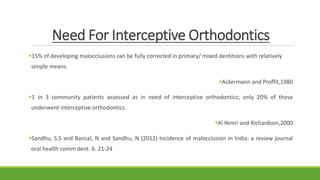
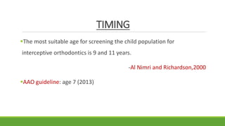
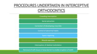
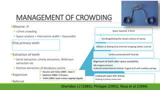
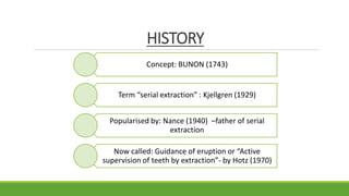
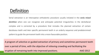
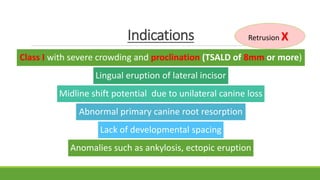
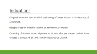
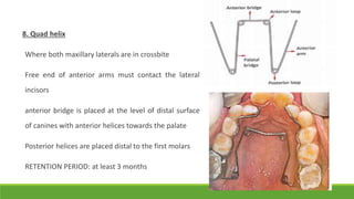
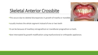
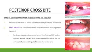
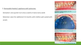
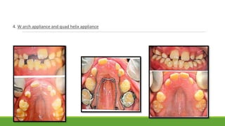
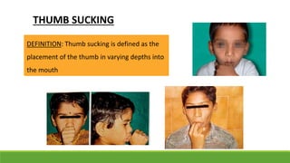
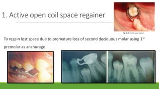
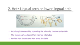
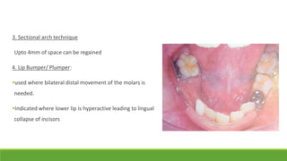
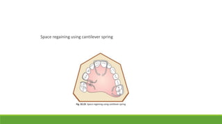
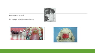
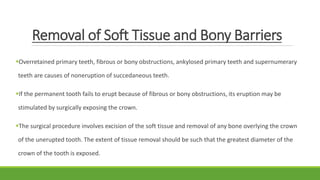
INTERCEPTIVE ORTHODONTICS
BY DR. SHRUTI SUDARSANAN
JUNIOR RESIDENT
DEPT OF PEDIATRIC AND PREVENTIVE DENTISTRY
INTRODUCTION
Early orthodontic intervention procedures are carried out
during the stage of mixed dentition when dentition is
forming, permanent teeth are in various stages of
development and the dental arches, dentoalveolar
processes, jaw bones and facial structures are growing.
DEFINITION
Interceptive orthodontics have been defined as that phase of the science and art of
orthodontics employed to recognize and eliminate potential irregularities and
malpositions in the developing dentofacial complex.
-American Association of Orthodontists (AAO 1969)
Interceptive orthodontics is defined as the action taken to preserve the integrity of
what appears to be normal occlusion at a specific time. -Graber (1966)
BENEFITS AND REASONS FOR EARLY
INTERVENTION
1. Premature loss of primary teeth- can cause a bizarre of
malocclusion. At later stages- complex mechanics required.
2. Early ortho interventions- does not impede normal growth
but guide the developing dentition into normal occlusion
3. Early treatment of habits is simpler than in later stage of life
4. Crowding gets worse if not intervened early
5. Severe rotations of teeth when corrected early are more
stable with less relapse
6. Early management of space in arch: possibility of arch development and
resolve minor crowding
(subjects treated with non-extraction treatment and normal craniofacial
structures show a naturally aesthetic profile.
7. Early occlusal guidance trains the tongue: act like functional appliance and
stimulate normal development of arches
8. Class II malocclusion, often half cusp (3.5mm or less) can be effectively
managed
*favourable growth pattern: low mandibular plane angle and horizontal growth:
more suitable for functional appliance treatment
9. Non compliance of adolescent years can be avoided
10. Early orthodontic treatment offers psychological benefits to children facing
bullying and teasing in school
Need For Interceptive Orthodontics
15% of developing malocclusions can be fully corrected in primary/ mixed dentitions with relatively
simple means.
Ackermann and Proffit,1980
1 in 3 community patients assessed as in need of interceptive orthodontics; only 20% of these
underwent interceptive orthodontics.
Al Nimri and Richardson,2000
Sandhu, S.S and Bansal, N and Sandhu, N (2012) Incidence of malocclusion in India: a review journal
oral health comm dent. 6. 21-24
TIMING
The most suitable age for screening the child population for
interceptive orthodontics is 9 and 11 years.
-Al Nimri and Richardson,2000
AAO guideline: age 7 (2013)
PROCEDURES UNDERTAKEN IN INTERCEPTIVE
ORTHODONTICS
Crowding interception
Serial extraction
Correction of developing cross bite
Control of abnormal habits
Space regaining
Muscle exercises
Interception of skeletal malrelation
Removal of soft tissue or bony barrier to enable eruption of teeth
1. CROWDING
Definition
Crowding is defined as a faulty relationship between the mesiodistal diameter of
teeth, jaw size and arch perimeter. Jaw size determine the available space for
teeth apices, arch perimeter determine the available space for teeth crown
while mesiodistal diameter falls in between them.
-Richardson 1999
Types of Crowding
• Tooth size and arch size discrepancy, with this ratio
being more often increased
• genetically determined
Primary crowding:
• By premature loss of primary molars
• Environmental in origin
Secondary Crowding
• Has both genetic and environmental contributions
• Main determinant: differential late jaw growth
Tertiary or late lower
incisor crowding
RICHTER’S SCALE FOR CROWDING (Clark et al 1977)
Minor
crowding
1-3 mm of
space
deficiency
Moderate
crowding
4-5 mm of
space
deficiency
Severe
crowding
6mm or more
of space
deficiency
LITTLE’S IRREGULARITY INDEX
0 Perfect alignment
l-3 Minimal irregularity
4-6 Moderate irregularity
7-9 Severe irregularity
10 Very severe irregularity
Will crowding resolve on its own?
Interdental spacing
Spacing in deciduous
anteriors: good sign
of alveolar growth
Eruption of
incisors lateral
shift of deciduous
canines create
space in the arch
Intercanine arch
width
Increases 6mm in
maxilla; 4mm in
mandible (from 2yrs
to age of maturity)
Hagberg (1994):
intercanine distance
28mm or more: little
risk of crowding
<26mm: crowding
may be associated
Inclination of
permanent incisors
Forward inclination
of permanent
incisors  increase
in arch
circumference
Tooth size ratio:
b/w permanent and
primary teethh
MANAGEMENT OF CROWDING
Observe: if:
 <2mm crowding
 Space analysis + intercanine width = favourable
Disk primary teeth
Extraction of teeth
 Serial extraction, timely extraction, Wilkinson
extraction etc
 Elective extraction of deciduous canine
Expansion
Referral
Space required: 2-4mm
Grinding/disking the mesial surfaces of canine
169Lbur or disking strip (minimal stripping; better control)
Surface protected with fluoride
Alignment of teeth after space availability
•By tongue pressure
•Laterals locked behind centrals: lingual arch with auxillary springs
Inadequate space after disking:
•Disking of primary molars later
-Sheridan J.J (1985), Philippe (1991), Rosa et al (1994)
2. SERIAL EXTRACTION
HISTORY
Concept: BUNON (1743)
Term “serial extraction” : Kjellgren (1929)
Popularised by: Nance (1940) –father of serial
extraction
Now called: Guidance of eruption or “Active
supervision of teeth by extraction”- by Hotz (1970)
Definition
Serial extraction is an Interceptive orthodontic procedure usually initiated in the early mixed
dentition when one can recognize and anticipate potential irregularities in the dentofacial
complex and is corrected by a procedure that includes the planned extraction of certain
deciduous teeth and later specific permanent teeth in an orderly sequence and predetermined
pattern to guide the permanent teeth into a more favourable position.
A program of selective or guided extraction of primary and sometimes permanent teeth
over a period of time, with the objective of relieving crowding and facilitating the
eruption of remaining teeth into improved positions. -AAO 2012
Indications
Class I with severe crowding and proclination (TSALD of 8mm or more)
Lingual eruption of lateral incisor
Midline shift potential due to unilateral canine loss
Abnormal primary canine root resorption
Lack of developmental spacing
Anomalies such as ankylosis, ectopic eruption
Retrusion X
Contraindications
Mild to moderate crowding (8mm or less)
Congenital absence of teeth providing space
Where extensive caries of permanent first molars requires their removal
deep or open bites
Class II/III malocclusion
Cleft lip and palate
Assessment
Facial photographs
Clinical examination
Occlusion study (models)
X rays- iopa, opg, ceph
Space Analysis For Serial Extraction
Conventional dental oriented
mixed dentition analysis:
Moyers mixed dentition
analysis
A facial oriented analysis,
incorporating the relations of
the incisor teeth to basal bone,
is preferable
Cephelaometric analysis
• Proportional facial analysis
• Steiner classification of facial
pattern
• Total space analysis (Levern
Merrifield)
• Steiner’s: to explain the need to
pts
• Tweed’s: to determine the
permanent tooth extraction
Procedure
1. Dewel’s method: (CD4) Removal of deciduous canine → Removal of deciduous 1st molars →
Removal of erupting 1st premolars.
◦ Usually followed in lower arch
2. Tweed’s method: (D4C) Removal of deciduous 1st molars → Removal of erupting 1st
premolars → Removal of deciduous canine.
◦ Followed in either arch
3. Nance method: (D4C) Removal of deciduous 1st molars → Removal of erupting 1st premolars
→ Removal of deciduous canine.
Advantages:
1. Further appliance therapy is minimised
2. Reduce the complexity of treatment needed
Disadvantages:
1. Serial extraction followed by fixed appliance
therapy in:
1. Deep bite cases
2. Ditching
2. Selectively used in class II (In alveolodental
protrusion used in maxilla only)
3. Pt cooperation affects treatment
4. Caries in 2nd PM may lead to their removal
5. Impacted canine
4. Wilkinson’s Extraction: elective extraction of all Permanent first molars
5. Elective extraction of 2nd molars - Richardson 1992
Timely Extraction- “Stemm”
Sequential removal of deciduous teeth
But no permanent teeth are removed
Indications
Gingival recession due to labial positioning of lower incisors + inadequacy of
arch length
Ectopic eruption of lateral incisors or permanent 1st molars
Crowding of 4mm or more: alignment of incisors after permanent canines have
erupted is difficult  EXTRACTION OF DECIDUOUS CANINE
3. CORRECTION OF
DEVELOPING CROSS BITE
DEFINITION
The maxillary dentition overlaps the mandibular dentition all
through its perimeter in anterior and buccal segment. This
relationship is called overjet. When the relationship is
reversed, it is termed as crossbite
“the best time to treat a crossbite is the first time it is seen”
ANTERIOR CROSSBITE INTERCEPTION
Anterior crossbite can be classified into:
Dento-alveolar anterior crossbite
Skeletal anterior crossbite
Functional anterior crossbite
Dentoalveolar Anterior Crossbite
often manifested as single tooth crossbite and usually occurs due to over retained deciduous
teeth
Treatment
1. Tongue blade therapy
◦ Simple one tooth crossbite
◦ Erupting stage
◦ Using lower tooth as fulcrum, locked tooth can
be pushed out by placing the tongue blade 450
behind the tooth
◦ Used 1-2hrs daily for 10-14 days
2. Lower inclined plane or Catalan’s appliance
Crossbite involving 1 or 2 teeth
Cemented lower acrylic inclined plane
Contoured and polished at 450 angle to long axis of lower incisor teeth
Steeper the angle , greater the force applied
Weekly review to monitor:
◦ More than 2 or 3 weeks: may open bite
◦ Labial movement of maxillary incisors
3. Double cantilever spring :
For single or multiple teeth in crossbite
Activate the springs 1.5-2mm every 2 weeks
24hr wear
For bite opening : posterior bite plane or glass ionomer turbo
on buccal teeth
4. Reverse Stainless steel crown: for single tooth crossbite
5. Composite inclines: to build a composite or compomer incline on the lower teeth
6. Hawley’s appliance using jackscrew:
to correct anterior crossbite involving all anteriors
Activation: quarter turn every day
7. 2 x 4 fixed appliance
8. Quad helix
Where both maxillary laterals are in crossbite
Free end of anterior arms must contact the lateral
incisors
anterior bridge is placed at the level of distal surface
of canines with anterior helices towards the palate
Posterior helices are placed distal to the first molars
RETENTION PERIOD: at least 3 months
Activation
1. extraoralactivation:openingthe helix:
anterior helix:posteriorexpansion(5mm on each molar side)
posteriorhelix:lateral movementof anterior arms anterior
expansion(1.5mm)
2. intraoralactivation:three prong plier:
anteriorexpansion
: posteriorbendis placedon palatal bridge by keepingsingle
prong towards the midline
Posteriorexpansion:anterior bendplace on anterior bridge by
keepingsingle prong anteriorly
Functional Anterior Crossbite
The presence of occlusal prematurities deflects the mandible into
a more forward path of closure. So this type of crossbite results
from the functional shift of the mandible.
These are commonly seen in pseudo Class III type of
malocclusion and are treated by eliminating the occlusal
prematurities (careful incisal grinding)
Skeletal Anterior Crossbite
This occurs due to skeletal discrepancies in growth of maxilla or mandible.
usually involves the whole segment instead of one or two teeth
It can be because of maxillary retrognathism or mandibular prognathism or both.
best intercepted by growth modification using myofunctional or orthopedic appliances.
POSTERIOR CROSS BITE
CAREFUL CLINICAL EXAMINATION AND IDENTIFYING THE ETIOLOGY
1. Occlusal equilibration: to correct crossbite caused by functional interference
2. Cross elastics: for correction of dental unilateral crossbite involving one or
two teeth
◦ Bands are adapted and cemented to teeth involved to which hook or
button is welded. The two teeth are engaged by cross elastic that run
on buccal of upper and lingual of lower molar or vice versa
3. Removable Hawley’s appliance with jackscrew:
Activation: one quarter turn once a week or twice every week
Retention: wear for additional 3-6 months with midline split sealed with
acrylic
4. W arch appliance and quad helix appliance
Skeletal Correction
Rapid maxillary expansion (RME): Removable expansion screw or with HYRAX type of expander
The rate of expansion: 1mm/day divided in two instalments: 2 quarter turns (0.5mm) in the morning and
2 in the evening
Expansion lasts for 7-10 days
Orthopaedic forces cause a mid line split of maxilla: midline diastema: successful RME therapy
5. CONTROL OF ABNORMAL HABITS
Definition:
Boucher OC defined habit as a tendency towards an act or an act that has become
a repeated performance, relatively fixed, consistent, easy to perform and almost
automatic
GRABER (1976): includes all habit under extrinsic factors
of general cause of malocclusion.
1. Thumb/digit sucking
2. Tongue thrusting
3. Lip/nail biting
4. Mouth breathing
5. Abnormal swallow
6. Speech defects
7. Postural defect
8. Psychogenic habits- bruxism
9. Defective occlusal habits
THUMB SUCKING
DEFINITION: Thumb sucking is defined as the
placement of the thumb in varying depths into
the mouth
• Normal Thumb Sucking
• Abnormal Thumb Sucking
• Psychological
• Habitual
• Nutritive sucking habits:
• Breastfeeding, bottle feeding
• Non-nutritive sucking habit:
• Thumb or finger sucking, pacifier
sucking
Classification of Thumb Sucking Habit
Type A:
•50 percent
•whole digit is placed inside the mouth with the pad of the thumb pressing over the
palate, while at the same time maxillary and mandibular oral contact is present.
Type B:
•13 to 24 percent
•thumb is placed into the oral cavity and at the same time maxillary and mandibular
contact is maintained.
Type C:
•18 percent of the children
•thumb is placed into the mouth just beyond the first joint and contacts hard palate and
the maxillary incisors, but there is no contact with mandibular anterior incisors.
Type D:
•6 percent of the children
•only a little portion of the thumb is placed into the mouth.
According to Subtelny (1973)
Theories and Concepts of Thumb Sucking
• Classical Freudian Theory (Sigmund Freud – 1919)
• Oral Drive Theory (Sears and Wise—1982) They suggest that the strength of the oral drive
is in part a function of how long a child continues to feed by sucking. It is not the
frustration of weaning that produces thumb sucking but in fact it is the prolonged nursing
that causes it.
• Rooting Reflex (Benjamin—1962) The rooting reflex is movement of the infant’s head and
tongue towards an object touching its cheeks. He suggested that thumb sucking arises
from the rooting and placing reflexes common to all mammalian infants during the first 3
months of life.
• Sucking Reflex (Ergel—1962) .
• Learning Theory (Davidson—1967)
SPACE REGAINING
If a primary molar is lost early and space maintainers are not used, a reduction in
arch length by mesial migration of the 1st molar is expected. In such cases the
space lost by mesial movement of the 1st molar can be regained by distalizing it.
Active open coil space regainer
Hotz lingual arch or lower lingual arch
sectional fixed appliances
Lip bumper/plumper
Free end loop space regainer
Split saddle/ split block space regainer
Sling shott space regainer
Jack screw or expansion screw
Anterior space regainer
Kloehn head gear
Jones jig/ pendulum appliance
1. Active open coil space regainer
To regain lost space due to premature loss of second deciduous molar using 1st
premolar as anchorage
2. Hotz Lingual arch or lower lingual arch
• Arch length increased by expanding the u loop by 2mm on either side
• The lingual arch posts are then inserted into tubes
• Review after 1 week and then every 4to 6wks
3. Sectional arch technique
Upto 4mm of space can be regained
4. Lip Bumper/ Plumper:
used where bilateral distal movement of the molars is
needed.
Indicated where lower lip is hyperactive leading to lingual
collapse of incisors
5. Free end Loop Space Regainer
Labial arch wire for retention and stability
Uprighting distal driving spring with helical
Activation in 2-3 weeks
Acrylic clearance in the direction and site of distal movement of
tooth
6. Split Saddle/ split block space regainer
Functional part consists of acrylic block
split buccolingually and joined by wire in
the form of buccal and lingual loop
Activated by periodic spreading of
loops.
7. Sling Shot Space Regainer
8. Jack screw
Space regaining using cantilever spring
Kloehn Head Gear
Jones Jig/ Pendulum appliance
MUSCLE EXERCISES
Interception of Skeletal Mal-relations
Removal of Soft Tissue and Bony Barriers
Overretained primary teeth, fibrous or bony obstructions, ankylosed primary teeth and supernumerary
teeth are causes of noneruption of succedaneous teeth.
If the permanent tooth fails to erupt because of fibrous or bony obstructions, its eruption may be
stimulated by surgically exposing the crown.
The surgical procedure involves excision of the soft tissue and removal of any bone overlying the crown
of the unerupted tooth. The extent of tissue removal should be such that the greatest diameter of the
crown of the tooth is exposed.
CONCLUSION
Thorough understanding of the development process of the dentition,
eruption timings and sequence, establishment of occlusion and its
features is fundamental to diagnosis of a case of potential malocclusion
The mixed dentition period is the greatest opportunity for occlusal
guidance and interception of malocclusion.
REFERENCES
1. Graber TM. Orthodontics: Principles and Practice, 3rd Edn. Philadelphia; 1972.
2. Kharbanda OP. Diagnosis and management of malocclusion, 2nd Edn, 2013.
3. Proffit WR. Contemporary orthodontics, 4th Edn, Elsevier publications.
4. S Gowri Shankar. Textbook of Orthodontics, 1st Edn, 2011.
5. Shobha Tondon. Textbook of Pedodontics, 2nd Edn. Paras Medical Publisher; 2009.
6. Nikhil Marwah, textbook Of Pedodontics
7. S.I. Bhalajhi, Orthodontics, The art and science
INTERCEPTIVE ORTHODONTICS

INTERCEPTIVE ORTHODONTICS

  • 1.
    INTERCEPTIVE ORTHODONTICS BY DR.SHRUTI SUDARSANAN JUNIOR RESIDENT DEPT OF PEDIATRIC AND PREVENTIVE DENTISTRY
  • 2.
    INTRODUCTION Early orthodontic interventionprocedures are carried out during the stage of mixed dentition when dentition is forming, permanent teeth are in various stages of development and the dental arches, dentoalveolar processes, jaw bones and facial structures are growing.
  • 3.
    DEFINITION Interceptive orthodontics havebeen defined as that phase of the science and art of orthodontics employed to recognize and eliminate potential irregularities and malpositions in the developing dentofacial complex. -American Association of Orthodontists (AAO 1969) Interceptive orthodontics is defined as the action taken to preserve the integrity of what appears to be normal occlusion at a specific time. -Graber (1966)
  • 4.
    BENEFITS AND REASONSFOR EARLY INTERVENTION 1. Premature loss of primary teeth- can cause a bizarre of malocclusion. At later stages- complex mechanics required. 2. Early ortho interventions- does not impede normal growth but guide the developing dentition into normal occlusion 3. Early treatment of habits is simpler than in later stage of life 4. Crowding gets worse if not intervened early
  • 6.
    5. Severe rotationsof teeth when corrected early are more stable with less relapse
  • 7.
    6. Early managementof space in arch: possibility of arch development and resolve minor crowding (subjects treated with non-extraction treatment and normal craniofacial structures show a naturally aesthetic profile. 7. Early occlusal guidance trains the tongue: act like functional appliance and stimulate normal development of arches
  • 8.
    8. Class IImalocclusion, often half cusp (3.5mm or less) can be effectively managed *favourable growth pattern: low mandibular plane angle and horizontal growth: more suitable for functional appliance treatment 9. Non compliance of adolescent years can be avoided 10. Early orthodontic treatment offers psychological benefits to children facing bullying and teasing in school
  • 9.
    Need For InterceptiveOrthodontics 15% of developing malocclusions can be fully corrected in primary/ mixed dentitions with relatively simple means. Ackermann and Proffit,1980 1 in 3 community patients assessed as in need of interceptive orthodontics; only 20% of these underwent interceptive orthodontics. Al Nimri and Richardson,2000 Sandhu, S.S and Bansal, N and Sandhu, N (2012) Incidence of malocclusion in India: a review journal oral health comm dent. 6. 21-24
  • 10.
    TIMING The most suitableage for screening the child population for interceptive orthodontics is 9 and 11 years. -Al Nimri and Richardson,2000 AAO guideline: age 7 (2013)
  • 11.
    PROCEDURES UNDERTAKEN ININTERCEPTIVE ORTHODONTICS Crowding interception Serial extraction Correction of developing cross bite Control of abnormal habits Space regaining Muscle exercises Interception of skeletal malrelation Removal of soft tissue or bony barrier to enable eruption of teeth
  • 12.
  • 13.
    Definition Crowding is definedas a faulty relationship between the mesiodistal diameter of teeth, jaw size and arch perimeter. Jaw size determine the available space for teeth apices, arch perimeter determine the available space for teeth crown while mesiodistal diameter falls in between them. -Richardson 1999
  • 14.
    Types of Crowding •Tooth size and arch size discrepancy, with this ratio being more often increased • genetically determined Primary crowding: • By premature loss of primary molars • Environmental in origin Secondary Crowding • Has both genetic and environmental contributions • Main determinant: differential late jaw growth Tertiary or late lower incisor crowding
  • 15.
    RICHTER’S SCALE FORCROWDING (Clark et al 1977) Minor crowding 1-3 mm of space deficiency Moderate crowding 4-5 mm of space deficiency Severe crowding 6mm or more of space deficiency
  • 16.
    LITTLE’S IRREGULARITY INDEX 0Perfect alignment l-3 Minimal irregularity 4-6 Moderate irregularity 7-9 Severe irregularity 10 Very severe irregularity
  • 17.
    Will crowding resolveon its own? Interdental spacing Spacing in deciduous anteriors: good sign of alveolar growth Eruption of incisors lateral shift of deciduous canines create space in the arch Intercanine arch width Increases 6mm in maxilla; 4mm in mandible (from 2yrs to age of maturity) Hagberg (1994): intercanine distance 28mm or more: little risk of crowding <26mm: crowding may be associated Inclination of permanent incisors Forward inclination of permanent incisors  increase in arch circumference Tooth size ratio: b/w permanent and primary teethh
  • 18.
    MANAGEMENT OF CROWDING Observe:if:  <2mm crowding  Space analysis + intercanine width = favourable Disk primary teeth Extraction of teeth  Serial extraction, timely extraction, Wilkinson extraction etc  Elective extraction of deciduous canine Expansion Referral Space required: 2-4mm Grinding/disking the mesial surfaces of canine 169Lbur or disking strip (minimal stripping; better control) Surface protected with fluoride Alignment of teeth after space availability •By tongue pressure •Laterals locked behind centrals: lingual arch with auxillary springs Inadequate space after disking: •Disking of primary molars later -Sheridan J.J (1985), Philippe (1991), Rosa et al (1994)
  • 19.
  • 20.
    HISTORY Concept: BUNON (1743) Term“serial extraction” : Kjellgren (1929) Popularised by: Nance (1940) –father of serial extraction Now called: Guidance of eruption or “Active supervision of teeth by extraction”- by Hotz (1970)
  • 21.
    Definition Serial extraction isan Interceptive orthodontic procedure usually initiated in the early mixed dentition when one can recognize and anticipate potential irregularities in the dentofacial complex and is corrected by a procedure that includes the planned extraction of certain deciduous teeth and later specific permanent teeth in an orderly sequence and predetermined pattern to guide the permanent teeth into a more favourable position. A program of selective or guided extraction of primary and sometimes permanent teeth over a period of time, with the objective of relieving crowding and facilitating the eruption of remaining teeth into improved positions. -AAO 2012
  • 22.
    Indications Class I withsevere crowding and proclination (TSALD of 8mm or more) Lingual eruption of lateral incisor Midline shift potential due to unilateral canine loss Abnormal primary canine root resorption Lack of developmental spacing Anomalies such as ankylosis, ectopic eruption Retrusion X
  • 23.
    Contraindications Mild to moderatecrowding (8mm or less) Congenital absence of teeth providing space Where extensive caries of permanent first molars requires their removal deep or open bites Class II/III malocclusion Cleft lip and palate
  • 24.
  • 25.
    Space Analysis ForSerial Extraction Conventional dental oriented mixed dentition analysis: Moyers mixed dentition analysis A facial oriented analysis, incorporating the relations of the incisor teeth to basal bone, is preferable Cephelaometric analysis • Proportional facial analysis • Steiner classification of facial pattern • Total space analysis (Levern Merrifield) • Steiner’s: to explain the need to pts • Tweed’s: to determine the permanent tooth extraction
  • 26.
    Procedure 1. Dewel’s method:(CD4) Removal of deciduous canine → Removal of deciduous 1st molars → Removal of erupting 1st premolars. ◦ Usually followed in lower arch 2. Tweed’s method: (D4C) Removal of deciduous 1st molars → Removal of erupting 1st premolars → Removal of deciduous canine. ◦ Followed in either arch 3. Nance method: (D4C) Removal of deciduous 1st molars → Removal of erupting 1st premolars → Removal of deciduous canine.
  • 29.
    Advantages: 1. Further appliancetherapy is minimised 2. Reduce the complexity of treatment needed Disadvantages: 1. Serial extraction followed by fixed appliance therapy in: 1. Deep bite cases 2. Ditching 2. Selectively used in class II (In alveolodental protrusion used in maxilla only) 3. Pt cooperation affects treatment 4. Caries in 2nd PM may lead to their removal 5. Impacted canine
  • 30.
    4. Wilkinson’s Extraction:elective extraction of all Permanent first molars 5. Elective extraction of 2nd molars - Richardson 1992
  • 31.
    Timely Extraction- “Stemm” Sequentialremoval of deciduous teeth But no permanent teeth are removed
  • 32.
    Indications Gingival recession dueto labial positioning of lower incisors + inadequacy of arch length Ectopic eruption of lateral incisors or permanent 1st molars Crowding of 4mm or more: alignment of incisors after permanent canines have erupted is difficult  EXTRACTION OF DECIDUOUS CANINE
  • 33.
  • 34.
    DEFINITION The maxillary dentitionoverlaps the mandibular dentition all through its perimeter in anterior and buccal segment. This relationship is called overjet. When the relationship is reversed, it is termed as crossbite “the best time to treat a crossbite is the first time it is seen”
  • 35.
    ANTERIOR CROSSBITE INTERCEPTION Anteriorcrossbite can be classified into: Dento-alveolar anterior crossbite Skeletal anterior crossbite Functional anterior crossbite
  • 36.
    Dentoalveolar Anterior Crossbite oftenmanifested as single tooth crossbite and usually occurs due to over retained deciduous teeth
  • 37.
    Treatment 1. Tongue bladetherapy ◦ Simple one tooth crossbite ◦ Erupting stage ◦ Using lower tooth as fulcrum, locked tooth can be pushed out by placing the tongue blade 450 behind the tooth ◦ Used 1-2hrs daily for 10-14 days
  • 38.
    2. Lower inclinedplane or Catalan’s appliance Crossbite involving 1 or 2 teeth Cemented lower acrylic inclined plane Contoured and polished at 450 angle to long axis of lower incisor teeth Steeper the angle , greater the force applied Weekly review to monitor: ◦ More than 2 or 3 weeks: may open bite ◦ Labial movement of maxillary incisors
  • 39.
    3. Double cantileverspring : For single or multiple teeth in crossbite Activate the springs 1.5-2mm every 2 weeks 24hr wear For bite opening : posterior bite plane or glass ionomer turbo on buccal teeth
  • 40.
    4. Reverse Stainlesssteel crown: for single tooth crossbite 5. Composite inclines: to build a composite or compomer incline on the lower teeth
  • 41.
    6. Hawley’s applianceusing jackscrew: to correct anterior crossbite involving all anteriors Activation: quarter turn every day
  • 42.
    7. 2 x4 fixed appliance
  • 43.
    8. Quad helix Whereboth maxillary laterals are in crossbite Free end of anterior arms must contact the lateral incisors anterior bridge is placed at the level of distal surface of canines with anterior helices towards the palate Posterior helices are placed distal to the first molars RETENTION PERIOD: at least 3 months
  • 45.
    Activation 1. extraoralactivation:openingthe helix: anteriorhelix:posteriorexpansion(5mm on each molar side) posteriorhelix:lateral movementof anterior arms anterior expansion(1.5mm) 2. intraoralactivation:three prong plier: anteriorexpansion : posteriorbendis placedon palatal bridge by keepingsingle prong towards the midline Posteriorexpansion:anterior bendplace on anterior bridge by keepingsingle prong anteriorly
  • 46.
    Functional Anterior Crossbite Thepresence of occlusal prematurities deflects the mandible into a more forward path of closure. So this type of crossbite results from the functional shift of the mandible. These are commonly seen in pseudo Class III type of malocclusion and are treated by eliminating the occlusal prematurities (careful incisal grinding)
  • 47.
    Skeletal Anterior Crossbite Thisoccurs due to skeletal discrepancies in growth of maxilla or mandible. usually involves the whole segment instead of one or two teeth It can be because of maxillary retrognathism or mandibular prognathism or both. best intercepted by growth modification using myofunctional or orthopedic appliances.
  • 48.
    POSTERIOR CROSS BITE CAREFULCLINICAL EXAMINATION AND IDENTIFYING THE ETIOLOGY 1. Occlusal equilibration: to correct crossbite caused by functional interference 2. Cross elastics: for correction of dental unilateral crossbite involving one or two teeth ◦ Bands are adapted and cemented to teeth involved to which hook or button is welded. The two teeth are engaged by cross elastic that run on buccal of upper and lingual of lower molar or vice versa
  • 49.
    3. Removable Hawley’sappliance with jackscrew: Activation: one quarter turn once a week or twice every week Retention: wear for additional 3-6 months with midline split sealed with acrylic
  • 50.
    4. W archappliance and quad helix appliance
  • 51.
    Skeletal Correction Rapid maxillaryexpansion (RME): Removable expansion screw or with HYRAX type of expander The rate of expansion: 1mm/day divided in two instalments: 2 quarter turns (0.5mm) in the morning and 2 in the evening Expansion lasts for 7-10 days Orthopaedic forces cause a mid line split of maxilla: midline diastema: successful RME therapy
  • 52.
    5. CONTROL OFABNORMAL HABITS Definition: Boucher OC defined habit as a tendency towards an act or an act that has become a repeated performance, relatively fixed, consistent, easy to perform and almost automatic
  • 54.
    GRABER (1976): includesall habit under extrinsic factors of general cause of malocclusion. 1. Thumb/digit sucking 2. Tongue thrusting 3. Lip/nail biting 4. Mouth breathing 5. Abnormal swallow 6. Speech defects 7. Postural defect 8. Psychogenic habits- bruxism 9. Defective occlusal habits
  • 55.
    THUMB SUCKING DEFINITION: Thumbsucking is defined as the placement of the thumb in varying depths into the mouth
  • 56.
    • Normal ThumbSucking • Abnormal Thumb Sucking • Psychological • Habitual • Nutritive sucking habits: • Breastfeeding, bottle feeding • Non-nutritive sucking habit: • Thumb or finger sucking, pacifier sucking Classification of Thumb Sucking Habit
  • 57.
    Type A: •50 percent •wholedigit is placed inside the mouth with the pad of the thumb pressing over the palate, while at the same time maxillary and mandibular oral contact is present. Type B: •13 to 24 percent •thumb is placed into the oral cavity and at the same time maxillary and mandibular contact is maintained. Type C: •18 percent of the children •thumb is placed into the mouth just beyond the first joint and contacts hard palate and the maxillary incisors, but there is no contact with mandibular anterior incisors. Type D: •6 percent of the children •only a little portion of the thumb is placed into the mouth. According to Subtelny (1973)
  • 58.
    Theories and Conceptsof Thumb Sucking • Classical Freudian Theory (Sigmund Freud – 1919) • Oral Drive Theory (Sears and Wise—1982) They suggest that the strength of the oral drive is in part a function of how long a child continues to feed by sucking. It is not the frustration of weaning that produces thumb sucking but in fact it is the prolonged nursing that causes it. • Rooting Reflex (Benjamin—1962) The rooting reflex is movement of the infant’s head and tongue towards an object touching its cheeks. He suggested that thumb sucking arises from the rooting and placing reflexes common to all mammalian infants during the first 3 months of life. • Sucking Reflex (Ergel—1962) . • Learning Theory (Davidson—1967)
  • 60.
    SPACE REGAINING If aprimary molar is lost early and space maintainers are not used, a reduction in arch length by mesial migration of the 1st molar is expected. In such cases the space lost by mesial movement of the 1st molar can be regained by distalizing it.
  • 61.
    Active open coilspace regainer Hotz lingual arch or lower lingual arch sectional fixed appliances Lip bumper/plumper Free end loop space regainer Split saddle/ split block space regainer Sling shott space regainer Jack screw or expansion screw Anterior space regainer Kloehn head gear Jones jig/ pendulum appliance
  • 62.
    1. Active opencoil space regainer To regain lost space due to premature loss of second deciduous molar using 1st premolar as anchorage
  • 63.
    2. Hotz Lingualarch or lower lingual arch • Arch length increased by expanding the u loop by 2mm on either side • The lingual arch posts are then inserted into tubes • Review after 1 week and then every 4to 6wks
  • 64.
    3. Sectional archtechnique Upto 4mm of space can be regained 4. Lip Bumper/ Plumper: used where bilateral distal movement of the molars is needed. Indicated where lower lip is hyperactive leading to lingual collapse of incisors
  • 65.
    5. Free endLoop Space Regainer Labial arch wire for retention and stability Uprighting distal driving spring with helical Activation in 2-3 weeks Acrylic clearance in the direction and site of distal movement of tooth
  • 66.
    6. Split Saddle/split block space regainer Functional part consists of acrylic block split buccolingually and joined by wire in the form of buccal and lingual loop Activated by periodic spreading of loops.
  • 67.
    7. Sling ShotSpace Regainer
  • 68.
  • 69.
    Space regaining usingcantilever spring
  • 70.
    Kloehn Head Gear JonesJig/ Pendulum appliance
  • 71.
  • 73.
  • 75.
    Removal of SoftTissue and Bony Barriers Overretained primary teeth, fibrous or bony obstructions, ankylosed primary teeth and supernumerary teeth are causes of noneruption of succedaneous teeth. If the permanent tooth fails to erupt because of fibrous or bony obstructions, its eruption may be stimulated by surgically exposing the crown. The surgical procedure involves excision of the soft tissue and removal of any bone overlying the crown of the unerupted tooth. The extent of tissue removal should be such that the greatest diameter of the crown of the tooth is exposed.
  • 76.
    CONCLUSION Thorough understanding ofthe development process of the dentition, eruption timings and sequence, establishment of occlusion and its features is fundamental to diagnosis of a case of potential malocclusion The mixed dentition period is the greatest opportunity for occlusal guidance and interception of malocclusion.
  • 77.
    REFERENCES 1. Graber TM.Orthodontics: Principles and Practice, 3rd Edn. Philadelphia; 1972. 2. Kharbanda OP. Diagnosis and management of malocclusion, 2nd Edn, 2013. 3. Proffit WR. Contemporary orthodontics, 4th Edn, Elsevier publications. 4. S Gowri Shankar. Textbook of Orthodontics, 1st Edn, 2011. 5. Shobha Tondon. Textbook of Pedodontics, 2nd Edn. Paras Medical Publisher; 2009. 6. Nikhil Marwah, textbook Of Pedodontics 7. S.I. Bhalajhi, Orthodontics, The art and science

Editor's Notes

  • #23 Straight profile with harmonious soft tissue pattern
  • #25 Mixed dentition analysis: carey’s analysis in lower arch and arch perimeter analysis in upper arch Opg: eruption status Ceph: skeletal relation
  • #27 Dewel’s method: deciduous canine- utilised for aligning crowded incisors The resorption of canine by a lateral incisor – indication to the same (age 8.5 yrs) Extraction of 1st deciduous molars: at 9.5 yrs : unerupted 1st PM will hv reached ½ the root length: incisor crowding gets resolved (when premolar erupts b4 canine) If canine erupts before pm(esp in lower arch): unfavourable Enucleate 1st pm with the 1st deciduous molar Extract 2nd deciduous molar followed by lingual arch: space for first premolar to move distally giving space for canine: when canine erupts – first pms are removed. Tweed’s method: 8yrs: all deciduous 1st molars removed Extract the 1st premolar along with the deciduous canine when it starts erupting
  • #28 Dewel’s method: deciduous canine- utilised for aligning crowded incisors The resorption of canine by a lateral incisor – indication to the same (age 8.5 yrs) Extraction of 1st deciduous molars: at 9.5 yrs : unerupted 1st PM will hv reached ½ the root length: incisor crowding gets resolved (when premolar erupts b4 canine) If canine erupts before pm(esp in lower arch): unfavourable Enucleate 1st pm with the 1st deciduous molar Extract 2nd deciduous molar followed by lingual arch: space for first premolar to move distally giving space for canine: when canine erupts – first pms are removed.
  • #29 Tweed’s method: 8yrs: all deciduous 1st molars removed Deciduous canine are maintained to hamper the eruption of permanent canine Extract the 1st premolar along with the deciduous canine when it starts erupting Advantages and disadvantages: Shobha tandon and balajhi
  • #30 Ditching: distoaxial inclination of canines and mesioaxial inclination of 2nd pM
  • #42 Schedule by Timms: Upto 15 yrsL 90 degree rotation in mrng and evening Over 15 yrs: 45 degree activation 4 tyms a day Schedule by zimring and isaacson: Growing pt 2 turns each day for 4-5 days, later 1 turn per day Non growing pts: 2 turns for 1-2 days and 1 turn per day for 5-7 days nd 1 turn every alternate day
  • #43 2 molar bands and 4 incisor bonded brackets: best choice for somewhat older mixed dentition patient with crowding, rotation and crossbite
  • #46 Posterior expansion: anterior bend place on anterior bridge by keeping single prong anteriorly
  • #49 1. Closing and opening of mandible is observed for functional shift and if found should be located and marked
  • #52 Diastema will spontaneously close with recoiling of stretched supracrestal fibres after expansion is stopped -children might experience pain in the nasal bone area
  • #53 THUMB SUCKING: presence upto 2 ½ to 3 years is normal Mouth breathing: obstructive: nasal obstruction: nasal polyp,tumor, chronic inflammatory conditions, dns Habitual- where mouth breathing persists as habit after removal of nasal obstruction -anatomic
  • #54 Useful Habits Should include all those habits of normal function such as correct tongue position proper respiration and deglutition. Harmful Habits All those that exert perverted stress against the teeth and dental arches, e.g. mouth breathing, tongue thrusting. Compulsive Habit Acquired as a fixation in the child to the extent that he retreats to the practice whenever his security is threatened. Noncompulsive Habit Children appear to undergo continuing behavior modification, which permit them to release certain undesirable habit patterns and form new ones which are socially accepted. Secondary habit is a habit that is due to a supplemental problem, e.g. large tongue causes tongue thrusting habit. Meaningful Habit Habit with a deep-rooted psychological problem. Empty Habit Meaningless habit that can be treated easily by a dentist using reminder therapy
  • #57 Normal Thumb Sucking The thumb sucking habit is considered normal during the first one and half years of life. Such a habit is usually seen to disappear as the child matures Abnormal Thumb Sucking When thumb sucking habit persists beyond the pre school period then it could be considered as an abnormal habit. If Psychological The habit may have a deep-rooted emotional factor involved and may be associated with neglect and loneliness experienced by the child. Habitual: The habit does not have a psychological bearing, however the child performs the act
  • #59 Classical Freudian Theory (Sigmund Freud – 1919) The psychoanalytic theory has proposed that a child goes through various distinct phases of psychological development. In oral phase, it is believed that the mouth is the erogenous zone. During this phase the child takes anything and everything to the oral cavity. It is believed that any kind of the deprivation of this activity will probably cause an emotionally inThe process of sucking is a reflex occurring in the oral stage of development and is seen even at 29 weeks of intrauterine life and may disappear during normal growth between the ages of 1 to 3½ years. It is the first coordinated muscular activity of the infant. Babies who are restricted from sucking due to disease or other factors become restless and irritable. This deprivation may motivate the infant to suck the thumb and finger for additional gratificationsecure individual. Sucking reflex:The process of sucking is a reflex occurring in the oral stage of development and is seen even at 29 weeks of intrauterine life and may disappear during normal growth between the ages of 1 to 3½ years. It is the first coordinated muscular activity of the infant. Babies who are restricted from sucking due to disease or other factors become restless and irritable. This deprivation may motivate the infant to suck the thumb and finger for additional gratification Learning Theory (Davidson—1967) This theory advocates that non-nutritive sucking stems from an adaptive response. The infant associates sucking with feelings like pleasure and hunger and recalls these events by sucking the suitable objects available, which is mainly thumb or finger
  • #62 R/G and study models needed: MOYERS MIXED DENTITION ANALYSIS: ESTIMATE OF AMOUNT TO BE REGAINED
  • #63 Molar banding round molar tube of 0.036’’ soldered or spot welded both buccally and lingually alginate impressioncast pored ss wire 0.036’’  bend into u shape following contour of distal surface of 1st pm… ends passing through buccal and lingual tubes 2-3mm beyond distal end of tube ss open coil 2-3mm longer than distance from anterior stop to molar tube Review after 1week  check for mobility of anchor teeth Review after 4-6 weeks: till molar is upright
  • #65 The lip plumper should be away by 2-3mm from inciosors Pressure exerted from lip is transferred on to the molars to upright them Lingually collapsed lower incisoors once relieved from lip pressure will tend to upright with pressure from tongue.
  • #68 Sling shot space regainer distalizes molar with a wire elastic holder with hooks as an alternative to spring which transmits a force against the tooth to be distalized. It is termed as sling shot appliance, as the forces to distalize tooth were produced by the elastic which was stretched on the middle of the lingual and the buccal surface of the molar to be moved. The child places new elastic between the hooks while the appliance is outside the mouth. It is slipped into place then the child’s fi ngers can guide the elastic into proper position. If the appliance is of a removable type, periodic checking should be done to evaluate whether the patient is using it or not, whether there is any distortion or breakage of the appliance or irritation of soft tissues. If the teeth are emerging underneath the appliance, the portion of the acrylic is cut off to give way for the teeth to erupt into position. In case of fi xed appliances, check for any breakage of the appliance at the soldered joints or band material. It is also checked that whether the appliance is loose due to dissolution of cement which may result in food lodgement and caries. The appliance is removed every 6 months or 1-year depending on the situation and the abutment tooth is checked for any caries or decalcifi cation. Polishing of the abutment is done followed by fl uoride application. Then the appliance is recemented in position. Regular radiographic examination of developing permanent teeth is also necessary. The appliance can be removed or discarded soon after the succedaneous teeth erupted into proper position in the oral cavity
  • #71 Ussr:unilateral spring space regainer